. 2025 Jan 29;31(1):28.
doi: 10.1186/s10020-025-01068-x.

Myricetin exposure reduces PC differentiation in vitro in primary human B cells

Affiliations

Myricetin exposure reduces PC differentiation in vitro in primary human B cells

Shabirul Haque et al. Mol Med. .

Abstract

Background: The process of B cell activation and plasma cell (PC) formation involves morphological, transcriptional, and metabolic changes in the B cell. Blocking or reducing PC differentiation is one approach to treat autoimmune diseases that are characterized by the presence of pathogenic autoantibodies. Recent studies have suggested the potential of myricetin, a natural flavonoid with anti-inflammatory and antioxidant properties, to block or reduce PC differentiation.

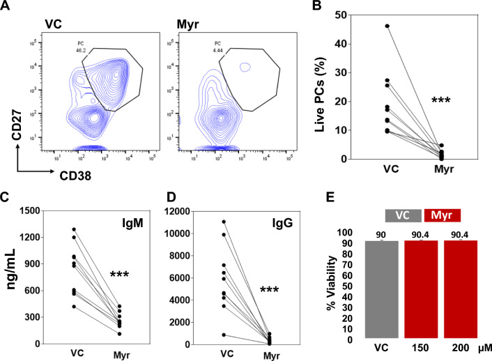
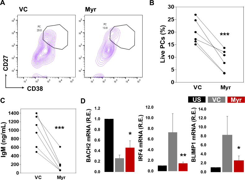
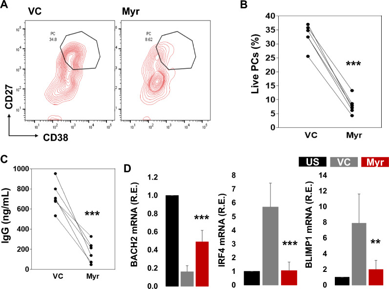
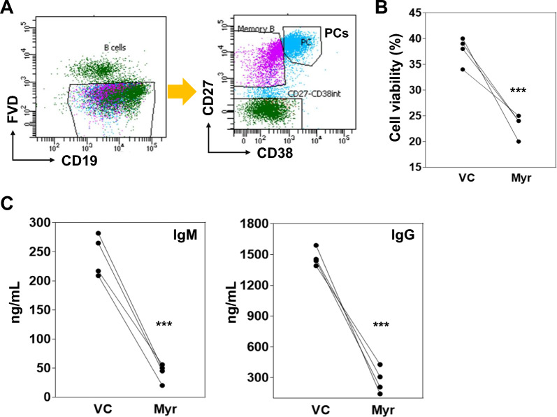
Methods: Primary human B cells were purified by using a human B cell isolation kit. B cell subsets such as IgG memory B cells, marginal zone B cells (MZ B cells), and naive B cells were isolated by flow cytometry and activated to induce PC differentiation. Quantification of PCs (CD27 + + , CD38 +) was obtained by flow cytometry. The expression of mRNA was measured by qPCR. Ig secretion in culture supernatant was measured by ELISA.

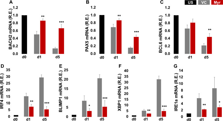
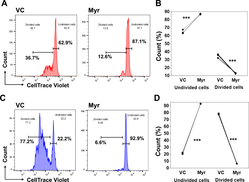
Results: Myricetin treatment significantly reduced PC differentiation in primary human B cells and all B cell subsets. Myricetin exposure reduced Ig production both IgM and IgG, in culture supernatants at day 5. Myricetin treatment led to augmented BACH2 expression and reduced IRF4, BLIMP1, and XBP1 expression compared to control cultures.

Conclusion: Myricetin treatment reduced PC differentiation and Ig secretion by primary human B cells. Targeting B cells in this way may be a therapeutic approach for some autoimmune diseases.

Keywords: B cell differentiation; Flavonoids; Myricetin; Plasma cells; SLE.


Conflict of interest statement

Declarations. Ethics approval and consent to participate: Not applicable. Consent for publication: All authors approved the final manuscript and the submission to this journal. Competing interests: Shabirul Haque PhD, is associated with Molecular Medicine and serving as Associate editor and Managing editor. Betty Diamond, MD is associated with Molecular Medicine as Editor-in-Chief.

Figures

Fig. 1
Fig. 1
Myricetin reduces PC differentiation in primary human B cells. A Total B cells (n = 10 healthy donors) were activated with R848, IL-2, IL-10, IL-21 in the presence of myricetin (200 µM) or VC. On day 5, B cells were harvested and stained to detect PCs. B Cumulative data was plotted from B cells of all the donors. C and D IgM and IgG concentration was determined in day 5 culture supernatants by ELISA. E Effect of myricetin (150, and 200 µM) on cell viability was determined by cell counter in day 1 culture. P value was calculated between VC and Myr. ***p < 0.001
Fig. 2
Fig. 2
Myricetin led to sustained BACH2 expression, and diminished the increase in IRF4, BLIMP1, and XBP1 expression in primary human B cells. Total B cells (n = 3 healthy donors) were activated with R848, IL-2, IL-10, IL-21 in the presence of myricetin (200 µM) or VC. Total RNA was isolated from B cells at d0 (unstimulated t = 0), day 1 culture and day 5 culture as indicated. Relative gene expression of BACH2 (A), PAX5 (B), BCL6 (C), IRF4 (D), BLIMP1 (E), XBP1 (F), and IRE1α (G) was evaluated by qPCR. *p < 0.05, **p < 0.01, ***p < 0.001
Fig. 3
Fig. 3
Myricetin reduces PCs in CD40L, CpG, IL10, IL21 activated B cells. A Total B cells (n = 10 healthy donors) were activated with CD40L, CpG, IL-10, and IL-21 in the presence of myricetin (200 µM) and vehicle control (VC) and cultured for PC differentiation. On day 5, cells were harvested and stained with CD19, CD27, and CD38 for live PC evaluation. B Overall, data was plotted from all the donors. C IgM and IgG concentration was estimated in day 5 culture supernatants by ELISA. D PC differentiation associated genes (BACH2, IRF4, and BLIMP1) expression was determined by qPCR. Relative expression was calculated using unstimulated (US) as control (black bar), VC (gray bar) and Myr (red bar). P value was calculated between VC and Myr. P value (*p < 0.05, **p < 0.01, ***p < 0.001)
Fig. 4
Fig. 4
Myricetin exposure reduced PC differentiation in naive B cells. A Naive B cells (n = 9 healthy donors) were activated with IL-21, and CD40L expressing feeder cells and treated with myricetin (200 µM) or VC. On day 6, cells were harvested, and live PCs were evaluated by flow cytometry. B Cumulative data was plotted from all the donors. C IgM and IgG concentration was quantified in day 6 culture supernatants by ELISA. D PC related gene expression detection, naive B cells were activated with R848, anti-IgM, IL-2, IL-10, and IL-21 for 24 h and treated with Myr (200 µM) or VC, and gene (BACH2, IRF4, and BLIMP1) expression was quantified by qPCR. P value VC vs. Myr, **p < 0.01, ***p < 0.001
Fig. 5
Fig. 5
Myricetin exposure reduced PC differentiation in MZ B cells. A MZ B cells (n = 6 healthy donors) were activated with R848, IL-2, IL-10, IL-21 and treated with myricetin (200 µM) or VC and cultured. On day 5, cells were harvested, and live PCs were evaluated by flow cytometer. B Cumulative data was plotted from all the donors. C IgM concentration was quantified in day 5 culture supernatants by ELISA. D To study PC differentiation related gene expression, MZ B cells were activated with only R848 for 24 h then relative gene expression of BACH2, IRF4, and BLIMP1 was quantified by qPCR. Relative expression was calculated using unstimulated (US) as control sample (black bar) vs. VC (gray bar) and Myr (red bar). P value *p < 0.05, **p < 0.01, ***p < 0.001
Fig. 6
Fig. 6
Myricetin treatment reduced PC differentiation in IgG memory B cells. A IgG memory B cells (n = 6 healthy donors) were activated with R848, IL2, IL-10, and IL-21 and treated with myricetin (200 µM) or VC. On day 5, cells were harvested and stained with CD19, CD27, CD38, and live dead dye, measured by flow cytometer. B Cumulative data was plotted. C IgG concentration was quantified within day 5 culture supernatants by ELISA. D IgG memory B cells were activated with R848, IL-10, and IL-21 for 24 h and gene (BACH2, IRF4, and BLIMP1) expression was evaluated by qPCR. Relative expression was calculated using unstimulated (US) as control sample (black bar) vs. VC (gray bar) and Myr (red bar). **p < 0.01, ***p < 0.001
Fig. 7
Fig. 7
Myricetin treatment reduced B cell proliferation. B cells (n = 3 healthy donors) were labelled with CellTrace Violet. B cells were activated with R848, IL2, IL-10, and IL-21 and treated with myricetin (200 µM) or VC. B cells were harvested on day 3 & 5 and stained with FVD eF660. A and B shows cell proliferation status of day 3 cultured B cells. C and D shows cell proliferation status of day 5 cultured B cells. B and D shows cumulative data plotted from undivided and divided B cells from day 3 & 5 culture respectively. P value ***p < 0.001
Fig. 8
Fig. 8
Myricetin treatment reduced Ig secretion by PCs. B cells (n = 4 healthy donors) were activated with R848, IL2, IL-10, and IL-21 for PC differentiation. A On day 5, PCs were sorted per gating strategy after staining with CD19, CD27, CD38, and live dead dye. B Sorted PCs were cultured in fresh culture medium containing stimulants and treated with either VC or myricetin (200 µM). Cell viability was measured after 24 h of treatment. C IgM and IgG concentration was quantified in culture supernatants of 24 h of treated conditions. P value was calculated between VC and myricetin group. **p < 0.01, ***p < 0.001

References

    1. Atisha-Fregoso Y, Toz B, Diamond B. Meant to B: B cells as a therapeutic target in systemic lupus erythematosus. J Clin Investig. 2021. 10.1172/JCI149095. - PMC - PubMed
    1. Benhamron S, et al. Regulated IRE1-dependent decay participates in curtailing immunoglobulin secretion from plasma cells. Eur J Immunol. 2014;44:867–76. - PubMed
    1. Büchter C, et al. Myricetin-mediated lifespan extension in Caenorhabditis elegans is modulated by DAF-16. Int J Mol Sci. 2013;14:11895–914. - PMC - PubMed
    1. Cho BO, et al. Anti-inflammatory activity of myricetin from Diospyros lotus through suppression of NF-κB and STAT1 activation and Nrf2-mediated HO-1 induction in lipopolysaccharide-stimulated RAW264.7 macrophages. Biosci Biotechnol Biochem. 2016;80:1520–30. - PubMed
    1. Finney J, Kelsoe G. Continuous culture of mouse primary B lymphocytes by forced expression of Bach2. J Immunol. 2021;207:1478–92. - PMC - PubMed

LinkOut - more resources