. 2025 Mar;11(2):e70234.
doi: 10.1002/vms3.70234.

Computed Tomographic Anatomy of the Head in Cockatiel (Nymphicus hollandicus)

Affiliations

Computed Tomographic Anatomy of the Head in Cockatiel (Nymphicus hollandicus)

Seyedmehran Kazemi et al. Vet Med Sci. 2025 Mar.

Abstract

Background: Nowadays, computed tomography (CT) scanning is one of the most practical and precise diagnostic imaging methods that can be utilized to evaluate the head in birds.

Objectives: This study aimed to present the normal anatomical data of the head of the cockatiel (Nymphicus hollandicus) using the CT method. In this research, the features of this bird's head were investigated in terms of bones, joints, muscles, sinuses and other constituent tissues.

Methods: The current retrospective cross-sectional study used carcasses of six adult cockatiels (Nymphicus hollandicus) (three males and three females) with an average age of 1-3 years and an average weight of 75-110 g. After preparing the CT images, the head of each parrot underwent gross anatomy studies.

Results: Based on the results, reconstructed CT images could identify most structures of the cockatiel (Nymphicus hollandicus) head. Parietal, mandible, occiput, maxillary, premaxillary, palatine, pterygoid, quadrate, temporal bones, epithelial membranes, external ear canal and bony labyrinth, ossicles and entoglossal bones, different parts of the infraorbital sinus, brain hemispheres and various parts of the eyeball and conchae of the nasal cavities were examined in CT images. The results related to the CT evaluation and anatomical examination of the cockatiel (Nymphicus hollandicus) head demonstrated a high correlation.

Conclusion: The results of this research can be employed as a reference and a suitable atlas for identifying anatomical features, examining different species of the cockatiel (Nymphicus hollandicus), teaching anatomy and interpreting CT scan images, as well as performing clinical examinations and treating this type of parrot.

Keywords: anatomy; cockatiel (Nymphicus hollandicus); computed tomography; head.


Conflict of interest statement

The authors declare no conflicts of interest.

Figures

FIGURE 1
FIGURE 1
Representative photographs of anatomic cross sections of the adult cockatiel (Nymphicus hollandicus) head. (a) Level of the eye, (b) level of the external acoustic meatus in the dorsal plane, (c) level of the rostral border of the orbital fossa and (d) level of the external acoustic meatus in the transverse plane. (a) (1) Rhamphotheca, (2) premaxilla bone, (3) maxilla bone, (4) left nasal cavity, (5) caudal nasal concha, (6) infraorbital sinus, (7) eye, (8) brain hemispheres, (9) falx cerebri, (10) occipital bone, (11) temporal bone. (b) (1) Rhamphotheca, (2) premaxilla bone, (3) palatine bone, (4) ethmomandibularis muscle, (5) pterygoideus muscle, (6) cerebellum, (7) bony labyrinth, (8) external acoustic meatus, (9) caudal nasal concha, (10) occipital bone. (c) (1) Fronto‐parietal bone, (2) eye, (3) infraorbital sinus, (4) pterygoideus muscle, (5) ethmomandibularis muscle, (6) hard palate, (7) eye, (8) caudal nasal concha, (9) lingual process of hyoid bone, (10) tongue, (11) choanal cleft, (12) mandible. (d) (1) Cerebrum, (2) falx cerebri, (3) occipital bone, (4) brain stem, (5) chiasma optic, (6) external acoustic meatus, (7) ethmomandibularis muscle, (8) pterygoideus muscle, (9) mandible, (10) hard palate, (11) lingual process of the hyoid bone. L, left; R, right.
FIGURE 2
FIGURE 2
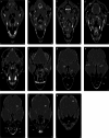
(a–n) Transverse computed tomography reconstruction images in the lateral plane of the normal skull of the cockatiel (Nymphicus hollandicus) from the rostrum to the caudal extremity of the nasal cavity. (1) Rostral diverticulum septum, (2) premaxillary bone, (3) rostral diverticulum, (4) paraglossum, (5) bony part of nasal septum, (6) mandible bone (pneumonized), (7) palate bone opening, (8) rostral nasal concha, (9) transverse canal, (10) maxillary process of palatal bone, (11) tongue, (12) middle nasal turbinate, (13) basal layer of middle nasal turbinate, (14) nasal cavity, (15) cartilaginous part of nasal septum, (16) nasopharyngeal airway, (17) lateral border of palatine bone, (18) periorbital process of infraorbital sinus, (19) jugal part of infraorbital sinus, (20) jugal arch, (21) glottis, (22) laryngeal protrusion, (23) arytenoid cartilages, (24) bronchial horn, (25) trachea, (26) choana of palatal bone, (27) ethmomandibular muscle, (28) periorbital part of the infraorbital sinus, (29) caudal nasal turbinate, (30) infraorbital sinus foramen, (31) infraorbital part of the infraorbital sinus, (32) eyeball, (33) epithelial membrane, (34) tracheal cartilage ring, (35) infraorbital septum, (36) cricoid cartilage, (37) procricoid cartilage, (38) scleral ossicles, (39) suborbital arch, (40) frontal bone (pneumonized), (41) pterygoid and quadrate muscles, (42) larynx, (43) zygomatic process of the squamosal bone, (44) quadrate bone (pneumatized), (45) quadrature part of infraorbital sinus, (46) postorbital part of infraorbital sinus, (47) external acoustic meatus, (48) cervicocephalic diverticulum, (49) brain stem, (50) bony labyrinth. L, left; R, right.
FIGURE 3
FIGURE 3
(a–f) Sagittal computed tomography reconstruction images (lateromedial plane) of the normal skull of the cockatiel (Nymphicus hollandicus) from the right aspect of the head to the inter‐orbital and nasal septum. (1) Scleral bones, (2) suborbital arch, (3) postorbital part of the infraorbital sinus, (4) quadrate bone (pneumonized), (5) external ear foramen, (6) mandible bone, (7) cervicocephalic diverticulum, (8) occipital bones (pneumonized), (9) infraorbital part of infraorbital sinus, (10) periorbital process, (11) epithelial membrane, (12) jugal portion of infraorbital sinus, (13) cervical vertebrae, (14) trachea, (15) encephalon of the brain, (16) caudal nasal turbinate, (17) middle nasal turbinate, (18) rostral nasal turbinate, (19) transverse canal, (20) premaxillary bone (pneumonized), (21) palate bone (pneumonized), (22) rostral diverticulum, (23) frontal bone (pneumonized), (24) nasal cavity, (25) nasopharyngeal airway, (26) larynx, (27) paraglossum, (28) basihyal, (29) bony part of nasal septum, (30) infraorbital septum, (31) nostril, (32) tracheal rings, (33) cartilaginous part of nasal septum, (34) tongue. Ca, caudal; Cr, cranial.
FIGURE 4
FIGURE 4
(a–k) Dorsal computed tomography reconstruction images (ventrodorsal plan) of a normal skull of the cockatiel (Nymphicus hollandicus) from the dorsal aspect of the skull to the larynx. (1) Basihyal, (2) arytenoid cartilage, (3) quadrature part of infraorbital sinus, (4) epithelial membrane, (5) quadrate bone, (6) larynx, (7) glottis, (8) pharynx, (9) pterygoid and quadrate muscle, (10) mandibular appendage, (11) oral cavity, (12) external acoustic meatus, (13) paraglossum, (14) pterygoid bone, (15) external ear foramen, (16) bony labyrinth, (17) jugal arch, (18) postorbital part of infraorbital sinus, (19) occipital bones (pneumonized), (20) suborbital arch, (21) palate bone, (22) ethmomandibular muscle, (23) infraorbital part of the infraorbital sinus, (24) nasopharyngeal canal, (26) scleral ossicles, (27) infraorbital septum, (28) cartilaginous part of the nasal septum, (29) palate foramen, (30) middle nasal turbinate, (31) rostral diverticulum, (32) cranial foramen of eyeball, (33) preorbital part of the infraorbital sinus, (34) rostral nasal turbinate, (35) nasal cavity, (36) bony part of nasal sinus, (37) infraorbital sinus foramen, (38) encephalon, (39) craniofacial flexion, (40) frontal bone. L, left; R, right.

Similar articles

Cited by

References

    1. Al‐Rubaie, N. I. , and Kadhim K.. 2023. “Anatomical Comparison of the Nasal Cavity in Adult Male and Female Cockatiel (Nymphicus hollandicus).” Acta Biomedica 94: 2023719. 10.23750/abm.v94i3.13443. - DOI
    1. Benedict, L. , Charles A., Brockington A., and Dahlin C. R.. 2022. “A Survey of Vocal Mimicry in Companion Parrots.” Scientific Reports 12: 202. 10.1038/s41598-022-24335-x. - DOI - PMC - PubMed
    1. Brühschwein, A. , Klever J., Wilkinson T., and Meyer‐Lindenberg A.. 2018. “DICOM Standard Conformance in Veterinary Medicine in Germany: A Survey of Imaging Studies in Referral Cases.” Journal of Digital Imaging 31: 13–18. 10.1007/s10278-017-9998-x. - DOI - PMC - PubMed
    1. Carril, J. , Tambussi C. P., Degrange F. J., Benitez Saldivar M. J., and Picasso M. B. J.. 2016. “Comparative Brain Morphology of Neotropical Parrots (Aves, Psittaciformes) Inferred From Virtual 3D Endocasts.” Journal of Anatomy 229: 239–251. 10.1111/joa.12325. - DOI - PMC - PubMed
    1. Carril, J. , Tambussi C. P., and Rasskin‐Gutman D.. 2021. “The Network Ontogeny of the Parrot: Altriciality, Dynamic Skeletal Assemblages, and the Avian Body Plan.” Evolutionary Biology 48: 41–53. 10.1007/s11692-020-09522-w. - DOI

Publication types

Grants and funding