

Trends in dentomaxillofacial radiology
- PMID: 39876885
- PMCID: PMC11755909
- DOI: 10.4329/wjr.v17.i1.97255
Trends in dentomaxillofacial radiology
Abstract
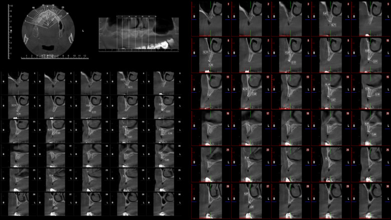
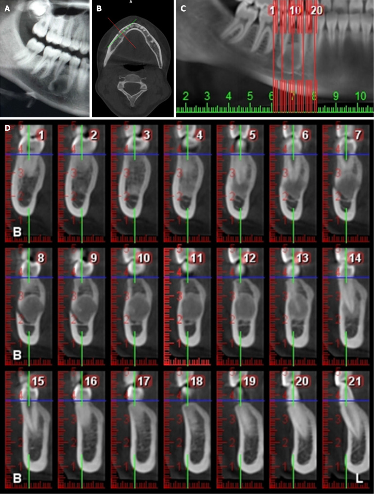
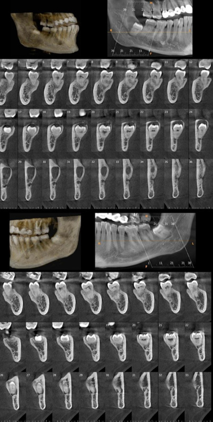
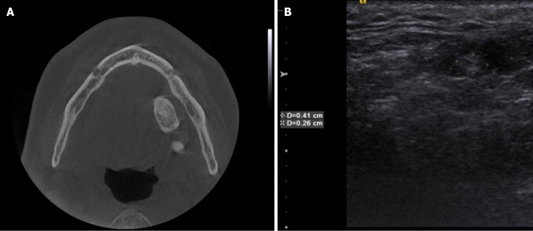
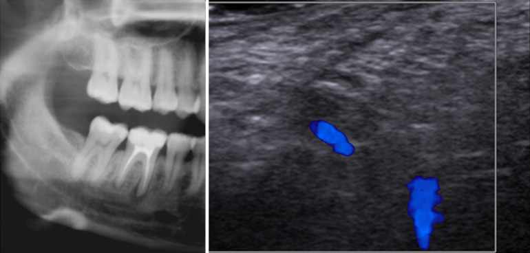
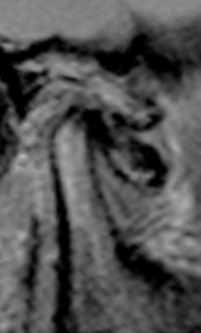
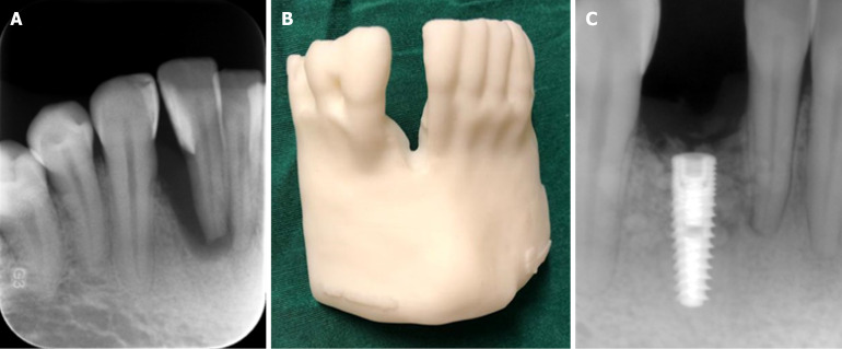
Oral and maxillofacial diagnostic imaging is of paramount importance in dental clinical diagnosis, treatment planning, and follow-up procedures. Periapical radiographic examination and numerous panoramic systems are used in routine clinical dental practice. Cone beam CT is widely used and currently the method of choice in oral and maxillofacial implantology, endodontics, maxillofacial surgery, periodontics, degenerative temporomandibular joint disease, orthodontics, airway studies, sleep disorders, and forensic dentistry. Another innovative laboratory research tool that offers three-dimensional (3D) detailed high-resolution images of in vitro teeth and neighboring structures with submicrometric accuracy is microcomputed tomography. Ultra-high radiation doses, long scanning times, and high costs preclude its routine clinical use. In response to the high demand for a technique that could provide real-time images using a cost-effective, rapid, user-friendly, and portable technique without ionizing radiation, some authors proposed ultrasound imaging methods as an alternative to X-ray imaging techniques. Ultrasonography can be used in the dentomaxillofacial region for various diagnostic purposes such as salivary gland and superficial tissue examination. Recently, dedicated dental magnetic resonance imaging with appropriate software, hardware, sequences, and field of view tailored to fit dentomaxillofacial anatomy was introduced. Lately, 3D printing technologies and their application in dentistry has attracted attention. During 3D printing a given material is added in successive layers to create a 3D object. The application of this technology has the potential to decrease operation time and minimize operator bias and the possibility of procedural errors. Another hot topic regarding dentomaxillofacial radiology is artificial intelligence, which is a field related to computer science dedicated to developing systems or machines that can perform tasks traditionally associated with human intelligence. It is obvious that further investigation and research in the field of dentomaxillofacial radiology will make great contributions to diagnostic imaging for various dental specialties.
Keywords: Artificial intelligence; Cone beam CT; Dental microcomputed tomography; Dentomaxillofacial radiology; Diagnostic imaging; Microcomputed tomography; Three-dimensional printing; Ultrasonography.
©The Author(s) 2025. Published by Baishideng Publishing Group Inc. All rights reserved.
Conflict of interest statement
Conflict-of-interest statement: The author reports no relevant conflicts of interest for this article.
Figures
References
-
- İncebeyaz B, Balkan EP, Deniz HA, Isayev A, Kamburoğlu K. Comparison of Dental Intraoral Digital Imaging Systems Using Simulation Phantoms. J Sci Tech Res. 2023;53
-
- Wenzel A. Bitewing and digital bitewing radiography for detection of caries lesions. J Dent Res. 2004;83:C72–C75. - PubMed
-
- Arai Y, Tammisalo E, Iwai K, Hashimoto K, Shinoda K. Development of a compact computed tomographic apparatus for dental use. Dentomaxillofac Radiol. 1999;28:245–248. - PubMed
Publication types
LinkOut - more resources
Full Text Sources
