. 2024 Nov 6;68(Spec Issue):e240085.
doi: 10.20945/2359-4292-2024-0085. eCollection 2024.

Prenatal exposure to nitrate alters uterine morphology and gene expression in adult female F1 generation rats

Affiliations

Prenatal exposure to nitrate alters uterine morphology and gene expression in adult female F1 generation rats

André Gilberto Cassiani et al. Arch Endocrinol Metab. .

Abstract

Objective: Nitrate is ubiquitously found in the environment and is one of the main components of nitrogen fertilizers. Previous studies have shown that nitrate disrupts the reproductive system in aquatic animals, but no study has evaluated the impact of nitrate exposure on the uterus in mammals. This study aimed to evaluate the impact of maternal exposure to nitrate during the prenatal period on uterine morphology and gene expression in adult female F1 rats.

Materials and methods: Pregnant Wistar rats were either treated with sodium nitrate 20 mg/L or 50 mg/L dissolved in drinking water from the first day of pregnancy until the birth of the offspring or were left untreated. On postnatal day 90, the uteri of female offspring rats were collected for histological and gene expression analyses. Morphometric analyses of the uterine photomicrographs were performed to determine the thickness of the layers of the uterine wall (endometrium, myometrium, and perimetrium) and the number of endometrial glands.

Results: The highest nitrate dose increased the myometrial thickness of the exposed female rats. Treatment with both nitrate doses reduced the number of endometrial glands compared with no treatment. Additionally, nitrate treatment significantly increased the expression of estrogen receptors and reduced the expression of progesterone receptors in the uterus.

Conclusion: Our results strongly suggest that prenatal exposure to nitrate programs gene expression and alters the uterine morphology in female F1 rats, potentially increasing their susceptibility to developing uterine diseases during adulthood.

Keywords: DOHaD; female rats; nitrate; uterus.


Conflict of interest statement

Disclosure: no potential conflict of interest relevant to this article was reported.

Figures
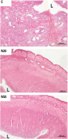
Figure 1
Figure 1. Histological sections of the uterine walls of adult female offspring rats exposed (N20 and N50) and not exposed (C) to nitrate during the prenatal period. Magnification: 100x. Hematoxylin and Eosin. Abbreviations: C, control group; L, uterine lumen; N20, nitrate 20 mg/L group; N50, nitrate 50 mg/L group.
Figure 2
Figure 2. Uterine wall thickness in female rats exposed or not exposed to nitrate during the prenatal period. Photomicrographs of histological slides were used to measure uterine thickness, obtained with a 5x objective, ensuring the visualization of the entire section. All values are expressed in micrometers (μm). The white markers within the box plots represent the median values, while the whiskers indicate the range of the data. *** P < 0.001 compared with the C group. Abbreviations: C, control group; N20, nitrate 20 mg/L group; N50, nitrate 50 mg/L group.
Figure 3
Figure 3. Myometrial and endometrial thickness of rats’ uteri exposed or not exposed to nitrate during the prenatal period. The measurement of myometrial (A) and endometrial (B) thickness was performed using photomicrographs of histological slides obtained with a 5x objective, ensuring the visualization of the entire section. All values are expressed in micrometers (μm). The white and black markers within the box plots represent the median values, while the whiskers indicate the range of the data. *** P < 0.001 compared with the C group. Abbreviations: C, control group; N20, nitrate 20 mg/L group; N50, nitrate 50 mg/L group.
Figure 4
Figure 4. Number of endometrial glands in female rats exposed or not exposed to nitrate during the prenatal period. The number of endometrial glands was measured in photomicrographs obtained using a 5x objective. The white markers within the box plots represent the median values, while the whiskers indicate the range of the data. *** P < 0.001 compared with the C group. Abbreviations: C, control group; N20, nitrate 20 mg/L group; N50, nitrate 50 mg/L group.
Figure 5
Figure 5. Gene expression of estrogen and progesterone receptors in uteri of rats exposed or not exposed to nitrate. The mRNA expression of (A) progesterone receptor (Pgr), (B) estrogen alpha receptor (Esr1), and (C) estrogen beta receptor (Esr2) was determined using real-time polymerase chain reaction (PCR), with Rpl19 mRNA serving as the internal control. The data are presented as mean ± standard deviation in arbitrary units (AU) (n = 4-6). The black markers within the box plots represent the mean values, while the whiskers indicate the range of thse data. * P < 0.05; *** p < 0.001 versus the C group. Abbreviations: C, control group; N20, nitrate 20 mg/L group; N50, nitrate 50 mg/L group.

References

    1. Kabir ER, Rahman MS, Rahman I. A review on endocrine disruptors and their possible impacts on human health. Environ Toxicol Pharmacol. 2015 Jul;40(1):241–258. doi: 10.1016/j.etap.2015.06.009. - DOI - PubMed
    1. Diamanti-Kandarakis E, Bourguignon JP, Giudice LC, Hauser R, Prins GS, Soto AM, et al. Endocrine-disrupting chemicals: an Endocrine Society scientific statement. Endocr Rev. 2009 Jun;30(4):293–342. doi: 10.1210/er.2009-0002. - DOI - PMC - PubMed
    1. Gonsioroski A, Mourikes VE, Flaws JA. Endocrine Disruptors in Water and Their Effects on the Reproductive System. Int J Mol Sci. 2020 Mar 12;21(6):1929–1929. doi: 10.3390/ijms21061929. - DOI - PMC - PubMed
    1. Beszterda M, Frański R. Endocrine disruptor compounds in environment: As a danger for children health. Pediatr Endocrinol Diabetes Metab. 2018;24(2):88–95. doi: 10.18544/PEDM-24.02.0107. - DOI - PubMed
    1. Gore AC, Martien KM, Gagnidze K, Pfaff D. Implications of prenatal steroid perturbations for neurodevelopment, behavior, and autism. Endocr Rev. 2014 Dec;35(6):961–991. doi: 10.1210/er.2013-1122. - DOI - PMC - PubMed

LinkOut - more resources