. 2025 Jan 17:31:e20240050.
doi: 10.1590/1678-9199-JVATITD-2024-0050. eCollection 2025.

Histopathological characterization of skin and muscle lesions induced by lionfish (Pterois volitans) venom in a murine experimental model

Affiliations

Histopathological characterization of skin and muscle lesions induced by lionfish (Pterois volitans) venom in a murine experimental model

Cecilia Díaz et al. J Venom Anim Toxins Incl Trop Dis. .

Abstract

Background: Fish venoms have been poorly characterized and the available information about their composition suggests they are uncomplicated secretions that, combined with epidermal mucus, could induce an inflammatory reaction, excruciating pain, and, in some cases, local tissue injuries.

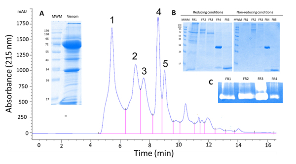
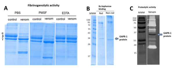
Methods: In this study, we characterized the 24-hour histopathological effects of lionfish venom in a mouse experimental model by testing the main fractions obtained by size exclusion-HPLC. By partial proteomics analysis, we also correlated these in vivo effects with the presence of some potentially toxic venom components.

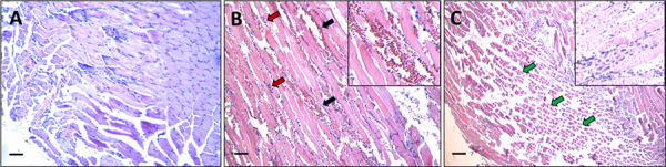
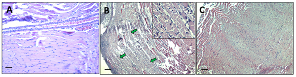
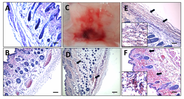
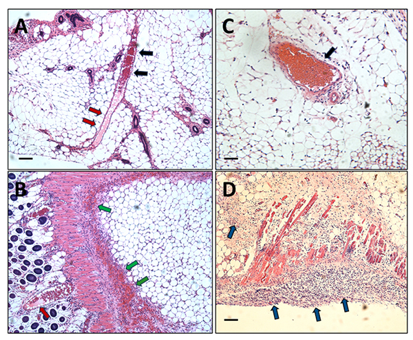
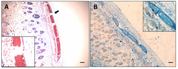
Results: We observed a strong lesion on the skin and evident necrosis in the skeletal muscle. None of the tissue-damaging effects were induced by the fraction containing cytolysins, membrane pore-forming toxins ubiquitously present in species of scorpionfish, stonefish, and lionfish, among others. On the contrary, injuries were associated with the presence of other components, which have remained practically ignored so far. This is the case of an abundant protein, present in venom, with homology to a Golgi-associated plant pathogenic protein 1-like (GAPR1), which belongs to the same protein superfamily as venom CRISPs and insect allergens.

Conclusion: This GAPR1-like protein and the hyaluronidase are probably responsible for the hemostasis impairment and hemorrhagic lesions observed in mouse skin, whereas muscle injuries can be indirectly caused by a combination of inflammatory and hemorrhagic events. More information is required to establish the components accountable for the myonecrotic effect.

Keywords: GAPR1; hyaluronidase; lionfish; myonecrosis; skin lesion; venom.


Conflict of interest statement

Competing interests: The authors declare that they have no competing interests.

Figures

Figure 1.
Figure 1.. Lionfish and collecting location. (A) Lionfish collected specimens, before removing dorsal spines and their utilization for venom extraction and characterization of the in vivo effects on mice. (B) Map of Costa Rica with the location of the Caribbean coast in the Gandoca-Manzanillo Wildlife Refuge (Province of Limón), where lionfish were collected. The image was modified from Google Maps.
Figure 2.
Figure 2.. Tissue damage of mice gastrocnemius muscle caused by venom after 24 hours. (A) Longitudinal section of control mouse gastrocnemius injected with distilled water. (B, C) Longitudinal sections of mouse gastrocnemius injected with venom extract showing alterations such as hemorrhage (black arrows), abundant inflammatory infiltrate (red arrows), and severe myonecrosis (green arrows). Figure insets show an area of amplification of micrographs B and C that evidences the toxicity provoked by the venom on mouse gastrocnemius. Scale bars represent 100 µm.
Figure 3.
Figure 3.. Size exclusion-HPLC of lionfish dorsal spine extract (including epidermal mucus), protein pattern, and hyaluronidase activity. SEC-HPLC chromatogram of the lionfish venom showing the main fractions (numbered from 1 to 5). (A) Figure inset: SDS-PAGE (under reducing conditions) of crude spine venom. (B) Figure inset: SDS-PAGE (under reducing and non-reducing conditions) of the crude venom extract's first five chromatographic fractions, FR1 to FR5. (C) Inset showing a zymography on hyaluronic acid of the first four fractions.
Figure 4.
Figure 4.. Tissue damage of mice gastrocnemius muscle induced by fraction 1 after 24 hours. (A) Longitudinal section of control muscle of mice injected with water. (B) Longitudinal section of mouse gastrocnemius injected with fraction 1 displaying severe damage of muscle fibers (green arrows), extensive leukocyte infiltrate, and almost absent hemorrhage. (C) In contrast, the mouse gastrocnemius muscle injected with fraction 2 showed no evidence of tissue injury. Figure inset in B shows an area of amplification that evidences the myofibrils fragmentation induced by fraction 1 in mouse gastrocnemius. Scale bars represent 100 µm.
Figure 5.
Figure 5.. Mouse skin lesions and hemorrhage caused by fractions 3, 4, and 5 after 24 hours. (A) Control skin of mice injected i.d. with water. (B) Skin of mice injected i.d. with fraction 2, where no hemorrhage was observed, only the presence of inflammatory infiltrate. (C) Macroscopic lesion observed in the skin of a mouse injected i.d. with fraction 3, similar to the ones observed with fractions 4 and 5 (not shown). (D) Skin of mice injected i.d. with fraction 3, (E) fraction 4, and (F) fraction 5 showing extensive hemorrhage (black arrows). Figure insets show an area of amplification of micrographs E and F that evidences the presence of erythrocytes outside the blood vessels and the inflammatory cells in the skin of mice injected with fractions 4 and 5, respectively. Scale bars represent 100 µm.
Figure 6.
Figure 6.. Red blood cell aggregation, presence of fibrinoid-like material in blood vessels, and inflammatory infiltrate in the skin of mice injected with fraction 4 and purified ~34 kDa GAPR1-like protein. (A) Skin of mice injected i.d. with fraction 4 showing capillary erythrocyte aggregation (black arrows), a hyaline material inside (red arrows), and (B) extensive hemorrhage (green arrows). (C) Skin of mice injected i.d. with purified GAPR1-like protein, showing the same pattern of red blood cell aggregation (black arrow) and (D) the presence of significant leukocyte infiltration (blue arrows). Scale bars represent 50 µm.
Figure 7.
Figure 7.. Skin of mice injected i.d. with fraction 3 and stained for the presence of fibrin with Martius, Scarlet, and Blue staining (MSB). (A) A clear pattern of red blood cell aggregation is observed in the hematoxylin and eosin staining and the rupture of the blood vessel basal membrane (black arrow) and erythrocyte extravasation. The inset shows a higher-power picture of red blood cell agglutination towards the capillary wall. Also, the morphology of the erythrocytes looks altered. (B) A hyaline substance present in the vessels stains blue (not red as expected for new fibrin deposits). Erythrocytes are stained in yellow color. Insets show an amplification of the same blood vessels. Blue staining of the hyaline material (normally observed for collagen staining) could correspond to pseudo-collagen materials, such as mature fibrin or amyloids. Scale bars represent 50 µm.
Figure 8.
Figure 8.. In vitro fibrinogenolytic activity induced by lionfish venom and ~34 kDa GAPR1-like protein characterization of potential proteolytic activity. (A) Fibrinogen was incubated with the extract for 3 hours in the absence or presence of two protease inhibitors (PMSF and EDTA). PMSF acts as a serine proteinase inhibitor, and EDTA is a cation-chelating agent that inhibits metalloproteinases. Fibrinogen β-chain is completely degraded, and the effect was only inhibited by EDTA. (B) Venom was separated by affinity chromatography in a Benzamidine-Sepharose column evidencing that, like serine proteases, GAPR1-like protein displays affinity to benzamidine. (C) Zymography using gelatin as a substrate to determine proteolytic activity of GAPR1-like protein, showing negative results.

Similar articles

References

    1. Smith WL, Stern JH, Girard MG, Davis MP. Evolution of venomous cartilaginous and ray-finned fishes. Integr Comp Biol. 2016;56(5):950–961. doi: 10.1093/icb/icw070. - DOI - PubMed
    1. Harris RJ. The piscine arsenal: an updated review of venomous fishes. Rev Fish Biol Fisheries. 2024;34(2):539–574. doi: 10.1007/s11160-023-09828-w.. - DOI
    1. Smith WL, Wheeler WC. Venom evolution widespread in fishes: a phylogenetic road map for the bioprospecting of piscine venoms. J Hered. 2006;97(3):206–217. doi: 10.1093/jhered/esj034.. - DOI - PubMed
    1. Warrell DA. Venomous bites, stings, and poisoning: An Update. Infect. Dis Clin North Am. 2019;33(1):17–38. doi: 10.1016/j.idc.2018.10.001.. - DOI - PubMed
    1. Ramos AD, Conceição K, Silva PI, Jr., Richardson M., Lima C, Lopes-Ferreira M. Specialization of the sting venom and skin mucus of Cathorops spixii reveals functional diversification of the toxins. Toxicon. 2012;59(6):651–665. doi: 10.1016/j.toxicon.2012.02.002.. - DOI - PubMed

LinkOut - more resources