. 2025 Jan 14:15:1522381.
doi: 10.3389/fimmu.2024.1522381. eCollection 2024.

Inflammatory microglia correlate with impaired oligodendrocyte maturation in multiple sclerosis

Affiliations

Inflammatory microglia correlate with impaired oligodendrocyte maturation in multiple sclerosis

J Q Alida Chen et al. Front Immunol. .

Abstract

Introduction: Remyelination of demyelinated axons can occur as an endogenous repair mechanism in multiple sclerosis (MS), but its efficacy varies between both MS individuals and lesions. The molecular and cellular mechanisms that drive remyelination remain poorly understood. Here, we studied the relation between microglia activation and remyelination activity in MS.

Methods: We correlated regenerative (CD163+) and inflammatory (iNOS+) microglia with BCAS1+ oligodendrocytes, subdivided into early-stage (<3 processes) and late-stage (≥3 processes) cells in brain donors with high or low remyelinating potential in remyelinated lesions and active lesions with ramified/amoeboid (non-foamy) or foamy microglia. A cohort of MS donors categorized as efficiently remyelinating donors (ERDs; n=25) or poorly remyelinating donors (PRDs; n=17) was included, based on their proportion of remyelinated lesions at autopsy.

Results and discussion: We hypothesized more CD163+ microglia and BCAS1+ oligodendrocytes in remyelinated and active non-foamy lesions from ERDs and more iNOS+ microglia with fewer BCAS1+ oligodendrocytes in active foamy lesions from PRDs. For CD163+ microglia, however, no differences were observed between MS lesions and MS donor groups. In line with our hypothesis, we found that INOS+ microglia were significantly increased in PRDs compared to ERDs within remyelinated lesions. MS lesions, more late-stage BCAS1+ oligodendrocytes were detected in active lesions with non-foamy or foamy microglia in comparison with remyelinated lesions. Although no differences were found for early-stage BCAS1+ oligodendrocytes between MS lesions, we did find significantly more early-stage BCAS1+ oligodendrocytes in PRDs vs ERDs in remyelinated lesions. Interestingly, a positive correlation was identified between iNOS+ microglia and the presence of early-stage BCAS1+ oligodendrocytes. These findings suggest that impaired maturation of early-stage BCAS1+ oligodendrocytes, encountering inflammatory microglia, may underlie remyelination deficits and unsuccessful lesion repair in MS.

Keywords: inflammation; microglia; multiple sclerosis; oligodendrocytes; remyelination.


Conflict of interest statement

The authors declare that the research was conducted in the absence of any commercial or financial relationships that could be construed as a potential conflict of interest. The author(s) declared that they were an editorial board member of Frontiers, at the time of submission. This had no impact on the peer review process and the final decision.

Figures
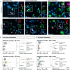
Figure 1
Figure 1
Microglia activation state comparisons in MS lesion types and donor groups. (A) Immunofluorescent double-stained images of CD163+ (regenerative) and iNOS+ (pro-inflammatory) with IBA1+ (microglia) in a remyelinated lesion, active non-foamy lesion, and active foamy lesion. Scale bar indicates 30 µm. Arrows indicate double-positive cells. Asterisks indicate CD163-IBA1+ or iNOS-IBA1+ cells. (B) Histological quantification of CD163+IBA1+ and iNOS+IBA1+ cells in remyelinated lesions, active non-foamy lesions, and active foamy lesions. (C) Histological quantification of CD163+IBA1+ and iNOS+IBA1+ cells per mm2 compared between ERDs and PRDs. Statistics were performed using negative binomial GLM test with Tukey’s post-hoc correction to compare between groups. Estimated marginal means reflect predicted mean of statistical model, with adjustment for multiple datapoints (lesions) per donor. *p<0.05; **p<0.01; ***p<0.001. N.s., not significant.
Figure 2
Figure 2
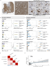
Early- and late-stage BCAS1+ cell comparisons in MS lesions and donors and association with microglia activation states. (A) Immunohistochemical image of BCAS1 staining in an active non-foamy lesion, and classification of early-stage versus late-stage BCAS1+ oligodendrocytes. White arrows indicate BCAS1+ cells. Scale bar in overview image indicates 150 µm. Scale bar of early- and late-stage BCAS+ oligodendrocyte classification indicates 30 µm. (B) Histological quantification of total BCAS1+, early-stage BCAS1+, and late-stage BCAS1+ cells per mm2 in remyelinated, active non-foamy, and active foamy lesions, and (C) compared between ERDs and PRDs. (D) Correlations between the number of early-stage BCAS1+ oligodendrocytes, late-stage BCAS1+ oligodendrocytes, CD163+IBA1+ microglia, and iNOS+IBA1+ microglia per mm2 shows (E) a positive correlation between iNOS+IBA1+ microglia per mm2 and early-stage BCAS1+ oligodendrocytes. Statistics were performed using negative binomial GLM test with Tukey’s post-hoc correction to compare between groups. Estimated marginal means reflect predicted mean of statistical model, with adjustment for multiple datapoints (lesions) per donor. Correlations were tested using Pearson’s correlation coefficient with Benjamini-Hochberg for multiple testing correction. *p<0.05; **p<0.01; ***p<0.001. N.s., not significant.

References

    1. Franklin RJM, Simons M. CNS remyelination and inflammation: From basic mechanisms to therapeutic opportunities. Neuron. (2022) 110:3549–65. doi: 10.1016/j.neuron.2022.09.023 - DOI - PubMed
    1. Chari DM. Remyelination in multiple sclerosis. Int Rev Neurobiol. (2007) 79:589–620. doi: 10.1016/S0074-7742(07)79026-8 - DOI - PMC - PubMed
    1. Lubetzki C, Zalc B, Williams A, Stadelmann C, Stankoff B. Remyelination in multiple sclerosis: from basic science to clinical translation. Lancet Neurol. (2020) 19:678–88. doi: 10.1016/S1474-4422(20)30140-X - DOI - PubMed
    1. Patrikios P, Stadelmann C, Kutzelnigg A, Rauschka H, Schmidbauer M, Laursen H, et al. . Remyelination is extensive in a subset of multiple sclerosis patients. Brain. (2006) 129:3165–72. doi: 10.1093/brain/awl217 - DOI - PubMed
    1. Lassmann H, Lucchinetti C. Remyelination multiple sclerosis. Multiple Sclerosis J. (1997) 3(2):133–8. doi: 10.1177/135245859700300213 - DOI - PubMed

MeSH terms