

Advantages and challenges of using arterial spin labelling MRI to monitor cerebral blood flow in multi-centre clinical trials of neurodegenerative disease: Experience from the RADAR study
- PMID: 39877419
- PMCID: PMC11773049
- DOI: 10.1016/j.cccb.2024.100376
Advantages and challenges of using arterial spin labelling MRI to monitor cerebral blood flow in multi-centre clinical trials of neurodegenerative disease: Experience from the RADAR study
Abstract
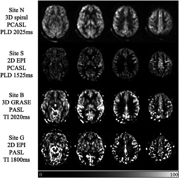
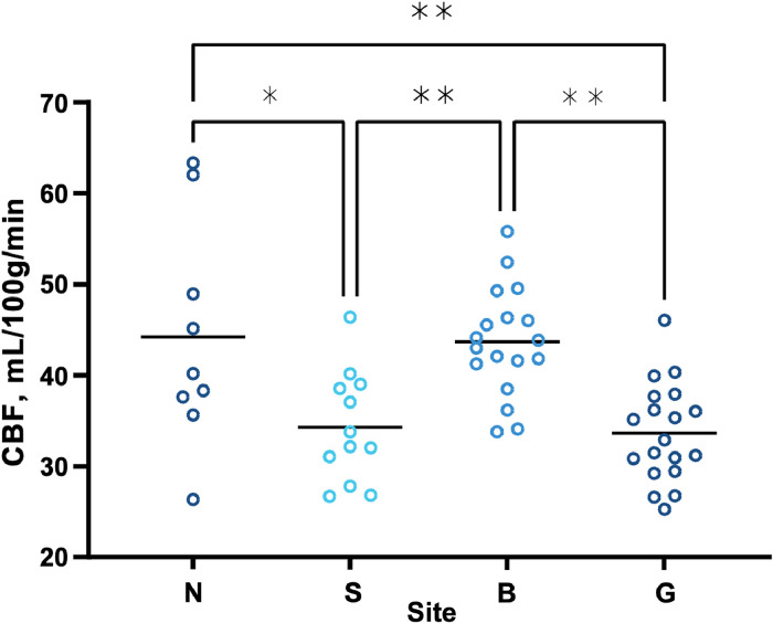
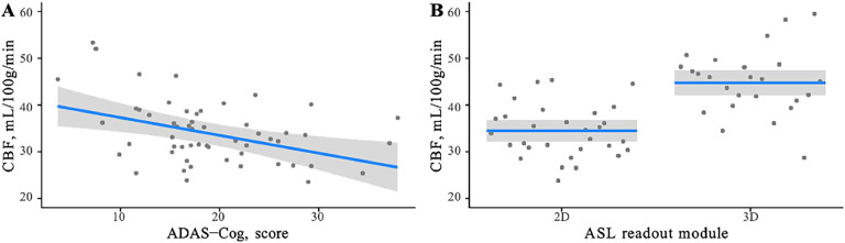
Arterial spin labelling (ASL) enables non-invasive quantification of regional brain perfusion using MRI. ASL was used in the Reducing Pathology in Alzheimer's Disease through Angiotensin TaRgeting (RADAR) multi-centre trial to pilot the assessment of the effects of the anti-hypertension drug losartan on cerebral blood flow (CBF). In the multi-centre setting, disparities in ASL implementation on scanners from different manufacturers lead to inherent differences in measured CBF and its associated parameters (e.g. spatial coefficient of variation (sCoV) of CBF, a proxy of arterial arrival times). In addition, differences in ASL acquisition parameter settings can also affect the measured quantitative perfusion values. In this study, we used data from the RADAR cohort as a case study to evaluate the site-dependent systematic differences of CBF and sCoV, and show that variations in the readout module (2D or 3D) and the post-labelling delay acquisition parameter introduced artifactual group differences. When accounting for this effect in data analysis, we show that it is still possible to combine ASL data across sites to observe the expected relationships between grey matter CBF and cognitive scores. In summary, ASL can provide useful information relating to CBF difference in multi-centre therapeutic trials, but care must be taken in data analysis to account for the inevitable inter-site differences in scanner type and acquisition protocol.
Keywords: Alzheimer's disease; Angiotensin; Arterial spin labelling; Blood pressure; Cerebral blood flow; Hypertension; MRI; Multi-centre randomised controlled trials.
© 2025 The Authors. Published by Elsevier B.V.
Conflict of interest statement
The authors declare the following financial interests/personal relationships which may be considered as potential competing interests: Patrick G Kehoe reports financial support was provided by Efficacy and Mechanism Evaluation Programme (NIHR). Henk-Jan Mutsaerts reports financial support was provided by Horizon Europe. Henk-Jan Mutsaerts and Jan Petr report financial support was provided by eScience Open eScience Call (OEC). Henk-Jan Mutsaerts and Jan Petr report financial support was provided by Dutch Heart Foundation. Henk-Jan Mutsaerts and Jan Petr report financial support was provided by Joint Program Neurodegenerative Disease (JPND). Henk-Jan Mutsaerts reports financial support was provided by Eurostars. If there are other authors, they declare that they have no known competing financial interests or personal relationships that could have appeared to influence the work reported in this paper.
Figures

Similar articles
-
Arterial spin labelling reveals prolonged arterial arrival time in idiopathic Parkinson's disease.Neuroimage Clin. 2014 Aug 1;6:1-8. doi: 10.1016/j.nicl.2014.07.014. eCollection 2014. Neuroimage Clin. 2014. PMID: 25379411 Free PMC article.
-
Longitudinal relation between blood pressure, antihypertensive use and cerebral blood flow, using arterial spin labelling MRI.J Cereb Blood Flow Metab. 2021 Jul;41(7):1756-1766. doi: 10.1177/0271678X20966975. Epub 2020 Dec 16. J Cereb Blood Flow Metab. 2021. PMID: 33325767 Free PMC article.
-
Arterial spin labelling MRI for brain tumour surveillance: do we really need cerebral blood flow maps?Eur Radiol. 2023 Nov;33(11):8005-8013. doi: 10.1007/s00330-023-10099-z. Epub 2023 Aug 11. Eur Radiol. 2023. PMID: 37566264 Free PMC article.
-
Advances in arterial spin labelling MRI methods for measuring perfusion and collateral flow.J Cereb Blood Flow Metab. 2018 Sep;38(9):1461-1480. doi: 10.1177/0271678X17713434. Epub 2017 Jun 9. J Cereb Blood Flow Metab. 2018. PMID: 28598243 Free PMC article. Review.
-
Arterial Spin Labeling (ASL) in Neuroradiological Diagnostics - Methodological Overview and Use Cases.Rofo. 2024 Jan;196(1):36-51. doi: 10.1055/a-2119-5574. Epub 2023 Jul 19. Rofo. 2024. PMID: 37467779 Review. English.
Cited by
-
Advantages and challenges of using arterial spin labelling MRI to monitor cerebral blood flow in multi-centre clinical trials of neurodegenerative disease: Comment.Cereb Circ Cogn Behav. 2025 Mar 18;8:100382. doi: 10.1016/j.cccb.2025.100382. eCollection 2025. Cereb Circ Cogn Behav. 2025. PMID: 40224382 Free PMC article.
References
-
- Global dementia observatory (GDO): world health organisation; 2020 [Available from: https://www.who.int/data/gho/data/themes/global-dementia-observatory-gdo.
-
- van Dyck C.H., Swanson C.J., Aisen P., Bateman R.J., Chen C., Gee M., et al. Lecanemab in early Alzheimer's disease. N. Engl. J. Med. 2023;388(1):9–21. - PubMed
-
- Launer L.J., Ross G.W., Petrovitch H., Masaki K., Foley D., White L.R., et al. Midlife blood pressure and dementia: the Honolulu-Asia aging study. Neurobiol. Aging. 2000;21(1):49–55. - PubMed
-
- Skoog I., Gustafson D. Update on hypertension and Alzheimer's disease. Neurol. Res. 2006;28(6):605–611. - PubMed
Grants and funding
LinkOut - more resources
Full Text Sources
