

Undesirable occupants of bone marrow creating a menace: A 4.5-year audit from a tertiary care centre in Eastern India
- PMID: 39877504
- PMCID: PMC11770638
- DOI: 10.60787/nmj.v65i6.574
Undesirable occupants of bone marrow creating a menace: A 4.5-year audit from a tertiary care centre in Eastern India
Abstract
Background: Bone marrow (BM) in addition to being the origin of primary hematological malignancies is also commonly involved in metastatic solid tumors. Bone marrow examination includes aspiration and biopsy, and it is a well-known procedure not only to diagnose hematological malignancies but also for staging and prognosis of various solid tumors. The presence of metastasis in the bone marrow is of grave prognostic significance and it is imperative to rule out marrow involvement in any malignancy where curative treatment is considered. The study's objectives were to evaluate the clinical, hematological, and biochemical characteristics of patients with BM metastases of solid tumors diagnosed by bone marrow (BM) aspiration and trephine biopsy and to find out the accuracy rate of diagnosing metastatic infiltration between bone marrow aspiration, trephine imprints, and trephine biopsy procedures.
Methodology: It was a 4.5-year retrospective hospital-based observational study where relevant clinical, biochemical, and hematological parameters including bone marrow aspirate and biopsy were analyzed and compiled from hospital medical records.
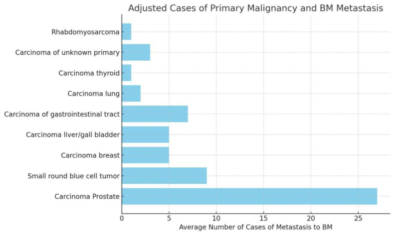
Results: The total number of BMA and trephine biopsies that came during the duration of 4.5 years were 3850 and 2980 respectively. Out of the 3850-bone marrow aspiration and 2980 trephine biopsies received in the dept of Hematology, 305 cases were referred to look for metastatic bone marrow infiltration. Out of these 305 cases, 69 cases showed the presence of metastatic deposits (12.6%). 45 patients (65.2%) were males, and 24 patients (34.7%) were females with M:F ratio of 1.8:1. Most common age group was 51-60 years (31.8%). The most common complaints were fever, body aches, weight loss, and weakness. Clinical examination revealed pallor in 38 out of 69 cases (55%) and organomegaly in 14 cases (20.2%). Microcytic hypochromic anemia (26%) was the most common finding on peripheral blood smear examination followed by pancytopenia (18.8%). The biochemical findings most commonly observed were raised LDH (60.8%), serum PSA (36.3%), and alkaline phosphatase (21.7%).
Conclusion: Trephine biopsy is a sensitive method for detecting marrow metastasis and should be done in all cases being investigated for this purpose. BMA alone may miss marrow metastases in almost half of cases. Trephine imprint cytology is more sensitive than BMA and can provide rapid diagnoses while waiting for trephine biopsy results.
Keywords: Bone Marrow Aspiration; Bone Marrow Imprint Smear; Bone Marrow Trephine Biopsy; Metastasis; Solid Tumors.
Copyright © 2024 Nigerian Medical Association.
Conflict of interest statement
none
Figures


Similar articles
-
Bone Marrow Metastasis of Solid Tumors: A Study of 174 Cases Over 2 Decades from a Single Institution in India.Indian J Hematol Blood Transfus. 2022 Jan;38(1):8-14. doi: 10.1007/s12288-021-01418-9. Epub 2021 Mar 6. Indian J Hematol Blood Transfus. 2022. PMID: 35125707 Free PMC article.
-
An Extensive Analysis and Comparison of Bone Marrow Aspiration and Bone Marrow Trephine Biopsy at a Tertiary Care Hospital in Jharkhand for Various Hematological and Non-hematological Illnesses.Cureus. 2024 Jun 19;16(6):e62661. doi: 10.7759/cureus.62661. eCollection 2024 Jun. Cureus. 2024. PMID: 39036242 Free PMC article.
-
Comparison of bone marrow aspirate cytology, touch imprint cytology and trephine biopsy for bone marrow evaluation.Hematol Rep. 2011 Oct 19;3(3):e22. doi: 10.4081/hr.2011.e22. Epub 2011 Oct 21. Hematol Rep. 2011. PMID: 22593814 Free PMC article.
-
Cytomorphological evaluation of non-haematopoietic malignancies metastasizing to the bone marrow.Am J Blood Res. 2023 Feb 15;13(1):1-11. eCollection 2023. Am J Blood Res. 2023. PMID: 36937461 Free PMC article. Review.
-
Flow Cytometric Analysis of Mechanically Disaggregated Bone Marrow Trephine Biopsies.Cytometry B Clin Cytom. 2018 Nov;94(6):935-940. doi: 10.1002/cyto.b.21646. Epub 2018 Oct 22. Cytometry B Clin Cytom. 2018. PMID: 30350400 Review.
References
-
- Bussard KM, Gay CV, Mastro AM. The bone microenvironment in metastasis; what is special about bone? Cancer Metastasis Rev 2008; 27:41–55. - PubMed
-
- Yu HH, Tsai YY, Hoffe SE. Overview of diagnosis and management of metastatic disease to bone. Cancer Control 2012; 19:84–91. - PubMed
-
- Kołda A, Helbig G, Kopińska A, Wichary R, Pająk J, Kyrcz- Krzemień S, Metastasis of solid tumors into bone marrow – Single center experience. Acta Haematol Pol 2017;48(20:130-134.10.1016/j.achaem.2017.05.001. - DOI
LinkOut - more resources
Full Text Sources
Research Materials
Miscellaneous