Review
. 2025 Jan 29;139(2):131-149.
doi: 10.1042/CS20240955.

Metabolically stable apelin analogs: development and functional role in water balance and cardiovascular function

Affiliations
Review

Metabolically stable apelin analogs: development and functional role in water balance and cardiovascular function

Pierre Couvineau et al. Clin Sci (Lond). .

Abstract

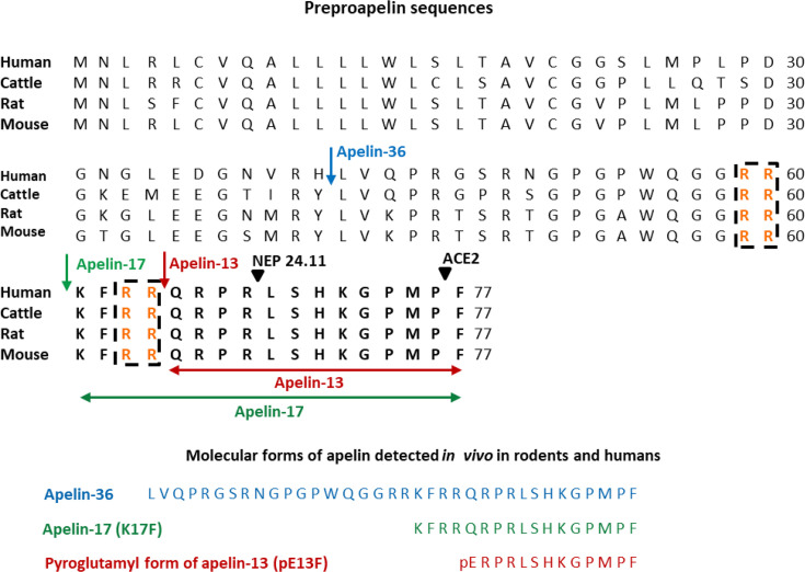
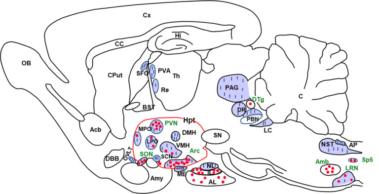
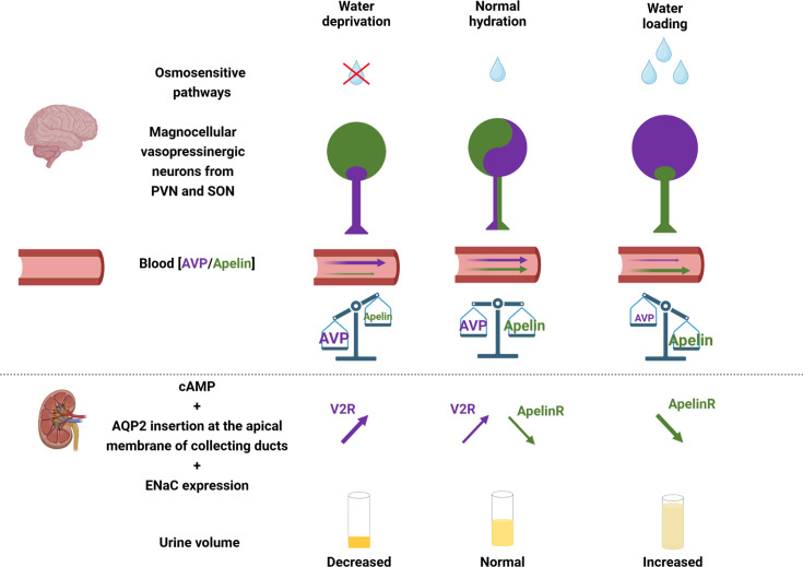
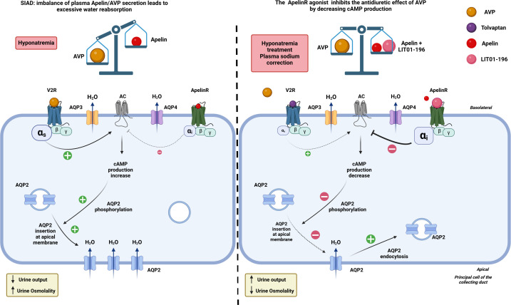
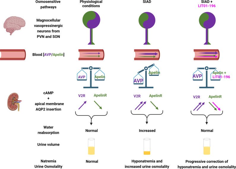
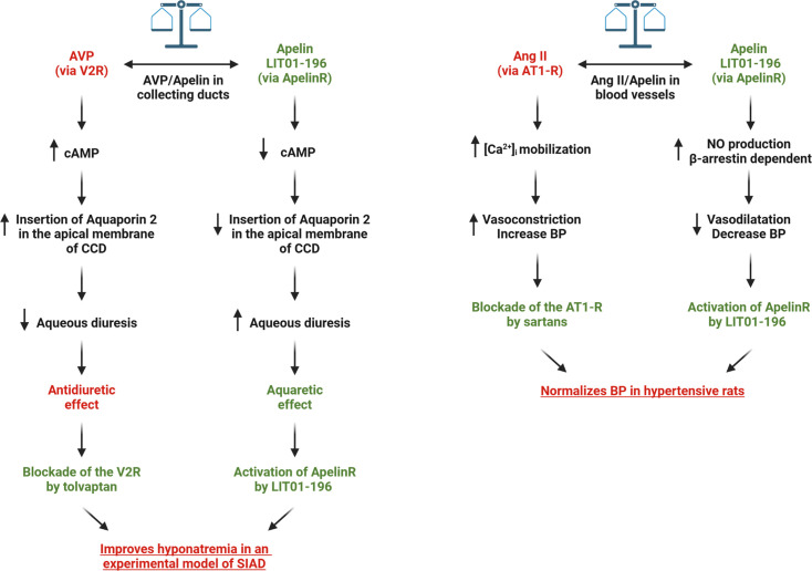
Apelin, a (neuro) vasoactive peptide, plays a prominent role in controlling water balance and cardiovascular functions. Apelin and its receptor co-localize with vasopressin in magnocellular vasopressinergic neurons. Apelin receptors (Apelin-Rs) are also expressed in the collecting ducts of the kidney, where vasopressin type 2 receptors are also present. Apelin and vasopressin interact at the brain and renal levels to maintain body fluid homeostasis by regulating diuresis in opposite directions. Apelin and angiotensin II have opposite effects on the regulation of blood pressure (BP). Angiotensin II, by binding to AT1 receptors present in VSMCs, induces intracellular calcium mobilization and vasoconstriction, while apelin, by binding to Apelin-R present on vascular endothelium, increases nitric oxide production and induces vasodilation. Apelin also plays a crucial role in the regulation of cardiac function. Apelin-deficient and Apelin-R-deficient mice develop progressive myocardial dysfunction with ageing and are susceptible to heart failure in response to pressure overload. Since the half-life of apelin is very short in vivo (in the minute range), several metabolically stable apelin analogs and non-peptidic Apelin-R agonists have been developed, with potential applications in diverse diseases. In this review, we highlight the interaction between apelin and vasopressin in the regulation of water balance and that between apelin and angiotensin II in the regulation of BP. Additionally, we underline the protective effects of apelin in cardiac function. Lastly, we discuss the beneficial effects of Apelin-R activation in different pathological states such as hyponatremia, hypertension, and heart failure.

Keywords: Apelin receptor; Aqueous diuresis; Blood pressure; Cardiac function;; G Protein-Coupled Receptor.


Conflict of interest statement

The authors declare that there are no competing interests associated with the manuscript.

Figures

Figure 1
Figure 1. Amino-acid sequences of the apelin precursor, preproapelin, in humans, cattle, rats, and mice, and the molecular forms of apelin detected in vivo.
The blue arrow indicates the beginning of the sequence of apelin-36, the green one that of the sequence of apelin-17 (K17F), which is strictly conserved in mammals, and the red one that of the apelin-13 sequence. The dibasic doublets (in orange) are framed by black dashed boxes. The black arrows show the cleavage sites by neutral endopeptidase 24.11 (NEP 24.11, EC 3.4.24.11) and angiotensin-converting enzyme 2 (ACE2, EC 3.4.17.23). The various molecular forms of apelin detected in vivo in humans and rodents: apelin-36, apelin-17, and the pyroglutamyl form of apelin-13 (pE13F). Figure adapted from [8] with permission from the copyright holders.
Figure 2
Figure 2. Distribution of apelineric cell bodies and nerve fibers in the adult rat brain.
Neuroanatomical distribution of apelin immunoreactive cell bodies and nerve fibers in a sagittal section of rat brain. Cell bodies are shown as dots and nerve fibers are shown as lines. Acb, nucleus accumbens; Amb, nucleus ambiguus; Amy, amygdala; AL, anterior lobe of the pituitary gland; AP, area postrema; Arc, arcuate nucleus; BST, nucleus of the layer of the stria terminalis; C, cerebellum; CC, corpus callosum; Cput, caudate putamen; Cx, cerebral cortex; DBB, diagonal band of Broca; DMH, dorsomedial nucleus of the hypothalamus; DR, dorsal raphe nucleus; DTg, dorsal tegmental nucleus; HI, hippocampus; Hpt, hypothalamus; IL, intermediate pituitary lobe; LC, locus coerulus; LPO, lateral preoptic area; LRN, lateral reticular nucleus; ME, median eminence; MPO, median preoptic nucleus; NL, neural lobe of the pituitary gland; NST, nucleus of the solitary tract; OB, olfactory bulb; OVLT, vascular organ of the lamina terminalis; PAG, periaqueductal grey matter; PBN, parabrachial nucleus; PVA, paraventicular nucleus of the thalamus; PVN, paraventicular nucleus of the hypothalamus; Re, reuniens nucleus of the thalamus; SCN, suprachiasmatic nucleus; SFO, subfornical organ; SN, substantia nigra; SON, supraotpic nucleus; Sp5, spinal nucleus of the trigeminal nerve; Th, thalamus; VMH, ventromedian nucleus of the hypothalamus. Figure adapted from [39] with permission from the copyright holders.
Figure 3
Figure 3. Apelin and vasopressin (AVP), the Yin and the Yang of water balance.
Under physiological conditions, apelin (green) and AVP (purple) are released by magnocellular vasopressinergic neurons at levels appropriate to plasma osmolality. In the renal collecting duct, AVP acts on V2R to increase cAMP production and the insertion of aquaporin 2 (AQP2) at the apical membrane, leading to water reabsorption and urine output decrease. Conversely, apelin, through its action on Apelin-R, has an opposing effect. Under physiological conditions, water reabsorption is adequate and plasma sodium concentrations are normal. Following dehydration, vasopressinergic neurons are strongly activated and AVP is released into the bloodstream more rapidly than it is synthesized, leading to a decrease in neuronal AVP content, while apelin accumulates in the neurons rather than being released. In contrast, under water loading, vasopressinergic neurons are inhibited, which stops AVP release in the blood circulation and results in neuronal AVP accumulation. Conversely, the release of apelin into the bloodstream increases rapidly, leading to a depletion of neuronal apelin content. Thereby, neuronal and plasma apelin levels are regulated by osmotic stimuli in an opposite direction to AVP. Figure adapted from [61] with permission from the copyright holders and made with Biorender©.
Figure 4
Figure 4. In an experimental model of SIAD, AVP secretion into the bloodstream is abnormally high in relation to plasma osmolality.
By binding to V2R in the collecting duct, AVP increases cAMP production, mobilizes aquaporin 2 (AQP2) to the apical membrane, and enhances water reabsorption, leading to hyponatremia. Administration of LIT01-196, a metabolically stable analogue of apelin-17 (K17F), stimulates Apelin-R located in the collecting ducts. This inhibits AVP-induced cAMP production, reduces the mobilization of AQP2 to the apical membrane, and decreases water reabsorption, thereby promoting diuresis. Moreover, Apelin-R activation reduces transepithelial amiloride-sensitive sodium current and decreases αENaC subunit expression, which in turn reduces sodium reabsorption. This leads to a progressive improvement in hyponatremia. Figure adapted from [66] with permission from the copyright holders and made with Biorender©.
Figure 5
Figure 5. Apelin and vasopressin regulation in SIAD and effects of LIT01-196 in this pathology.
In normal conditions of hydration, apelin and AVP, co-localized in magnocellular vasopressinergic neurons, are released in the blood circulation, and the balance between apelin and AVP allows both peptides by activating at the kidney level the V2R and the Apelin-R to induce normal water reabsorption and urine volume and ensure normal natremia. In SIAD, there is a high amount of AVP released in the blood circulation relative to osmolality, whereas plasma apelin levels are slightly modified, and the AVP/apelin balance is not reached, increasing water reabsorption, decreasing aqueous diuresis, and leading to hyponatremia. In SIAD, treatment with LIT01-196 allows to re-establish the balance between AVP and apelin by the supplementary activation of the Apelin-Rby LIT01-196. This counteracts vasopressin-induced water reabsorption and correct hyponatremia. AVP, arginine-vasopressin; ENaC, epithelial sodium channel; PVN, paraventicular nucleus of the hypothalamus; SON, supraotpic nucleus; V2R, AVP type 2 receptor. Figure adapted from [66] with permission from the copyright holders and made with Biorender©.
Figure 6
Figure 6. Potential therapeutic effects of LIT01-196 in SIAD and hypertension.
By activating ApelinR in the collecting duct, LIT01-196 would counteract the overactivation of V2R by decreasing cAMP production, leading to a decrease in the insertion of aquaporin 2 in the apical membrane, resulting in the increase in aqueous diuresis. Together with tolvaptan that inhibits V2R, LIT01-196 would improve hyponatremia in an experimental model of SIADH. In the other hand, by activating ApelinR in the endothelial cells of blood vessel, LIT01-196 would counteract the overactivation of AT1-R by increasing NO production in a β-arrestin-dependent manner, leading to a vasodilatation and a decrease in blood pressure. Together with sartans that block AT1-R, LIT01-196 would normalizes blood pressure in hypertensive rats. The figure was made with Biorender©. Ang II, angiotensin II; AT1-R, angiotensin II type 1 receptor; AVP, arginine-vasopressin; BP, blood pressure; CCD, cortical collecting duct; NO, nitric oxide; SIAD, syndrome of inappropriate antidiuresis; SIADH, syndrome of inappropriate antidiuretic hormone; V2R, AVP type 2 receptor.

Similar articles

References

    1. O’Dowd B.F., Heiber M., Chan A., Heng H.H., Tsui L.C., Kennedy J.L., et al. A human gene that shows identity with the gene encoding the angiotensin receptor is located on chromosome 11. Gene. 1993;136:355–360. doi: 10.1016/0378-1119(93)90495-o. - DOI - PubMed
    1. Devic E., Rizzoti K., Bodin S., Knibiehler B., Audigier Y. Amino acid sequence and embryonic expression of msr/APJ, the mouse homolog of Xenopus X-msr and human APJ. Mech. Dev. 1999;84:199–203. doi: 10.1016/s0925-4773(99)00081-7. - DOI - PubMed
    1. De Mota N., Lenkei Z., Llorens-Cortès C. Cloning, pharmacological characterization and brain distribution of the rat apelin receptor. Neuroendocrinology. 2000;72:400–407. doi: 10.1159/000054609. - DOI - PubMed
    1. O’Carroll A.M., Selby T.L., Palkovits M., Lolait S.J. Distribution of mRNA encoding B78/APJ, the rat homologue of the human APJ receptor, and its endogenous ligand apelin in brain and peripheral tissues. Biochim. Biophys. Acta. 2000;1492:72–80. doi: 10.1016/s0167-4781(00)00072-5. - DOI - PubMed
    1. Tatemoto K., Hosoya M., Habata Y., Fujii R., Kakegawa T., Zou M.X., et al. Isolation and characterization of a novel endogenous peptide ligand for the human APJ receptor. Biochem. Biophys. Res. Commun. 1998;251:471–476. doi: 10.1006/bbrc.1998.9489. - DOI - PubMed

MeSH terms