Multicenter Study
. 2025 Apr 7;74(5):740-751.
doi: 10.1136/gutjnl-2024-333353.

Low-coverage whole genome sequencing of low-grade dysplasia strongly predicts advanced neoplasia risk in ulcerative colitis

Affiliations
Multicenter Study

Low-coverage whole genome sequencing of low-grade dysplasia strongly predicts advanced neoplasia risk in ulcerative colitis

Ibrahim Al Bakir et al. Gut. .

Abstract

Background: The risk of developing advanced neoplasia (AN; colorectal cancer and/or high-grade dysplasia) in ulcerative colitis (UC) patients with a low-grade dysplasia (LGD) lesion is variable and difficult to predict. This is a major challenge for effective clinical management.

Objective: We aimed to provide accurate AN risk stratification in UC patients with LGD. We hypothesised that the pattern and burden of somatic genomic copy number alterations (CNAs) in LGD lesions could predict future AN risk.

Design: We performed a retrospective multicentre validated case-control study using n=270 LGD samples from n=122 patients with UC. Patients were designated progressors (n=40) if they had a diagnosis of AN in the ~5 years following LGD diagnosis or non-progressors (n=82) if they remained AN-free during follow-up. DNA was extracted from the baseline LGD lesion, low-coverage whole genome sequencing performed and data processed to detect CNAs. Survival analysis was used to evaluate CNAs as predictors of future AN risk.

Results: CNA burden was significantly higher in progressors than non-progressors (p=2×10⁻⁶ in discovery cohort) and was a very significant predictor of AN risk in univariate analysis (OR=36; p=9×10⁻⁷), outperforming existing clinical risk factors such as lesion size, shape and focality. Optimal risk prediction was achieved with a multivariate model combining CNA burden with the known clinical risk factor of incomplete LGD resection. Within-LGD lesion genetic heterogeneity did not confound risk prediction.

Conclusion: Measurement of CNAs in LGD is an accurate predictor of AN risk in inflammatory bowel disease and is likely to support clinical management.

Keywords: CANCER PREVENTION; COLORECTAL CANCER; GENETIC INSTABILITY; INFLAMMATORY BOWEL DISEASE; ULCERATIVE COLITIS.


Conflict of interest statement

Competing interests: The authors are in discussions about potential commercialisation and clinical translation of the findings described here. AH has served as consultant, advisory board member or speaker for AbbVie, Arena, Atlantic, Bristol-Myers Squibb, Celgene, Celltrion, Falk, Galapogos, Lilly, Janssen, MSD, Napp Pharmaceuticals, Pfizer, Pharmacosmos, Roche, Shire and Takeda. KC has an investigator-led research grant from Phathom Pharmaceuticals. TAG and A-MB are named as coinventors on a patent application that describe a method for TCR sequencing (GB2305655.9), and TAG is named on a method to measure evolutionary dynamics in cancers using DNA methylation (GB2317139.0). TAG has received honorarium from Genentech and DAiNA therapeutics.

Figures

Figure 1
Figure 1. Study design. Schematic showing the detection and removal of low-grade dysplasia during routine IBD surveillance, with progressors developing high-grade dysplasia and/or colorectal cancer within 5 years (top), and non-progressors remaining free from these (bottom). CRC, colorectal cancer; IBD, inflammatory bowel disease.
Figure 2
Figure 2. Representative endoscopic and histological images with copy number profiles. Endoscopy images (upper right) and H&E (upper left) with genome-wide copy number profile below for representative non-progressor (A) and progressor (B) patients.
Figure 3
Figure 3. Genomic alterations in non-progressor (NP) vs progressor LGD in discovery cohort. (A) Heatmap of genome-wide copy number alterations for lesions in the discovery LGD cohort, sorted by percent genome altered. Sporadic adenomas included below. (B) Violin plots showing the number of altered genomic segments in progressor and NP lesions. (C) Genome-wide CNA frequency for NP (top) and progressor (P, bottom) patients. Stars indicate significant arm level differences at adjusted p<0.01 level. For patients with multiregion analysis, the most highly altered sample per patient was included. LGD, low-grade dysplasia; MSI, microsatellite instability; PGA, percent genome altered; WGD, whole genome doubling.
Figure 4
Figure 4. Model performance for prediction of future HGD/CRC. (A) ORs for univariate models considering genomic score, UC-CaRE and PSC variables. ‘Unresected LGD’ is defined as either endoscopically invisible LGD (which was detected on histological examination of biopsy only and so by definition cannot be completely resected) or LGD specifically noted to have unclear resection margins. (B) AUC values for genomic score alone at 5 years post-LGD resection and PPV/NPV over time in validation data (all 55 patients included at baseline) with 95% bootstrap CIs (see the Methods section ‘Prediction accuracy’ for more details). Kaplan-Meier progression-free survival for the discovery (C) and validation (D) cohorts using a univariate genomic score model. (E) ORs for multivariate model chosen using stepwise selection. (F) AUC values for multivariate model at 5 years post-LGD resection and PPV/NPV over time in validation data (n=37 patients included at baseline with data on incomplete LGD resection). (G) Kaplan-Meier curves for validation data; high-risk (>0.5 risk) patients determined by multivariate model prediction. AUC, area under the curve; CRC, colorectal cancer; HGD, high-grade dysplasia; LGD, low-grade dysplasia; MSI, microsatellite instability; NPV, negative predictive value; PPV, positive predictive value; PSC, primary sclerosing cholangitis; UC, ulcerative colitis.
Figure 5
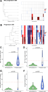
Figure 5. Phylogenetic analysis of multiregion data. Phylogenetic trees (left) and matched copy number profiles (right) for (A) a representative non-progressor (NP) lesion and (B) a representative progressor lesion. Tree statistics for lesions in the discovery cohort that had at least two regions sequenced were maximum tree length (C), minimum tree length (D), clonal CNA (E) and average subclonal CNA (F). CNA, copy number alteration; LGD, low-grade dysplasia.

Update of

References

    1. Odze RD, Goldblum J, Noffsinger A, et al. Interobserver variability in the diagnosis of ulcerative colitis-associated dysplasia by telepathology. Mod Pathol. 2002;15:379–86. doi: 10.1038/modpathol.3880534. - DOI - PubMed
    1. Eaden JA, Abrams K, McKay H, et al. Inter-observer variation between general and specialist gastrointestinal pathologists when grading dysplasia in ulcerative colitis. J Pathol. 2001;194:152–7. doi: 10.1002/path.876. - DOI - PubMed
    1. Nasreddin N, Jansen M, Loughrey MB, et al. Poor Diagnostic Reproducibility in the Identification of Nonconventional Dysplasia in Colitis Impacts the Application of Histologic Stratification Tools. Mod Pathol. 2024;37:S0893-3952(23)00324-1. doi: 10.1016/j.modpat.2023.100419. - DOI - PubMed
    1. Choi C-HR, Rutter MD, Askari A, et al. Forty-Year Analysis of Colonoscopic Surveillance Program for Neoplasia in Ulcerative Colitis: An Updated Overview. Am J Gastroenterol. 2015;110:1022–34. doi: 10.1038/ajg.2015.65. - DOI - PMC - PubMed
    1. Siegel CA, Schwartz LM, Woloshin S, et al. When should ulcerative colitis patients undergo colectomy for dysplasia? Mismatch between patient preferences and physician recommendations. Inflamm Bowel Dis. 2010;16:1658–62. doi: 10.1002/ibd.21233. - DOI - PMC - PubMed

Publication types

LinkOut - more resources