. 2025 Jan 29;15(1):3708.
doi: 10.1038/s41598-024-83530-0.

Mitochondrial dysfunction-driven AMPK-p53 axis activation underpins the anti-hepatocellular carcinoma effects of sulfane sulfur

Affiliations

Mitochondrial dysfunction-driven AMPK-p53 axis activation underpins the anti-hepatocellular carcinoma effects of sulfane sulfur

Xue Zheng et al. Sci Rep. .

Abstract

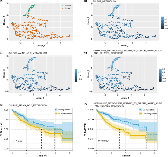
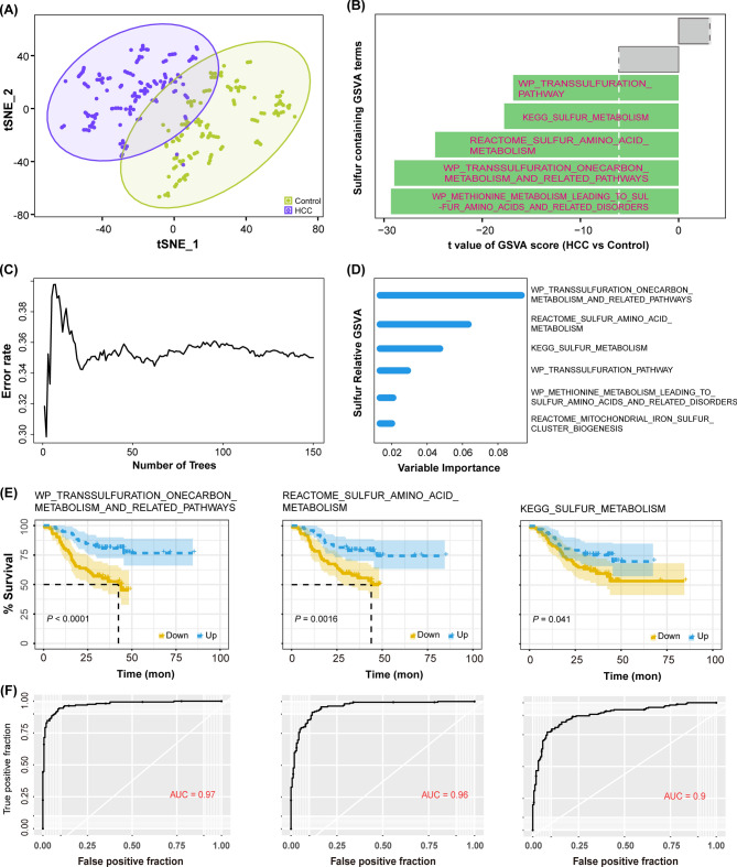
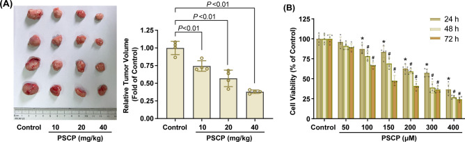
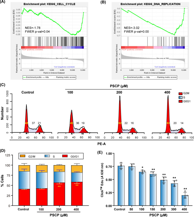
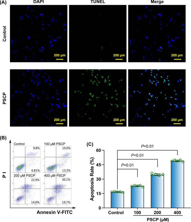
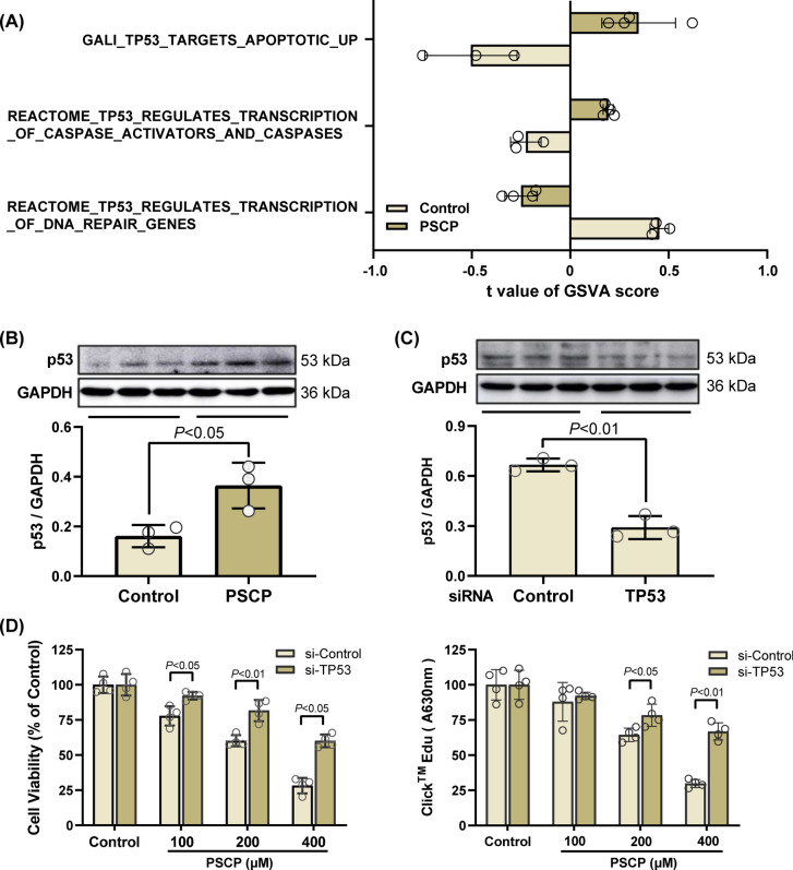
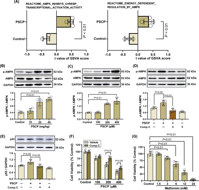
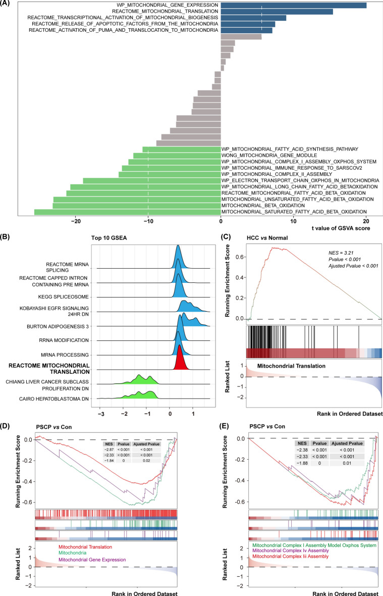
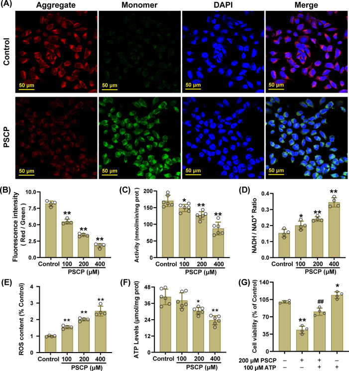
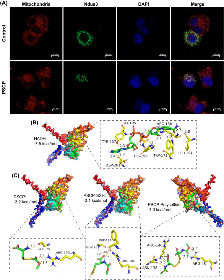
Hepatocellular carcinoma (HCC) is the most prevalent form of primary liver cancer, notoriously refractory to conventional chemotherapy. Historically, sulfane sulfur-based compounds have been explored for the treatment of HCC, but their efficacy has been underwhelming. We recently reported a novel sulfane sulfur donor, PSCP, which exhibited improved chemical stability and structural malleability. This study aimed to investigate the effects of PSCP on HCC and elucidate the underlying mechanisms. We utilized bioinformatics algorithms for clustering, function enrichment, feature screening and survival analysis on proteomic data from the Cancer Proteome Atlas (CPTAC) and transcriptomic data from the Cancer Genome Atlas (TCGA). The impact of PSCP on HCC was assessed in vitro and in vivo, focusing on the expression and activity of p53 and AMP-activated protein kinase (AMPK), as well as mitochondrial function. The molecular target of PSCP was identified using Autodock, and binding interactions were visually analyzed. Sulfur metabolism was found to be reprogrammed in HCC, with downregulation of sulfur-related pathways correlating with poor patient prognosis. PSCP treatment significantly inhibited HCC tumor growth in an allograft model, reduced cell viability and proliferation, and induced apoptosis. PSCP potently increased p53 expression and induced AMPK phosphorylation in SNU398 HCC cells. AMPK suppression diminished PSCP-induced p53 upregulation. PSCP also impaired mitochondrial function by inhibiting mitochondrial respiratory complex I, with Ndus3 likely being the target of PSCP's action. Supplementation with ATP significantly countered PSCP-induced SNU398 cell injury. Our findings suggest that the reprogramming of sulfur-related metabolic pathways is pivotal in HCC. PSCP presents as a promising therapeutic strategy by activating the AMPK-p53 signaling axis.

Keywords: AMPK; Hepatocellular carcinoma; Metabolic reprogramming; Mitochondrial complex; Sulfane sulfur; p53.


Conflict of interest statement

Declarations. Competing interests: The authors declare no competing interests. Ethical approval: This study was approved by the Animal Ethics Committee of Guangzhou Medical University (Approval Number: GY2020-099). Accordance statement: All procedures were strictly adhered to the guidelines of the Animal Ethics Committee of Guangzhou Medical University. ARRIVE statement: All procedures were conducted in full compliance with the ARRIVE guidelines.

Figures

Fig. 1
Fig. 1
Analysis of sulfur and related metabolic pathways using TCGA transcriptomic data. (A) Cluster analysis of RNA expression of HCC (n = 374) and control para-cancerous (n = 50) tissues with the Umap dimensionality reduction algorithm. (BD) Umap visualization of sulfur metabolism (B), sulfur amino acid metabolism (C), and methionine metabolism leading to sulfur amino acid and related disorders (D). The color gradient represents GSVA scores, with deeper blue indicating higher pathway enrichment. (EF) Cox regression analysis was conducted to assess the impact of each phenotype on the survival of HCC patients (n = 187).
Fig. 2
Fig. 2
Examination of sulfur and related metabolism using CPTAC proteomic data. (A) Protein expression of HCC (n = 160) and control para-cancerous (n = 160) tissues was analyzed using GSVA, followed by t-SNE clustering. (B) Utilizing the t-values generated by the limma algorithm, a diverging bar chart was constructed to depict the GSVA scores of sulfur-related metabolisms. (CD) Random Survival Forest analysis was performed to evaluate the effects of sulfur-related metabolism phenotypes on the survival of HCC patients. (E) Cox regression analysis was conducted to observe the impact of each phenotype group (Down 80 versus Up 80) on the survival of HCC patients. (F) ROC curves were plotted, and the AUC was calculated to evaluate the diagnostic efficacy for HCC.
Fig. 3
Fig. 3
Effects of PSCP on the growth of HCC in vivo and in vitro. H22 HCC cells were subcutaneously implanted into nude mice. Four days post-implantation, mice were administered varying doses of PSCP intraperitoneally once daily for three weeks. (A) Upon sacrifice, the allografts from the mice were imaged, and their relative tumor volumes were measured. (B) SNU398 HCC cells were treated with increasing concentrations of PSCP for various time periods, and the cell viability was assessed using the CCK-8 assay. Data are expressed as mean ± SD (n = 4–5). *P < 0.01 vs. Control group. #P < 0.01 vs. 24 h within the same group.
Fig. 4
Fig. 4
Influence of PSCP on cell cycle progression and proliferation in HCC cells. (AB) The GSEA algorithm was used to identify enriched pathways related to cell cycle (A) and DNA replication (B) in PSCP-treated HCC cells. (CE) Following a 24-h treatment with PSCP, cell cycle was analyzed using flow cytometry after PI staining (C and D), and cell proliferation was measured with the EdU incorporation assay (E). Data are presented as mean ± SD (n = 4). *P < 0.05, **P < 0.01 vs. Control group.
Fig. 5
Fig. 5
Impact of PSCP on apoptosis in HCC cells. (A) SNU398 HCC cells were exposed to 200 μM PSCP for 24 h, and the TUNEL assay followed by fluorography was utilized to assess apoptosis. (BC) After treatment with PSCP at concentrations ranging from 100 to 400 μM for 48 h, SNU398 HCC cells were stained with Annexin V and PI, and then analyzed for apoptosis rate using flow cytometry. Data are represented as mean ± SD, n = 5.
Fig. 6
Fig. 6
Inhibitory effects of PSCP on HCC were mediated through the p53 pathway (A) The enriched KEGG pathways related to p53 were identified using the GSVA algorithm in SNU398 HCC cells treated with PSCP. (B) The expression of p53 protein in SNU398 HCC cells exposed to 200 μM PSCP was measured using Western blotting. (C) RNA interference was conducted to reduce p53 expression in SNU398 HCC cells. (D) Scramble control and TP53 knockdown SNU398 HCC cells were treated with 0 ~ 400 μM PSCP for 48 h, cell viability and proliferation were assessed using the CCK-8 assay and the EdU incorporation assay, respectively. Data are expressed as mean ± SD, n = 3–4.
Fig. 7
Fig. 7
Roles of AMPK in PSCP-induced anti-HCC effects. (A) The enriched KEGG pathways associated with AMPK were identified in PSCP-treated SNU398 HCC cells using the GSVA algorithm. (BC) Proteins were extracted from the mouse allografts (B) or SNU398 HCC cells (C) treated with PSCP, and the levels of phosphorylated (p) and total AMPK were measured with Western blot. (DE) The effects of pretreatment with 2 μM Comp. C (an AMPK inhibitor) for 2 h on the phosphorylation of AMPK (D) and the upregulation of p53 (E) induced by 200 μM PSCP for 48 h were examined. (F) After SNU398 HCC cells were treated with PSCP in the absence or presence of Comp. C pretreatment, cell viability was measured using the CCK-8 assay. (G) The effects of increasing concentrations of Metformin (an AMPK agonist) on SNU398 cell viability were assessed as described in (F). Data are expressed as mean ± SD, n = 3–4.
Fig. 8
Fig. 8
Enhanced mitochondrial function in HCC tissues and PSCP-treated HCC cells. (A) Using the t-values generated by the limma algorithm, a diverging bar chart was constructed to represent the GSVA scores of mitochondrial function. (BC) The GSEA algorithm was applied to identify the top ten enriched pathways in HCC tissues (B) and the mitochondrial translation set was displayed (C). (DE) Following treatment of SNU398 HCC cells with PSCP, the GSEA algorithm was employed to analyze the enriched gene sets related to mitochondrial function (D) and complex activity (E).
Fig. 9
Fig. 9
PSCP-induced impairment of mitochondrial function in HCC cells. (A) Following a 24-h treatment with 100 μM PSCP, MMP in SNU398 HCC cells was observed with JC-1 staining assay and fluorography. (B) The MMP levels were quantified by measuring the fluorescence ratio of red to green using Image J software. (CF) After treatment with PSCP (100 ~ 400 μM) for 24 h, the activity of mitochondrial complex I (C), the NADH/NAD+ ratio (D), intracellular ROS (E), and intracellular ATP content (F) were measured in SNU398 HCC cells. (G) After SNU398 HCC cells were treated with PSCP in the absence or presence of ATP, the cell viability was measured using the CCK-8 assay. Data are expressed as mean ± SD (n = 4 ~ 5). *P < 0.05, **P < 0.01 vs. Control group.
Fig. 10
Fig. 10
Interaction of PSCP with mitochondrial complex I. (A) Following a 24-h treatment of SNU398 HCC cells with 200 μM PSCP, the expression and mitochondrial translocation of Ndus3 were observed using confocal fluorescence microscopy. (B) The interaction of NADH with the Ndus3 protein was analyzed using Autodock, and the visualization of the specific types of interactions was achieved through the PyMOL software. (C) The interaction of PSCP, as well as its degradation products (PSCP-SSH and PSCP-Polysulfide) with the Ndus3 protein was analyzed and visualized using the same methods as in (B).

Similar articles

References

    1. Llovet, J. M. et al. Hepatocellular carcinoma. Nat. Rev. Dis. Primers7, 6. 10.1038/s41572-020-00240-3 (2021). - PubMed
    1. Yang, C. et al. Evolving therapeutic landscape of advanced hepatocellular carcinoma. Nat. Rev. Gastroenterol. Hepatol.20, 203–222. 10.1038/s41575-022-00704-9 (2023). - PubMed
    1. Li, N. et al. Deficient DNASE1L3 facilitates neutrophil extracellular traps-induced invasion via cyclic GMP-AMP synthase and the non-canonical NF-kappaB pathway in diabetic hepatocellular carcinoma. Clin. Transl. Immunol.11, e1386. 10.1002/cti2.1386 (2022). - PMC - PubMed
    1. Philips, C. A. et al. Hepatocellular carcinoma in 2021: An exhaustive update. Cureus13, e19274. 10.7759/cureus.19274 (2021). - PMC - PubMed
    1. Weisberger, A. S. & Pensky, J. Tumor-inhibiting effects derived from an active principle of garlic (Allium sativum). Science126, 1112–1114. 10.1126/science.126.3283.1112-a (1957). - PubMed

Publication types

MeSH terms

Substances