. 2025 Feb;638(8049):126-132.
doi: 10.1038/s41586-024-08362-4. Epub 2025 Jan 29.

Copper-dependent halogenase catalyses unactivated C-H bond functionalization

Affiliations

Copper-dependent halogenase catalyses unactivated C-H bond functionalization

Chen-Yu Chiang et al. Nature. 2025 Feb.

Abstract

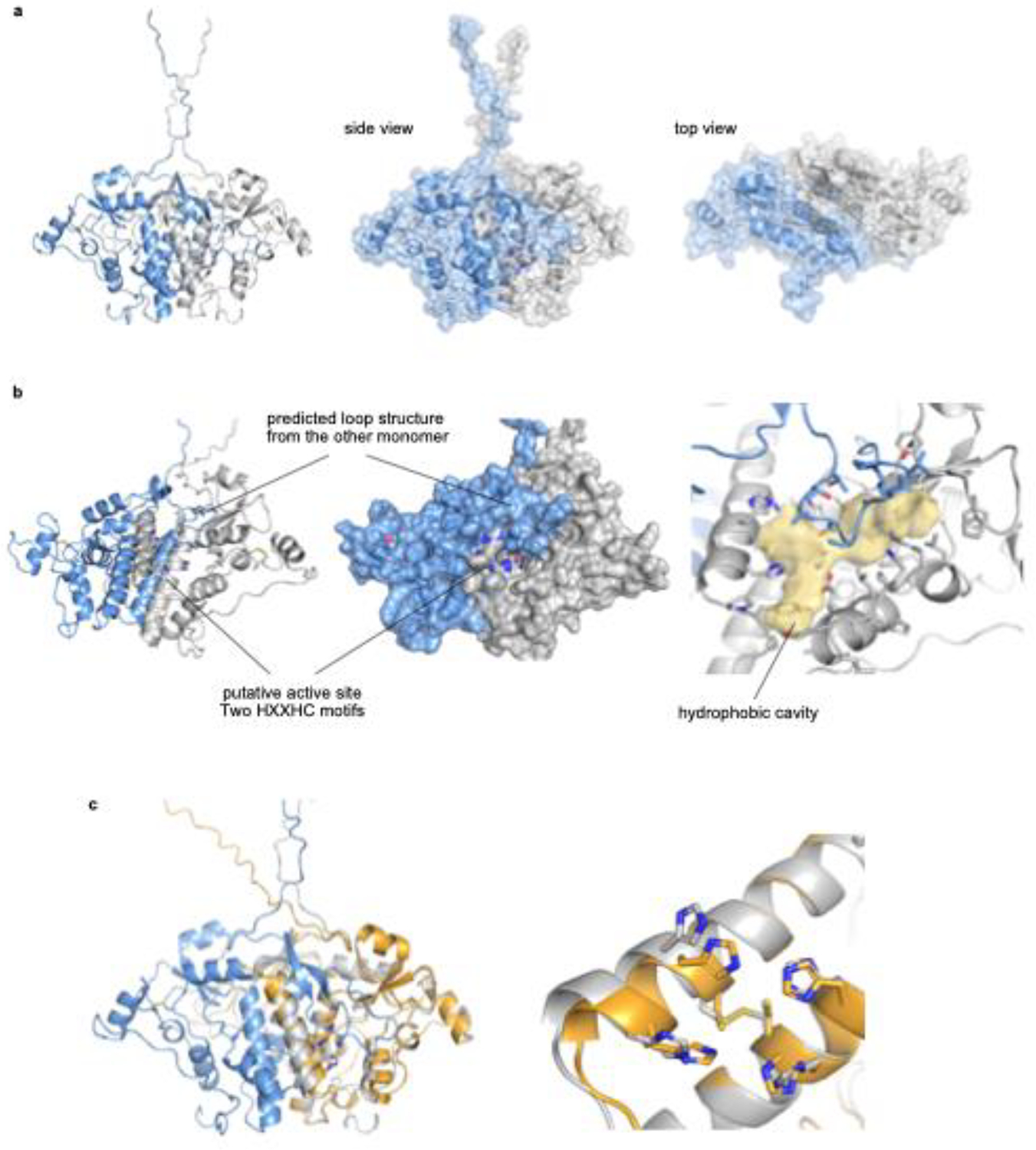
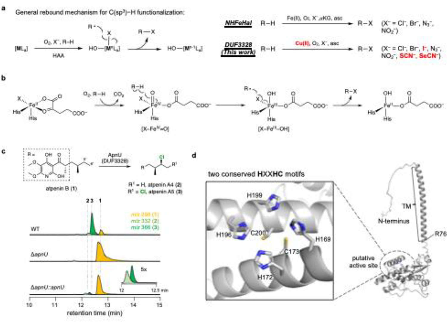
Carbon-hydrogen (C-H) bonds are the foundation of essentially every organic molecule, making them an ideal place to do chemical synthesis. The key challenge is achieving selectivity for one particular C(sp3)-H bond1-3. In recent years, metalloenzymes have been found to perform C(sp3)-H bond functionalization4,5. Despite substantial progresses in the past two decades6,7, enzymatic halogenation and pseudohalogenation of unactivated C(sp3)-H-providing a functional handle for further modification-have been achieved with only non-haem iron/α-ketoglutarate-dependent halogenases, and are therefore limited by the chemistry possible with these enzymes8. Here we report the discovery and characterization of a previously unknown halogenase ApnU, part of a protein family containing domain of unknown function 3328 (DUF3328). ApnU uses copper in its active site to catalyse iterative chlorinations on multiple unactivated C(sp3)-H bonds. By taking advantage of the softer copper centre, we demonstrate that ApnU can catalyse unprecedented enzymatic C(sp3)-H bond functionalization such as iodination and thiocyanation. Using biochemical characterization and proteomics analysis, we identified the functional oligomeric state of ApnU as a covalently linked homodimer, which contains three essential pairs-one interchain and two intrachain-of disulfide bonds. The metal-coordination active site in ApnU consists of binuclear type II copper centres, as revealed by electron paramagnetic resonance spectroscopy. This discovery expands the enzymatic capability of C(sp3)-H halogenases and provides a foundational understanding of this family of binuclear copper-dependent oxidative enzymes.


Conflict of interest statement

Competing interests: The authors declare no competing interests.

Figures

Extended Data Fig. 1|
Extended Data Fig. 1|. In vivo study of uncharacterized halogenase family DUF3328.
a, Biosynthetic gene cluster comparison of harzianopyridone (har) cluster and atpenin (apn) cluster. Harzianopyridone is a structural homolog to atpenin but lacking chlorines on the alkyl chain. There are three additional genes in apn cluster but not in har cluster, which are apnS, apnT, and apnU. Since there is no known halogenase in apn cluster, one of the additional gene might be responsible for chlorination on substrate 1. b, Gene knockout study of apnS, apnT, and apnU. Complementary gene expression of apnU and apnT in its mutant strain are indicated as ΔapnU::apnU and ΔapnT::apnT, respectively. Selected EIC(+) chromatograms are shown.
Extended Data Fig. 2|
Extended Data Fig. 2|. Multiple sequence alignment of selected DUF3328 enzymes.
a, Multiple sequence alignment of DUF3328 enzymes. Conserved His and Cys residues are bolded. Two conserved HXXHC motifs are highlighted. The numbering of Cys residue is based on full-length ApnU sequence. C210 and C237, which are involved in the formation of intermolecular disulfide bond, are conserved in all selected DUF3328 enzyme sequences. b, Proposed function of selected DUF3328 enzymes in the biosynthesis of fungal secondary metabolites. The reaction includes chlorination (CctP2), hydroxylation (CctR and PhomYa), macrocyclization (AprY, UstYa/Yb, and PhomYb), desaturation (PhomYb, PhomYc, and PhomYe), and transacylation (CctO). All functions are proposed according to in vivo experiment, either heterologous expression or gene inactivation study.
Extended Data Fig. 3|
Extended Data Fig. 3|. Biochemical characterization of soluble ApnU and refolded ApnU.
a, SDS−PAGE of soluble ApnU expressed in E. coli and refolded ApnU from inclusion body. Target band is indicated by black arrow. b, Activity assays using soluble ApnU under different conditions. To provide evidence of product formation, EIC(+) spectra were shown in the enzymatic condition. The reaction is done in a 50-μL scale with 10 μM enzyme, 100 μM 1, 100 mM MES (pH6.0), and 5 mM sodium ascorbate at room temperature overnight. c, Activity assay using refolded ApnU with different reductants or hydrogen peroxide. The reaction condition is as followed: 10 μM refolded enzyme, 100 μM 1, 10 μM CuSO4, 100 mM MES (pH6.0), and 5 mM reductant (unless otherwise specified) at room temperature for 1 hour. d, Product profile of refolded ApnU in different buffer and pH value. e, Product profile of refolded ApnU with different concentration of NaCl. The bar chart represents the mean from triplicate experiments. Error bars represent one standard deviation. f, Activity assay of refolded ApnU with or without adding glutathione redox pair (GSH/GSSG) during refolding process. This result highlights the necessity of adding the redox pair during refolding process to obtain soluble and functional enzyme.
Extended Data Fig. 4|
Extended Data Fig. 4|. Metal requirement for the activity of ApnU.
a, In vitro assay using refolded ApnU with 1mM or 10μM metal ion. λ = 335nm UV spectra are shown. b, In vitro assay using dimer ApnU (with CHis6-tag) with 1mM or 10μM metal ion. The reaction is done in a 50-μL scale, and the reaction condition is as followed: 10 μM enzyme, 100 μM 1, 100 mM MES (pH6.0), 5 mM sodium ascorbate and specified metal concentration at room temperature for 1 hr.
Extended Data Fig. 5|
Extended Data Fig. 5|. Investigation of enzymatic C(sp3)−H functionalization catalyzed by ApnU with different anion source.
a, Side products formation under standard condition (NaCl) and no anion source condition (no salt). Additional peaks appeared to be comparable with 5 under no salt condition. *: proposed hydroxylated product with m/z 314 and 296 ([M−H2O+H+]). **: proposed hydroxylated and desaturated product with m/z 312. b, Detailed chromatograms of λ = 335 nm and EIC(+) for each corresponding functionalized product. *: the peak was observed in control (without salt). In the NaOCN trace, although EIC(+) spectrum can observe a corresponding adduct, the UV signal is below detection limit. The assay is done in a 50-μL scale, and the reaction condition is as followed: 10 μM refolded enzyme, 500 μM 1, 100 mM MES (pH6.0), 10 μM CuSO4, 5 mM anion source and 5 mM sodium ascorbate at room temperature for 1 hr.
Extended Data Fig. 6|
Extended Data Fig. 6|. Dimer characterization and Mutagenesis.
a, FPLC chromatograms of refolded ApnU purified by anion exchange chromatography (AEX) or size exclusion chromatography (SEC). b, Reducing SDS−PAGE, non-reducing SDS−PAGE, and native PAGE for peak I and II in AEX. r: refolded ApnU (before FPLC purification). c, Activity assays of each fraction purified from FPLC. d, Denatured protein mass spectrum for peak I in AEX chromatogram. e, The UV−vis spectrum of 100 μM dimer ApnU (without His6 tag). The holo enzyme was prepared by adding 2 or 4 equivalent numbers of CuSO4 (based on dimer concentration). There is no obvious absorption around 600 nm, which is the characteristic absorption band of ligand-to-metal charge transfer (LMCT) from thiolate to cupric center. f, Mutagenesis study on all cysteine residues and four histidine residues in the putative active site. Activity assay result of each mutant was shown. The assay is done in a 50-μL scale, and the reaction condition is as followed: 10 μM enzyme, 100 μM 1, 100 mM MES (pH6.0), 10 μM CuSO4, and 5 mM sodium ascorbate at room temperature for 1 hr. g, Non-reducing SDS−PAGE analysis of each mutant. h, Non-reducing SDS−PAGE analysis of dimer ApnU with different reducing reagent. Incubation of 5 μM dimer ApnU and 1 mM reducing agent for 30 minutes before subjecting to gel analysis. The monomer (M) and dimer (D) species are indicated by the black arrow.
Extended Data Fig. 7|
Extended Data Fig. 7|. Peptide mapping experiment.
a, Overall workflow. b, LC−QTOF MS analysis of ApnU dimer. Only the mass spectra are shown. The deconvoluted mass spectrum revealed three masses, corresponding to the three different dimer species: 14N−14N, 14N−15N, and 15N−15N.
Extended Data Fig. 8|
Extended Data Fig. 8|. Predicted ApnU dimer.
The predicted homodimer structure was constructed based on the disulfide bond pairing result. Truncated sequence (NΔ75-ApnU) was used for prediction. All structures are visualized by PyMOL. a, The overall structure of homodimer. b, The putative active site was covered by a loop (I210−Q233) from the counterpart chain, forming a putative substrate binding pocket. A hydrophobic cavity closed to active site can be simulated by KVFinder-web. c, Structural alignment of monomer and homodimer model. There is no significant change in overall structure except for both N-terminal and C-terminal disorder loops. Looking into the active site, the formation of intramolecular disulfide bond (C173−C200) in homodimer structure causes the movement of histidine residue in the HXXHC motifs. Blue and gray: chain A and chain B of homodimer. Orange: monomer.
Extended Data Fig. 9|
Extended Data Fig. 9|. Metal quantification of dimer ApnU.
a, ICP−MS metal quantification of each metal by titrating 1 equivalent or 2 equivalents of metal ion with dimer ApnU (no His6-tag). b, ICP−MS metal quantification of copper titration with dimer ApnU (no His6-tag). The equivalent numbers are based on the number of active site (two active site per dimer). All values reported here are per active site.
Extended Data Fig. 10|
Extended Data Fig. 10|. EPR spectroscopic analysis.
a, X-band EPR spectra of Cu(II) reconstituted ApnU by using two reconstitution methods. Top: EPR spectrum of Cu(II)-ApnU reconstituted by method I; Bottom: EPR spectrum of Cu(II)-ApnU reconstituted by method II. For both spectra, the spin concentrations of the total Cu(II) signals obtained by double integration of the signals are also shown. As for the top spectrum, the ApnU concentration was 117 μM, thus the ratio of Cu(II):ApnU (active site) is 2.36. For the bottom spectrum the ApnU concentration was 170 μM, thus the ratio is 2.06. For the reconstitution methods, see Method section. b, Zoom-in view of the g = 2.05 resonance showing a partially resolved super-hyperfine feature, which is most likely resulted from 14N super-hyperfine due to Cu-His ligation.
Figure 1|
Figure 1|. Discovery of a new halogenase.
a, General rebound mechanism for C(sp3)−H functionalization in metalloenzymes. Mechanistically, M can be any metal that is able to activate C(sp3)−H bonds, and X can be an X-type ligand such as halides or pseudohalides coordinated to the metal center. The copper-dependent halogenase, which belongs to DUF3328 family, described in this work catalyzes halogenation on unactivated C(sp3)−H bonds. b, Proposed halide rebound mechanism of non-heme iron/ α-ketoglutarate dependent halogenase (NHFeHal). c, Gene knockout study of apnU designated as the ΔapnU strain (based on P. oxalicum FO-125). Reintegration of apnU into the genome of the ΔapnU strain represented as ΔapnU::apnU was carried out as a genetic complementation experiment. Selected EIC(+) chromatograms are shown. d, Predicted structure of ApnU by AlphaFold2. Transmembrane region (TM) is highlighted. Two conserved HXXHC motifs form a putative metal binding site likely binds metal ion(s) that is different from iron. Disulfide mapping indicated two cysteines form a disulfide bond and not involved in metal binding. Structure is visualized by PyMOL.
Figure 2|
Figure 2|. Biochemical characterization of ApnU.
a, Reactions catalyzed by ApnU in the presence of 1. b, Activity assays to determine the metal requirement for refolded ApnU. Selected metal traces are shown. More information is provided in Extended Data Fig. 4. Activity assays were performed in 50-μL reactions. A typical reaction condition is the following unless otherwise specified: 10 μM refolded enzyme, 100 μM 1, 10 μM CuSO4, 100 mM MES (pH6.0), and 5 mM sodium ascorbate (asc) at room temperature for 1 hr. The estimated conversion for chlorination of 1 to 3 and 4 is ~ 50%, as quantified by UV absorbance (λ = 335 nm). c, Activity assays of refolded ApnU to determine the reaction cofactors and cosubstrates. d, Effects of using different reductants or hydrogen peroxide (5 mM) in ApnU-catalyzed reaction. e, Enzymatic C(sp3)−H bond functionalization catalyzed by ApnU using different anions (X) in the reaction. Reaction conditions: 10 μM refolded enzyme, 500 μM 1, 10 μM CuSO4, 5 mM anion source, 100 mM MES (pH6.0), and 5 mM sodium ascorbate at room temperature for 1 hr. The relative conversions are shown (all normalized to NaCl conversion as 100%). All data points are shown, and the bar chart represents the mean from triplicate experiments. Error bars represent one standard deviation. Structurally characterized atpenin analogs (6-8) are listed in the box. f, Substrate tolerance of ApnU. Functionalized DIF-3 products were detected by LC−MS in the presence of Cl, I, and SCN. See Supplementary Information for more details.
Figure 3|
Figure 3|. Characterization of functional ApnU reveals a disulfide bond-linked homodimer.
a, FPLC chromatogram of refolded ApnU purified by anion exchange chromatography (AEX). Only peak I showed chlorination activity. b, Deconvoluted mass of active fraction isolated from peak I in AEX chromatogram. c, Predicted homodimer structure of ApnU by considering the experimentally determined disulfide bond pairings. The structure is shown as an Alphafold2 predicted structure revised by Rosetta. Structure is visualized by PyMOL. Six cysteine residues are labeled. For simplicity, the structure starts at residue 90. See Extended Data Fig. 8 for more information. d, Mapping disulfide bonds using digested peptides. ○: 14N−14N, ●: 15N−15N, : 14N−15N, : 15N−14N, ■/□: other peptide fragments. Detailed MS1 and MS2 spectra are provided in Supplementary Information.
Figure 4|
Figure 4|. Electron paramagnetic resonance (EPR) characterization of ApnU and proposed halogenation mechanism.
a, X-band EPR spectra of Cu(II)-loaded ApnU and the ascorbate reduced form measured at 15K. (A) The EPR spectrum of Cu(II)-loaded ApnU (black) and its corresponding spectral simulation (red) with two magnetically non-interacting Cu(II) sites. The individual Cu(II) site simulations are shown in (C) and (D). The simulation parameters are Site_1 (green): g = [2.24, 2.06, 2.00]; σ(g) = [0.02, 0.015, 0.015]; A[Cu(II)] = [520, 20, 20] MHz; Site_2 (blue): g = [2.29, 2.08, 2.04]; σ(g) = [0.02, 0.015, 0.015]; A[Cu(II)] = [490, 20, 20] MHz. The Az[Cu(II)] values of the two copper sites are indicated in A. (B) The EPR spectrum of the ascorbate reduced ApnU showing the disappearance of the Cu(II) EPR signals. b, Proposed mechanism of binuclear copper-dependent halogenation by ApnU. X is Cl (native) or nonnative ligands including Br, I, N3, NO2, SCN, and SeCN.

Similar articles

Cited by

References

    1. Hartwig JF & Larsen MA Undirected, Homogeneous C–H Bond Functionalization: Challenges and Opportunities. ACS Cent. Sci 2, 281–292 (2016). - PMC - PubMed
    1. Hong B, Luo T & Lei X Late-Stage Diversification of Natural Products. ACS Cent. Sci 6, 622–635 (2020). - PMC - PubMed
    1. Liu B, Romine AM, Rubel CZ, Engle KM & Shi B-F Transition-Metal-Catalyzed, Coordination-Assisted Functionalization of Nonactivated C(sp3)–H Bonds. Chem. Rev 121, 14957–15074 (2021). - PMC - PubMed
    1. Ren X & Fasan R Engineered and artificial metalloenzymes for selective C–H functionalization. Curr. Opin. Green Sustain. Chem 31, 100494 (2021). - PMC - PubMed
    1. Li F, Zhang X & Renata H Enzymatic C–H functionalizations for natural product synthesis. Curr. Opin. Chem. Biol 49, 25–32 (2019). - PMC - PubMed

LinkOut - more resources