. 2025 Mar;639(8055):727-734.
doi: 10.1038/s41586-024-08498-3. Epub 2025 Jan 29.

CARD domains mediate anti-phage defence in bacterial gasdermin systems

Affiliations

CARD domains mediate anti-phage defence in bacterial gasdermin systems

Tanita Wein et al. Nature. 2025 Mar.

Abstract

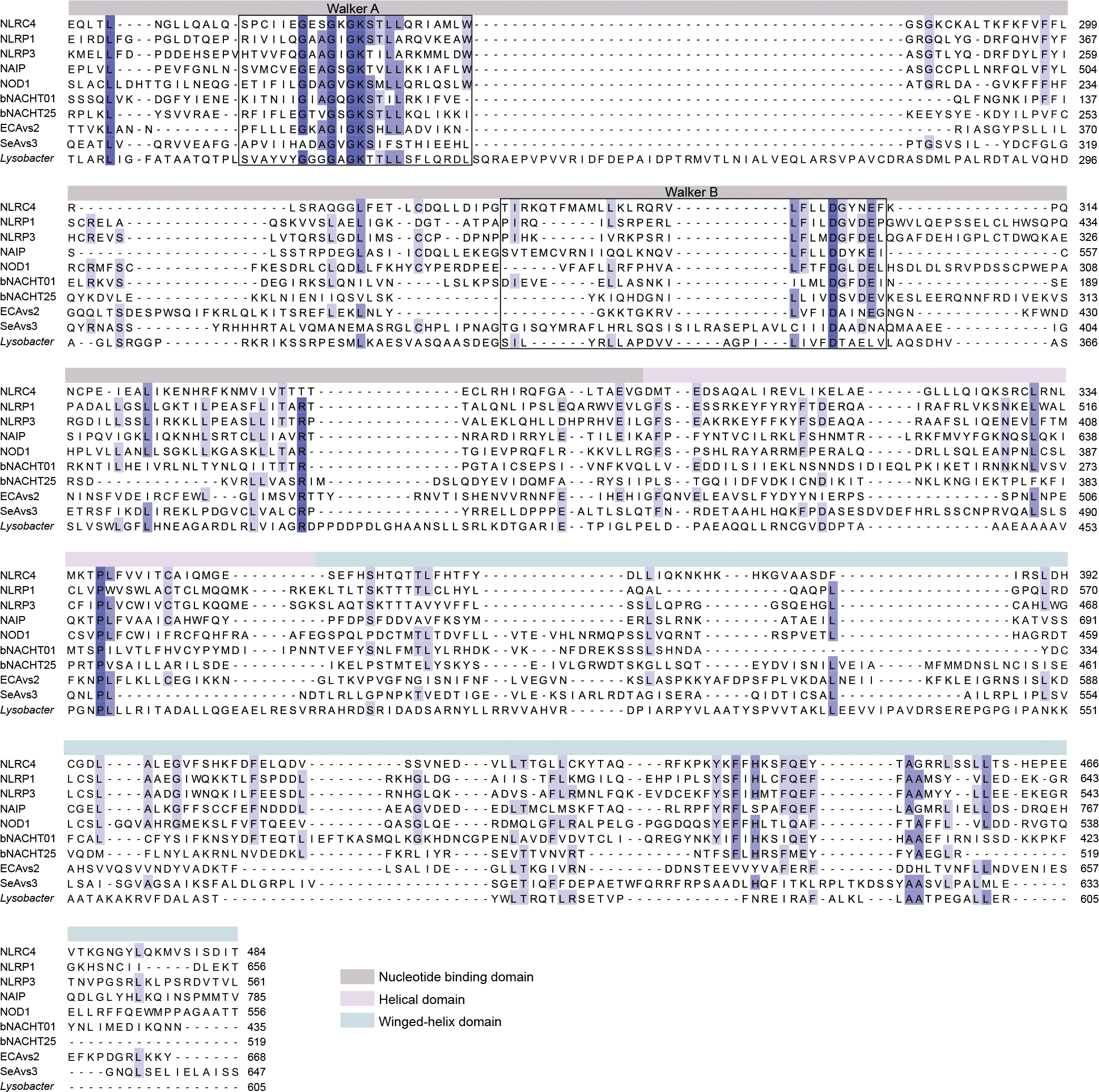
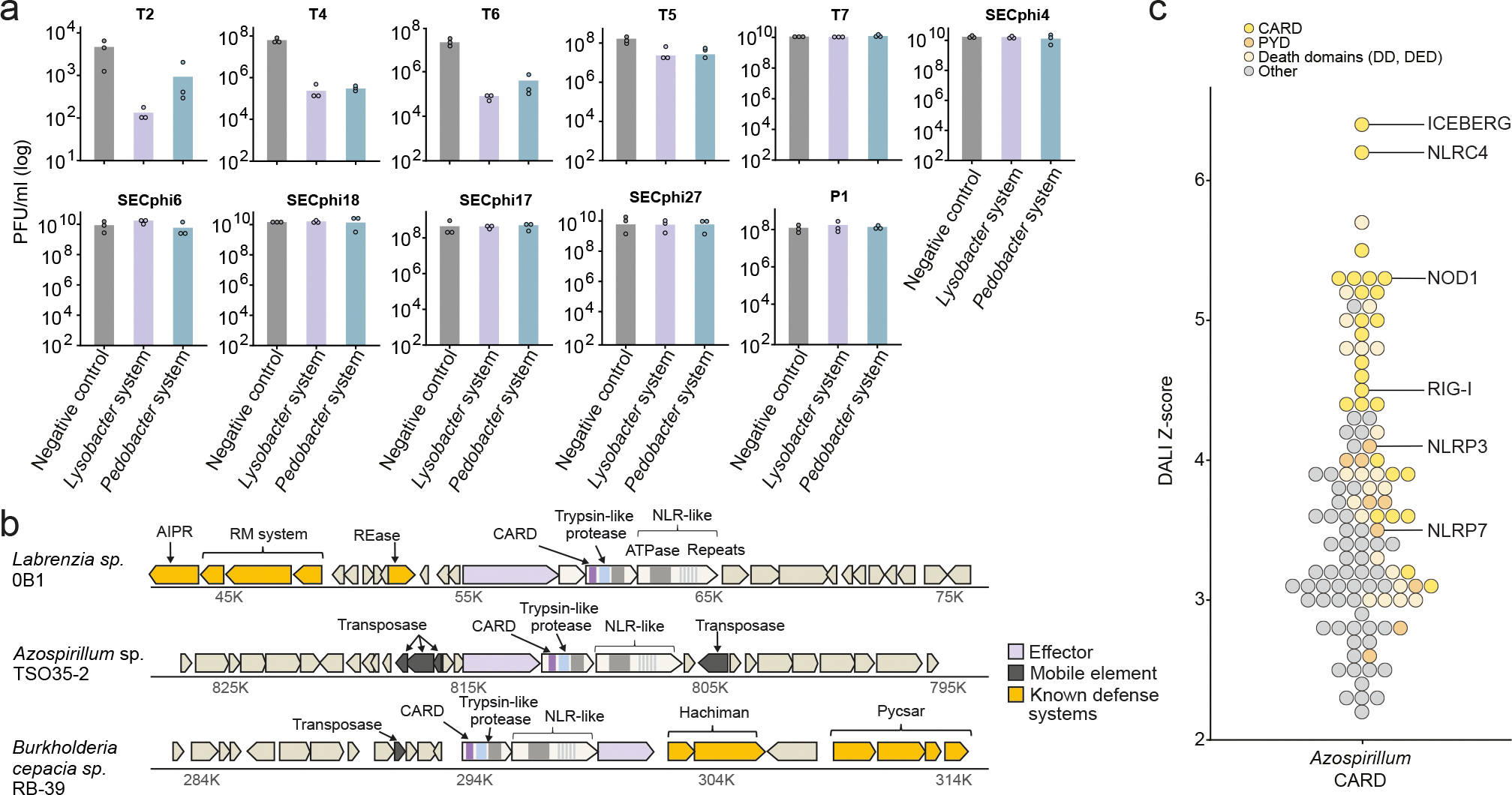
Caspase recruitment domains (CARDs) and pyrin domains are important facilitators of inflammasome activity and pyroptosis1. Following pathogen recognition by nucleotide binding-domain, leucine-rich, repeat-containing (NLR) proteins, CARDs recruit and activate caspases, which, in turn, activate gasdermin pore-forming proteins to induce pyroptotic cell death2. Here we show that CARD domains are present in defence systems that protect bacteria against phage. The bacterial CARD domain is essential for protease-mediated activation of certain bacterial gasdermins, which promote cell death once phage infection is recognized. We further show that multiple anti-phage defence systems use CARD domains to activate a variety of cell death effectors, and that CARD domains mediate protein-protein interactions in these systems. We find that these systems are triggered by a conserved immune-evasion protein used by phages to overcome the bacterial defence system RexAB3, demonstrating that phage proteins inhibiting one defence system can activate another. Our results suggest that CARD domains represent an ancient component of innate immune systems conserved from bacteria to humans, and that CARD-dependent activation of gasdermins is shared in organisms across the tree of life.


Conflict of interest statement

Competing interests: R.S. is a scientific cofounder and advisor of BiomX and Ecophage. The other authors declare no competing interests.

Figures

Extended Data Fig. 1.
Extended Data Fig. 1.. Anti-phage defense of tagged Lysobacter gasdermin system.
Efficiency of plating of phages infecting E. coli cells that express the WT Lysobacter gasdermin system, as well as systems in which gasdermin was N-terminally fused to an HA-tag or to GFP. Negative control is a strain in which GFP is expressed instead of the gasdermin system. Data represent plaque-forming units (PFU) per ml. Average of three independent biological replicates, with individual data points overlaid.
Extended Data Fig. 2.
Extended Data Fig. 2.. Multiple sequence alignment of the Nucleotide binding oligomerization domain (NOD) module of STAND ATPases.
Shown are NOD modules from human proteins (NLRC4, NCBI accession: AAH31555; NLRP1, AAG30288; NLRP3, AAL33911; NAIP, A55478; NOD1, AAD29125) and bacterial proteins (bNACHT01, NCBI accession: WP_015632533.1; bNACHT25, WP_001702659.1; ECAvs2, WP_063118745.1; SeAvs3, WP_126523998.1), including the ATPase protein from the Lysobacter gasdermin system (IMG gene ID 2841794910). The depth of shading indicates degree of residue conservation.
Extended Data Fig. 3.
Extended Data Fig. 3.. Predicted structures of CARD domains.
AlphaFold2 prediction of N-terminal CARD domains from the proteases homologous to the protease of the Lysobacter gasdermin system (Supplementary Table 1).
Extended Data Fig. 4.
Extended Data Fig. 4.. Systems homologous to the Lysobacter gasdermin system.
a. Efficiency of plating of phages infecting E. coli cells that express the Lysobacter or Pedobacter defense systems. Negative control is a strain in which GFP is expressed instead of the gasdermin system. Data represent plaque-forming units (PFU) per milliliter. Average of three independent biological replicates, with individual data points overlaid. b. Representative instances of homologous systems in their genomic neighborhood. Genes known to be involved in anti-phage defense are shown in yellow (RM, restriction-modification; Pycsar, pyrimidine cyclase system for anti-phage resistance; AIPR, abortive infection phage resistance; REase, restriction endonuclease). c. A plot of DALI Z-scores of protein structures similar to the Azospirillum CARD domain structure.
Extended Data Fig. 5.
Extended Data Fig. 5.. Lysobacter NLR-like protein encodes a CARD domain at the C-terminus.
a. An AlphaFold2 model of the Lysobacter NLR-like ATPase protein. b. An AlphaFold2 model of the C-terminal domain of the NLR-like ATPase. The human ICEBERG CARD domain (PDB ID 1DGN) is shown for comparison. c. A plot of DALI Z-scores of protein structures similar to the Lysobacter NLR-like CARD domain structure predicted by AlphaFold2. d. Deletion of the C-terminus of the NLR-like gene in the Lysobacter gasdermin system leads to toxicity. Bacteria expressing the WT or CARD-deleted Lysobacter gasdermin system were plated in 10-fold serial dilution on LB-agar plates in conditions that repress expression (1% glucose) or induce expression (0.2% arabinose).
Extended Data Fig. 6.
Extended Data Fig. 6.. Bacterial CARD domains mediate protein-protein interactions in multiprotein anti-phage defense complexes.
a, b, AlphaFold2-Multimer model of the trypsin-like protease and the NLR-like protein from the Lysobacter gasdermin system. The model confidence score for protein-protein interactions is depicted below. c, Predicted Aligned Error (PAE) of the AlphaFold2-Multimer predicted interactions between the trypsin-like protease and the NLR-like protein. d, AlphaFold2-Multimer models of the trypsin-like protease and the NLR-like protein from systems homologous to the Lysobacter gasdermin system. The model confidence score for protein-protein interactions is depicted below each model. Percent sequence identity between the Lysobacter NLR/protease and the respective protein is presented for each model. e, Western blot analyses of the experiments presented in Figure 3h. The FLAG-tagged NLR-like protein was expressed together with either the full-length HA-tagged protease, the tagged protease in which the CARD domain was deleted or only with the HA-tagged CARD domain. Left panel, anti-FLAG beads were used to immunoprecipitate the FLAG-tagged NLR-like protein, and anti-HA antibody was used for western blotting. Right panel, anti-HA beads were used to immunoprecipitate the HA-tagged proteins, and anti-FLAG antibody used for western blotting. Representative of three replicates. f, Control experiments showing specificity of pulldown. Shown is SDS-PAGE with Coomassie stain analysis, with the following immunoprecipitation results: FLAG-tagged NLR-like protein from Lysobacter that was co-expressed with RFP; HA-tagged trypsin-like protease; HA-tagged CARD-deleted protease; and HA-tagged CARD domain from Lysobacter that was co-expressed with GFP g, Cell lysates of the samples used for immunoprecipitations shown in Figure 3h, presented here as control. h, Cell lysates of the samples used for immunoprecipitations shown in panel f, presented here as control.
Extended Data Fig. 7.
Extended Data Fig. 7.. Expression of RIIB proteins activate gasdermin-mediated defense.
a Bacteria expressing RIIB homologs together with a negative control or the Lysobacter gasdermin system were plated in 10-fold serial dilution on LB-agar plates in conditions that repress expression (1% glucose) or induce expression (0.2% arabinose and 0.1 mM IPTG). b. Transformation efficiency assays of plasmids encoding rIIB or RFP into cells that contain the Lysobacter or Pedobacter systems on a pBAD plasmid. Bars represent the average of three biological replicates with individual data points overlaid. c. Phylogenetic tree of phage RIIB homologs (Supplementary Table 4). Names of phages from which RIIB homologs were experimentally tested are indicated. d. Bacteria co-expressing RIIB homologs together with the Lysobacter gasdermin system or a negative control were plated in 10-fold serial dilution on LB-agar plates in conditions that repress expression (1% glucose) or induce expression (0.2% arabinose and 1 mM IPTG). Numbering of RIIB homologs corresponds to the numbering on the tree in panel c. e. Western blot analyses of N-terminally HA-tagged gasdermin co-expressed with RIIB homologs, before (0 minutes) and 80 minutes after RIIB expression was induced. GroEL was used as a loading control. f. Genomic neighborhoods of two homologous phage CARD-only proteins with 57.5% sequence identity. g. DALI Z-scores of protein structures similar to the AlphaFold2 model of the phage CARD-only protein from Acinetobacter phage Acj9.
Extended Data Fig. 8.
Extended Data Fig. 8.. Toxic genes of phage origin do not activate the Lysobacter gasdermin system.
a. Bacteria co-expressing the Lysobacter gasdermin system with toxic genes of phage origin that are unrelated to RIIB (Supplementary Table 3) were plated in 10-fold serial dilution on LB-agar plates in conditions that repress expression (1% glucose) or induce expression (0.2% arabinose and 1 mM IPTG). Negative control represents cells expressing GFP instead of the gasdermin system. b. Expression of toxic phage genes does not lead to gasdermin cleavage. Western blot analyses of N-terminally HA-tagged gasdermin co-expressed with toxic genes of phage origin, before (0 minutes) and 80 minutes after the expression of the toxic gene was induced. GroEL was used as a loading control.
Figure 1.
Figure 1.. Single cell analysis of gasdermin-mediated defense in bacteria.
a. Time-lapse microscopy of live E. coli cells expressing GFP (green) mixed with cells expressing the Lysobacter gasdermin system (black). Cells were infected with phage T6 at an MOI of 2 in the presence of propidium iodide (PI) and incubated at room temperature on an agar pad. Overlay images of phase contrast, green channel (GFP), and magenta channel (PI) are presented (scale bar = 0.5 μm). b. Phage replication assay. Plaque-forming units of phage T6 were sampled from the supernatant of E. coli cells containing an empty vector as a negative control or expressing the gasdermin system. Data show measured phage titer divided by the titer measured prior to infection. Cells were infected at an MOI of 0.01. Bars represent the average of three biological replicates with individual data points overlaid. c. Gasdermin cleavage during phage infection. Western blot analysis of N-terminally HA-tagged gasdermin following infection by phage T6 at an MOI of 2, in the WT or protease-mutated Lysobacter gasdermin system. d. Time-lapse microscopy of live cells expressing the WT or mutated Lysobacter gasdermin system in which gasdermin was N-terminally fused to GFP. Cells were infected with phage T6 in the presence of propidium iodide (PI) and incubated at room temperature on an agar pad. Overlay images of phase contrast, green channel (GFP), and magenta channel (PI) are presented.
Figure 2.
Figure 2.. CARD domains in the bacterial gasdermin system.
a. Domain architecture of the gasdermin defense system from Lysobacter enzymogenes. b. AlphaFold2 prediction of the structure of the protease with the CARD domain. c. Crystal structure of the CARD domain encoded in the N-terminus of the trypsin-like protease of the Lysobacter gasdermin system. Also shown, for comparison, are the human NLRC4 CARD domain (PDB ID 6MKS), the human ICEBERG CARD (PDB ID 1DGN) and the CARD domain from human NOD1 (PDB ID 2DBD). Topology diagrams comparing the Lysobacter CARD domain and the CARD domain of NLRC4 are presented to the right. d. DALI Z-scores of protein structures similar to the structure of the Lysobacter CARD domain. e. The CARD domain is essential for gasdermin-mediated defense. Efficiency of plating of phages infecting E. coli cells that express either the WT gasdermin system or the system in which the CARD domain (residues 1–91) was deleted. Data represent plaque-forming units (PFU) per ml. Average of three independent biological replicates, with individual data points overlaid. Negative control is a strain in which GFP is expressed instead of the gasdermin system. f. Liquid culture growth of E. coli cells with WT or CARD-deleted gasdermin system, infected by phage T6 at room temperature. Bacteria were infected at time 0 at an MOI of 2 or 0.02. Three independent biological replicates are shown for each MOI, and each curve represents an individual replicate. g. Gasdermin cleavage depends on the presence of the CARD domain. Western blot analyses of N-terminally HA-tagged gasdermin following infection by phage T6 at an MOI of 2, in cells encoding the WT or CARD-deleted Lysobacter gasdermin system. h. Time-lapse microscopy of live E. coli cells expressing the WT or CARD-deleted Lysobacter gasdermin system in which gasdermin was N-terminally fused to GFP. Cells were infected with phage T6 in the presence of propidium iodide (PI) and incubated at room temperature on an agar pad. Overlay images of phase contrast, green channel (GFP), and magenta channel (PI) are presented.
Figure 3.
Figure 3.. CARD domains in multiple bacterial defense systems.
a. Domain architecture of operons homologous to the Lysobacter gasdermin system. b. Defense profiles of the Lysobacter and the Pedobacter operons transformed into E. coli under an inducible promoter. Fold defense was measured using serial dilution plaque assays, comparing the efficiency of plating (EOP) of phages on the system-containing strain with the EOP on a control strain that lacks the system. Data represent an average of three biological replicates (see Extended Data Fig. 4a). c. EOP of phages infecting E. coli cells that express either the WT, mutated or CARD-deleted system from Pedobacter rhizosphaerae. Data represent PFU per ml. Average of three independent biological replicates, with individual data points overlaid. Negative control is a strain in which GFP is expressed instead of the defense system. d. Domain architecture of operons in which structural homologs of the Lysobacter and Pedobacter proteases were detected. e. Multiple sequence alignment of the CARD domain from the protease of the Lysobacter, Pedobacter and Azospirillum systems. Shading indicates residue conservation and secondary structure elements are based on the Lysobacter CARD domain. f. The crystal structure of the Azospirillum protease CARD domain (crystal form 2) confirms homology with Lysobacter CARD domain. g, Schematic of the co-immunoprecipitation experiments performed to examine interactions between the protease and NLR-like protein. FLAG-tagged NLR-like protein was co-expressed with either the full-length HA-tagged protease, the tagged protease in which the CARD domain was deleted, or with the HA-tagged CARD domain only. h, The CARD domain mediates protein-protein interactions. Co-immunoprecipitation of HA-tagged trypsin protease and FLAG-tagged NLR-like protein from Lysobacter analyzed by SDS-PAGE and Coomassie stain. Anti-FLAG beads were used to immunoprecipitate the FLAG-tagged NLR-like protein (left panel) and anti-HA beads were used to immunoprecipitate the HA-tagged trypsin-like protease, the CARD-deleted tagged protease, or the HA-tagged CARD domain (right panel). For both panels a representative image of three biological replicates is shown.
Figure 4.
Figure 4.. A phage protein that activates gasdermin-mediated defense.
a. T6 mutants that escape gasdermin-mediated defense. Data represent PFU per ml of phages infecting cells expressing the Lysobacter gasdermin system or control cells expressing GFP instead. Bars represent the average of three biological replicates with individual data points overlaid. b. Positions mutated in the rIIB gene of phage T6 escaper phages. Mutant numbers correspond to the numbers in panel A. c. Phage RIIB activates gasdermin aggregation in the absence of phage infection. Time-lapse microscopy of live cells co-expressing RIIB with the WT or CARD-deleted Lysobacter gasdermin system, in which gasdermin was N-terminally fused to GFP. Cells were visualized at room temperature on an agar pad in the presence of propidium iodide (PI). Overlay images of phase contrast, green channel (GFP), and magenta channel (PI) are presented. d. Gasdermins are cleaved when the system is co-expressed with RIIB. Western blot analyses of N-terminally HA-tagged gasdermin following induction of RIIB expression, in the WT or CARD-deleted Lysobacter gasdermin system. e. EOP of T6 mutant phages infecting cells that express the Pedobacter defense system. Bars represent the average of three biological replicates with individual data points overlaid. f. EOP of phages infecting E. coli cells that express either the Lysobacter gasdermin system or the RexAB system from the phage Lambda. Data represent PFU per ml. Average of three independent biological replicates, with individual data points overlaid. Negative control is a strain in which GFP is expressed instead of the defense system.

Update of

References

    1. Broz P & Dixit VM Inflammasomes: mechanism of assembly, regulation and signalling. Nat. Rev. Immunol. 16, 407–420 (2016). - PubMed
    1. Man SM & Kanneganti T-D Converging roles of caspases in inflammasome activation, cell death and innate immunity. Nat. Rev. Immunol. 16, 7–21 (2016). - PMC - PubMed
    1. Wong S, Alattas H & Slavcev RA A snapshot of the λ T4rII exclusion (Rex) phenotype in Escherichia coli. Curr. Genet. 67, 739–745 (2021). - PubMed
    1. Jorgensen I, Rayamajhi M & Miao EA Programmed cell death as a defence against infection. Nat. Rev. Immunol. 17, 151–164 (2017). - PMC - PubMed
    1. Broz P, Pelegrín P & Shao F The gasdermins, a protein family executing cell death and inflammation. Nat. Rev. Immunol. 20, 143–157 (2020). - PubMed

Additional references

    1. Doron S et al. Systematic discovery of antiphage defense systems in the microbial pangenome. Science 359, eaar4120 (2018). - PMC - PubMed
    1. Millman A et al. Bacterial Retrons Function In Anti-Phage Defense. Cell 183, 1551–1561.e12 (2020). - PubMed
    1. Bernheim A et al. Prokaryotic viperins produce diverse antiviral molecules. Nature 589, 120–124 (2021). - PMC - PubMed
    1. Lee TS et al. BglBrick vectors and datasheets: A synthetic biology platform for gene expression. J. Biol. Eng. 5, 12 (2011). - PMC - PubMed
    1. Mazzocco A, Waddell TE, Lingohr E, and Johnson RP Enumeration of bacteriophages using the small drop plaque assay system. Bacteriophages: Methods and Protocols (Humana Press; ), 81–85 (2009). - PubMed

LinkOut - more resources