Review
. 2025 Jan 29;23(1):61.
doi: 10.1186/s12951-025-03102-w.

Prominosomes - a particular class of extracellular vesicles containing prominin-1/CD133?

Affiliations
Review

Prominosomes - a particular class of extracellular vesicles containing prominin-1/CD133?

Jana Karbanová et al. J Nanobiotechnology. .

Abstract

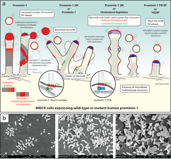
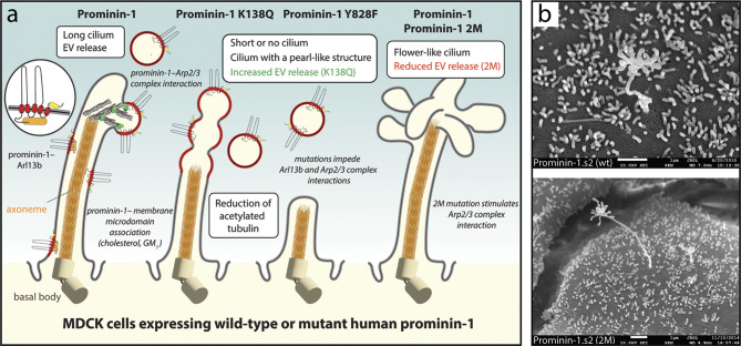
Extracellular membrane vesicles (EVs) offer promising values in various medical fields, e.g., as biomarkers in liquid biopsies or as native (or bioengineered) biological nanocarriers in tissue engineering, regenerative medicine and cancer therapy. Based on their cellular origin EVs can vary considerably in composition and diameter. Cell biological studies on mammalian prominin-1, a cholesterol-binding membrane glycoprotein, have helped to reveal new donor membranes as sources of EVs. For instance, small EVs can originate from microvilli and primary cilia, while large EVs might be produced by transient structures such as retracting cellular extremities of cancer cells during the mitotic rounding process, and the midbody at the end of cytokinesis. Here, we will highlight the various subcellular origins of prominin-1+ EVs, also called prominosomes, and the potential mechanism(s) regulating their formation. We will further discuss the molecular and cellular characteristics of prominin-1, notably those that have a direct effect on the release of prominin-1+ EVs, a process that might be directly implicated in donor cell reprogramming of stem and cancer stem cells. Prominin-1+ EVs also mediate intercellular communication during embryonic development and adult homeostasis in healthy individuals, while disseminating biological information during diseases.

Keywords: CD133; Cell signaling; Cilium; Ectosome; Exosome; Intercellular communication; Lipid droplet; Microvillus; Midbody; Stem cell.


Conflict of interest statement

Declarations. Ethics approval and consent to participate: Not applicable. Consent for publication: Not applicable. Competing interests: The authors declare no competing interests.

Figures

Fig. 1
Fig. 1
Small prominin-1+ ectosomes are released from microvilli on the apical plasma membrane of epithelial cells. a, b MDCK cells stably transfected with wild-type (wt) or mutants of human prominin-1.s1 splice variant were characterized in terms of morphology and EV release. Their effects on microvilli microvillar architecture and dynamics are illustrated (a). Scanning electron microscopy (SEM) micrographs of MDCK cells either non-transfected (MDCK) or expressing prominin-1 wt and the 2M mutant are shown (b). The 2M mutation affects the GM1-binding site at the extracellular N-terminus of prominin-1 (yellow rounded box), while the Y819F mutation impairs the phosphorylation of tyrosine (Y) 819 (or Y828F in the s2 variant) in the cytoplasmic C-terminal domain. The latter post-translational modification stimulates interactions with the Arp2/3 complex (green) and the PI3K (cyan). Cholesterol-rich membrane microdomains (outlined in red) could be generated either by a gradient of membrane cholesterol towards the tips of microvilli, or by the coalescence of small prominin-1-containing membrane microdomains with both mechanisms leading to the budding of ectosomes (left). Expression of human prominin-1 increases the number of microvilli and leads to branched and/or clustered microvilli, a phenotype stimulated by the 2M mutation, which also promotes the formation of microvilli with numerous membrane constrictions and reduces the release of prominin-1+ ectosomes (middle). The “pearling” state is reminiscent of the phenotype observed upon cholesterol depletion (CD) by means of mβCD treatment. The Y819F (or Y828F) mutation results in short microvilli without any increase in prominin-1+ ectosome release (right). The main phenotypes (white boxes) and potential causes (in italics) are indicated. Illustration (a) and micrographs (b) are based on results presented in Refs [54, 83, 200, 221, 234]. For technical details see Ref [200]. Scale bars are indicated
Fig. 2
Fig. 2
Small prominin-1+ ectosomes are released from the primary cilium on the apical plasma membrane of epithelial cells. a, b MDCK cells stably transfected with wild-type (wt) or mutants of human prominin-1.s2 splice variant were characterized in terms of morphology and EV release. The effects on ciliary architecture and dynamics are illustrated (a). SEM micrographs of prominin-1 wt and 2M mutant are shown (b). Mutants K138Q and Y828F affect the Arl13b/HDAC6-binding site or the phosphorylation of tyrosine (Y) 828 in the first cytoplasmic loop or C-terminal domain of prominin-1, respectively. The latter post-translational modification favors the interaction with the Arl13b (orange) and Arp2/3 complex (green). Mutant 2M affects the GM1-binding site (yellow rounded box) in prominin-1. Cholesterol-rich prominin-1-containing membrane microdomains are highlighted in red, and their coalescence can lead to the budding of prominin-1+ ectosomes (left). Expression of human prominin-1 increases primary cilia length, while the K138Q and Y828F mutants reduce cilia length. In the case of the K138Q mutant, some cells show longer cilia with numerous membrane constrictions, and the release of prominin-1+ ectosomes is increased (middle). Although rarely, the expression of prominin-1 wt and 2M mutant can create primary cilia with abundant membrane extensions at their tips (right). The main phenotypes (white boxes) and potential causes (in italics) are indicated. Illustration (a) and micrographs (b) are based on results presented in Refs [57, 140, 142, 221]. For technical details see Refs [140, 200]. Scale bars are indicated
Fig. 3
Fig. 3
Dividing neural progenitor cells release the prominin-1+ midbody and small ectosomes derived therefrom. a Various EVs are released into the CSF during brain development. In neuroepithelial (NE) cells, small ectosomes (prominosomes) may originate from the central part of the midbody, where prominin-1 is concentrated in nascent buds. Large prominin-1+ectosomes lacking tubulin and anillin, an actin-binding protein associated with the contractile ring of the midbody, are also released. Bilateral membrane fissions on both flanks of the central part of the midbody (red dashed line) lead to its release as secreted midbody remnant and constitutes another source of large prominosomes containing tubulin and anillin. b Various fates of the secreted midbody remnant. After a second abscission-like fission (step i, red dashed line), the midbody remnant is released into the brain’s ventricular system and can be transported through the CSF flow to distant cells (ii). Potentially, small and/or large prominosomes continue to bud from the secreted midbody remnant (iii), and/or its extracellular degradation could also occur (iv). Alternatively, the secreted midbody remnant can remain near the cell surface (ii’) or be endocytosed by a phagocytosis-like process (iii’). Actin coats around the endocytosed secreted midbody remnant might slow down its degradation (iv). The midbody could be internalized by distant cells after transport via the CSF. The main events (white boxes) and the potential fate of a given EV (italics) are indicated. Illustrations are based on results presented in Refs [57, 343, 349, 350, 354, 355] and adapted from Ref [55]
Fig. 4
Fig. 4
Prominin-1 is released into the extracellular milieu in association with various types of prominosomes. a, a’ In FEMX-I melanoma cells, prominin-1 is distributed over the cell surface during interphase, while the cell extremities contain mitochondria and are enriched in lipid droplets. Small prominosomes are released as exosomes and/or ectosomes and display all characteristics of cholesterol-rich membrane microdomains (red segment). As the cell enters mitosis, the rounding process leads to the retraction of their extremities involving the disassembly of focal adhesion. In some cases, the retraction of a cell extremity is not fully accomplished (dashed arrow) resulting in the release of large prominosomes containing lipid droplets and mitochondria. The so-called extracellular lipidosomes can be taken up by other cells (not illustrated) or may contribute indirectly to the asymmetric distribution of certain organelles between daughter cells. b-d Micrographs of extracellular lipidosomes observed either by SEM (b) or confocal laser scanning microscopy after triple staining of wild-type (WT) melanoma cells with fluorescently conjugated WGA to highlight glycoconjugates and the fluorescent dyes BODIPY™ 493/503 and MitoView™ Fix 640, which label lipid droplets and mitochondria, respectively (c, d). e Silencing of prominin-1 (PROM1) leads to a redistribution of lipid droplets from the cell extremities to the perinuclear regions (not shown), accompanied by a reduction or complete loss of lipid droplets on the resulting large EVs. The distribution and number of mitochondria are unaffected. f In hematopoietic stem and progenitor cells (or cancer cells), prominin-1 is endocytosed en route to late endosomes/MVBs, which will either fuse with the plasma membrane to release their prominin-1+ ILVs as exosomes, or fuse with lysosomes and initiate degradation. The ubiquitination of prominin-1 and its interactions with TSG101 or syntenin-1 (and their interacting partners such as syndecan, ALIX or other proteins of the ESCRT-1 or ESCRT-III complex) may contribute to the sorting of prominin-1 into ILVs. The main events (white boxes) and potential causes (in italics) are indicated. Illustrations are based on results presented in Refs [11, 170, 181, 241, 358, 373]. Scale bars are indicated
Fig. 5
Fig. 5
Banana-like shapes of prominin proteins predicted by AlphaFold. a-e Models of mammalian prominin-1 (a-c) and prominin-2 (d), and Drosophila melanogaster and Caenorhabditis elegans homologues (e) are displayed. A lateral view human prominin-1.s1 (UniProt number 043490) is presented with its five transmembrane (TM) domains, three extracellular (EC) and intracellular (IC) domains. The predicted disulfide-bridges formed between thiol groups of cysteine (C) residues are indicated. The sequence and position of the potential GM1 ganglioside-binding site (GM1, cyan rounded box) and a facultative small exon (exon 4, red box) present in the s2 splice variant, but absent in s1 variant, are shown. This small 9-amino-acid residue encoding exon is located between the potential GM1-binding site and the first transmembrane segment (TM1, blue rounded box) of prominin-1 (b). The position of all asparagine (N) residues found in the consensus N-glycosylation site (Asn-X-Ser/Thr-Y sequons, where X and Y ≠ proline residue) are indicated (c). The predicted tertiary structure of prominin-2 (Q8N271) is very close to that of prominin-1 (d). The non-mammalian prominin-like protein found in Drosophila melanogaster (P82295) and Caenorhabditis elegans (Q19188) also exhibits a banana-like shape, both displaying a stronger curvature than the mammalian ones (e). Note that an additional transmembrane segment (TM1) is predicted for the Drosophila prominin-like protein in place of a signal peptide, suggesting that this concave side of prominin will face the cellular membrane. Illustrations are based on data presented in the AlphaFold Protein Structure Database [417, 418]

References

    1. Yáñez-Mó M, Siljander PR, Andreu Z, Zavec AB, Borras FE, Buzas EI, et al. Biological properties of extracellular vesicles and their physiological functions. J Extracell Vesicles. 2015;4:27066. - PMC - PubMed
    1. Welsh JA, Goberdhan DCI, O’Driscoll L, Buzas EI, Blenkiron C, Bussolati B, et al. Minimal information for studies of extracellular vesicles (MISEV2023): From basic to advanced approaches. J Extracell Vesicles. 2024;13(2):e12404. - PMC - PubMed
    1. Raposo G, Stoorvogel W. Extracellular vesicles: exosomes, microvesicles, and friends. J Cell Biol. 2013;200(4):373–83. - PMC - PubMed
    1. Colombo M, Raposo G, Théry C. Biogenesis, secretion, and intercellular interactions of exosomes and other extracellular vesicles. Annu Rev Cell Dev Biol. 2014;30:255–89. - PubMed
    1. Meldolesi J. Exosomes and Ectosomes in Intercellular Communication. Curr Biol. 2018;28(8):R435–44. - PubMed

LinkOut - more resources