. 2025 Jan 29;23(1):59.
doi: 10.1186/s12951-024-03087-y.

Anti-inflammatory coupled anti-angiogenic airway stent effectively suppresses tracheal in-stents restenosis

Affiliations

Anti-inflammatory coupled anti-angiogenic airway stent effectively suppresses tracheal in-stents restenosis

Yanan Zhao et al. J Nanobiotechnology. .

Abstract

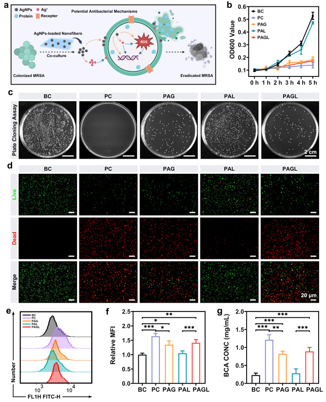
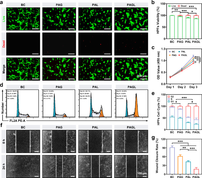
Excessive vascularization during tracheal in-stent restenosis (TISR) is a significant but frequently overlooked issue. We developed an anti-inflammatory coupled anti-angiogenic airway stent (PAGL) incorporating anlotinib hydrochloride and silver nanoparticles using advanced electrospinning technology. PAGL exhibited hydrophobic surface properties, exceptional mechanical strength, and appropriate drug-release kinetics. Moreover, it demonstrated a remarkable eradication effect against methicillin-resistant Staphylococcus aureus. It also displayed anti-proliferative and anti-angiogenic properties on human umbilical vein endothelial cells and lung fibroblasts. PAGL was implanted into the tracheae of New Zealand rabbits to evaluate its efficacy in inhibiting bacterial infection, suppressing the inflammatory response, reducing angiogenesis, and attenuating excessive fibroblast activation. RNA sequencing analysis revealed a significant downregulation of genes associated with fibrosis, intimal hyperplasia, and cell migration following PAGL treatment. This study provides insight into the development of airway stents that target angiogenesis and inflammation to address problems associated with TISR effectively and have the potential for clinical translation.

Keywords: Airway stent; Anlotinib; Silver nanoparticles; Tracheal in-stents restenosis.


Conflict of interest statement

Declarations. Ethics approval and consent to participate: All animal experiments were granted approval by the animal care committee of the First Affiliated Hospital of Zhengzhou University (2023042602). Consent for publication: Written informed consent for publication was obtained from all participants. Competing interests: The authors declare no competing interests.

Figures

Scheme 1
Scheme 1
Schematic illustration of the nanofiber-covered airway stent carrying anlotinib hydrochloride (AL) and silver nanoparticles (AgNPs) against tracheal in-stents restenosis (TISR). (a-b) The preparation process of the PAGL nanofiber-covered airway stent and its application to the trachea of New Zealand rabbits. (c) Unlike the conventional self-expanding metallic stent (SEMS) that results in TISR, the PAGL nanofiber-coated airway stent can remold the inflammatory and hyperplastic tracheal microenvironment to prevent TISR. (d) The PAGL nanofiber-coated airway stent effectively prevents TISR by eradicating methicillin-resistant Staphylococcus aureus (MRSA), suppressing the inflammatory response, inhibiting the uncontrolled angiogenesis, and subsequent excessive ECM deposition. Created in BioRender. Liu, Y. (2024) https://BioRender.com/m52f909
Fig. 1
Fig. 1
Physicochemical properties, mechanical properties, and drug-release behavior of nanofiber films covered on the stent surfaces. (a) TEM image of AgNPs. (b) FTIR spectra and (c) Raman spectra of PLA, PAG, PAL, and PAGL nanofiber films, respectively. (d) SEM images of PLA, PAG, PAL, and PAGL nanofiber films, respectively. (e) EDS elemental mapping results of PAGL nanofiber films. (f) The fitted curve of fiber diameter distribution for PLA, PAG, PAL, and PAGL, respectively. (g) EDS spectra of PAGL nanofiber films. (h) Water contact angle images and statistical analyses. (i) Typical stress-strain curves of all nanofiber films. (j) AgNPs release profile from PAG and PAGL for 0–21 days at 37 °C, respectively. (k) AL release profile from PAG and PAGL for 0–21 days at 37 °C, respectively. Scale bar: 10 nm, 500 nm, and 2 μm
Fig. 2
Fig. 2
Antibacterial performance of the nanofiber films covered on the stent surface. (a) Mechanism diagram for the inhibition of bacterial growth by the nanofiber films. (b) The proliferation of MRSA. (c) Representative colony formation images of MRSA. (d) Representative live/dead bacterial assay of MRSA. (e-f) The fluorescence intensity histograms and relative mean fluorescence intensity (MFI) of the intracellular ROS levels in MRSA. (g) Protein leakage analysis of MRSA. Scale bar: 20 μm and 2 cm. Significantly different: *P < 0.05, **P < 0.01, and ***P < 0.001
Fig. 3
Fig. 3
In vitro anti-angiogenic effect of the nanofiber films covered on the stent surface. (a) Schematic illustration of anti-angiogenic mediated by PAGL. (b) Statistical analysis of HUVECs viability. (c) Cell proliferation of HUVECs over three consecutive days by CCK-8 assay. (d) Flow cytometry to detect cell apoptosis in HUVECs stained with Annexin V/PI. (e) Scratch assay and Transwell assay of HUVECs. (f) The proportion of apoptosis based on the cell apoptosis assay for HUVECs. (g) Quantitative results of the wound closure rate. (h) Quantitative results of the migrating HUVECs number. (i) Representative VEGFR2 immunofluorescence staining of HUVECs. (j) Statistical analysis of the relative mean fluorescence intensity (MFI) for VEGFR2 in HUVECs. Scale bar: 40, 100, and 200 μm. Significant differences: *P < 0.05, **P < 0.01, and ***P < 0.001
Fig. 4
Fig. 4
In vitro anti-hyperplasia of nanofiber films covered on the stent surface. (a) Representative live/dead cell staining of HPFs. (b) Statistical analysis of HPFs viability. (c) Cell proliferation of HPFs over three consecutive days by CCK-8 assay. (d) Flow cytometry of cell cycle for HPFs stained with PI. (e) Statistical analysis of cell cycle percentage for HPFs. (f) Scratch assay of HUVECs. (g) Quantitative results of wound closure rate. Scale bar: 100 and 200 μm. Significant differences: *P < 0.05, **P < 0.01, and ***P < 0.001
Fig. 5
Fig. 5
Airway stent placement and evaluation of postoperative TISR. (a) Schematics of the procedural events in the New Zealand rabbit models treated with airway stent placement. (b) Diagram of DSA fluoroscopic monitoring images of the airway stent placement procedure in New Zealand rabbits. (c) Airway volume rendering technique (VRT) images, stent VRT images, and sagittal CT images four weeks after airway stent placement. (d) H&E and Masson’s Trichrome staining of the tracheal tissues. (e) Airway stenting time for each group. (f) Tracheal ventilation ratio in rabbits in each group four weeks after airway stent placement. (g) Statistical analysis of tracheal epithelial thickness. (h) Statistical analysis of collagen deposition. Scale bar: 100 μm. Significant differences: *P < 0.05, **P < 0.01, and ***P < 0.001
Fig. 6
Fig. 6
Airway stent suppresses the inflammation response and angiogenesis. (a) Bacterial plate cloning representation of the diluted bacterial contents. (b) Statistical analysis of the bacterial contents. (c) Correlation analysis of bacterial contents and granulation tissue formation between the Ctrl and PAG groups. (d-g) Expression levels of IL-8, MCP-1, IL-6, and TNF-α in the tracheal tissue. (h) Immunofluorescence staining of CD31 in the tracheal tissue. (i) Double immunofluorescence staining of Ki67 and Caspase-3 in the tracheal tissue. (j) Immunohistochemical staining of α-SMA and Sirius red staining in the tracheal tissue. (k) Statistical analysis of relative mean fluorescence intensity (MFI) for CD31. (l) Statistical analysis of the relative MFI for Ki67. (m) Statistical analysis of the relative MFI for Caspase-3. (n) The relative mRNA expression of VEGFR2, α-SMA, and Col IIIa1, respectively. Scale bar: 50 μm, 100 μm, and 1 cm. Significant differences: *P < 0.05, **P < 0.01, and ***P < 0.001
Fig. 7
Fig. 7
Biological function analysis of PAGL treatment. (a) A heatmap illustrating the differentially expressed genes (DEGs) identified in the PAGL and Ctrl groups (n = 3). (b-c) Gene Ontology enrichment analysis of the identified DEGs in the Ctrl and PAGL groups, respectively. (d) Gene Set Enrichment Analysis results are based on the fold-change in gene expression between the PAGL and Ctrl groups. (e) Multiscale embedded gene co-expression network analysis. (f) Three clusters were obtained using MEGENAR package. (g) Overlap of cluster 3 genes with DEGs and Angiogenesis-related genes. Sixteen genes: SIRT1, OVOL2, VEGFA, TSPAN12, EIF2AK3, KCNJ8, GJC1, MIA3, SENP2, ADIPOR2, SLC12A2, SGCB, PTN, ACE, GAPDH, and TGFBR1. (h) Protein-protein interaction analysis

Similar articles

References

    1. Guibert N, Saka H, Dutau H. Airway stenting: Technological advancements and its role in interventional pulmonology. Respirology. 2020;25:953–62. - PubMed
    1. Murgu SD, Egressy K, Laxmanan B, Doblare G, Ortiz-Comino R, Hogarth DK. Central Airway obstruction: Benign strictures, Tracheobronchomalacia, and malignancy-related obstruction. Chest. 2016;150:426–41. - PubMed
    1. Li L, Zhang X, Shi J, Chen Y, Wan H, Herth FJ, Luo F. Airway stents from now to the future: a narrative review. Respiration. 2023;102:439–48. - PubMed
    1. Avasarala SK, Freitag L, Mehta AC. Metallic endobronchial stents: a contemporary resurrection. Chest. 2019;155:1246–59. - PubMed
    1. Lin L-Q, Chen D-F, Wu H-K, Chen Y, Zhong C-H, Chen X-B, Tang C-L, Zhou Z-Q, Li S-Y. Long-term efficacy and safety of the Dumon stent for treatment of benign airway stenosis. Ther Adv Respir Dis 2023, 17. - PMC - PubMed

MeSH terms

LinkOut - more resources