. 2025 Apr 2;33(4):1466-1484.
doi: 10.1016/j.ymthe.2025.01.042. Epub 2025 Jan 28.

Systematic empirical evaluation of individual base editing targets: Validating therapeutic targets in USH2A and comparison of methods

Affiliations

Systematic empirical evaluation of individual base editing targets: Validating therapeutic targets in USH2A and comparison of methods

Yuki Tachida et al. Mol Ther. .

Abstract

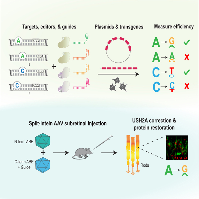
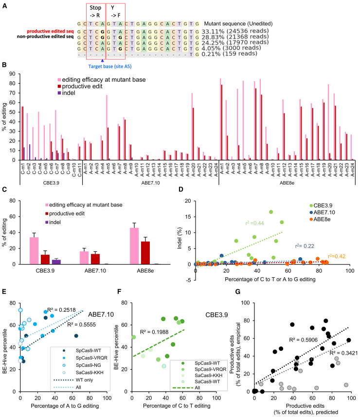
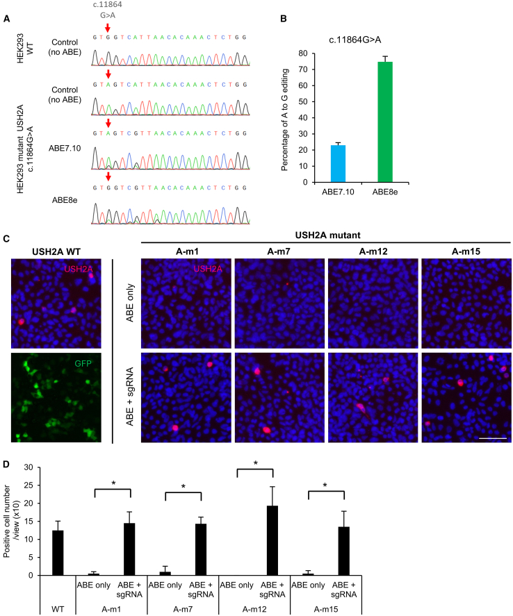
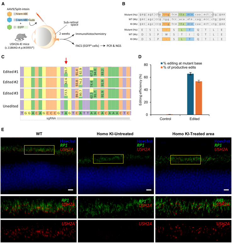
Base editing shows promise for the correction of human mutations at a higher efficiency than other repair methods and is especially attractive for mutations in large genes that are not amenable to gene augmentation therapy. Here, we demonstrate a comprehensive workflow for in vitro screening of potential therapeutic base editing targets for the USH2A gene and empirically validate the efficiency of adenine and cytosine base editor/guide combinations for correcting 35 USH2A mutations. Editing efficiency and bystander edits are compared between different target templates (plasmids vs. transgenes) and assays (next-generation sequencing vs. Sanger), as well as comparisons between unbiased empirical results and computational predictions. Based on these observations, practical assay recommendations are discussed. Finally, a humanized knockin mouse model was created with the best-performing target, the nonsense mutation c.11864G>A p.(Trp3955∗). Split-intein AAV9 delivery of editing reagents resulted in the restoration of USH2A protein and a correction rate of 65% ± 3% at the mutant base pair and of 52% ± 3% excluding bystander amino acid changes. This efficiency is higher than that seen in a retinal gene editing program testing in a clinical trial. These results demonstrate the effectiveness of this overall strategy to identify and test base editing reagents with the potential for human therapeutic applications.

Keywords: AAV; USH2A; Usher syndrome; adenine base editor; base editing; cytosine base editor; photoreceptors; retina; retinitis pigmentosa.


Conflict of interest statement

Declaration of interests Patent US20230159913A1 was issued based on the reported work. J.C. has received consulting payments from Applied Genetic Technologies Corporation, Beam Therapeutics, Biogen, Gensight Biologics, Octant Bio, Wave Life Sciences, and Vedere. Q.L. has received consulting payments from Editas Medicine and Entrada Therapeutics. Y.T. received salary support from his employer, Daiichi Sankyo Co., Ltd., while acting as a visiting scientist at Mass Eye and Ear. D.R.L. is a consultant and equity owner of Beam Therapeutics, Prime Medicine, Pairwise Plants, Chroma Medicine, and Nvelop Therapeutics, companies that use or deliver genome editing or genome engineering agents. J.M.L. is currently an employee of Prime Medicine.

Figures

None
Graphical abstract
Figure 1
Figure 1
Experimental approach (A) Schema describing the in silico filtering of candidate USH2A mutations from LOVD and HGMD to be tested for correction by base editing. (B) Numbering convention used for describing base editing target locations. The bottom strand runs 5′ to 3′ and contains the target A or C to be edited (red) and the PAM sequence (blue).
Figure 2
Figure 2
System for evaluating base editing on multiple target mutations (A) Schematic showing the transgene consisting of universal primers and a tandem array of 51 bp human USH2A sequences containing pathogenic mutations. (B) Strategy for establishment of the transgenic, stable HEK293 cell lines, using a donor vector (pAAVS1-puro-DNR) targeting the AAVS1 site by HDR. (C) Experimental protocol for establishing the transgene stable cell line; and base editing of candidate sites in transient transfected plasmids and stable transgenes. (D) Comparison of editing efficiency of the target sites on a transiently transfected plasmid vs. on a stably integrated transgene, using Sanger sequencing. (E) Similarity between base editing efficiency quantification using Sanger sequencing vs. NGS amplicon sequencing. (F) Comparison of the editing efficiency in the Dnmt1 locus in the stable cell line vs. mouse retina. Labels (e.g., C1) represent the position of the edited cytosine in the protospacer sequence, shown above.
Figure 3
Figure 3
Editing efficiency of base editor/guide combinations on all target mutations, measured using Sanger sequencing with EditR quantification, in stably transfected cell lines with USH2A target mutations (A) C-to-T editing efficiency of CBE and (B) A-to-G editing efficiency of ABE7.10 and ABE8e are shown, with mean ± SD (n = 3). (C) Average editing efficiency of all CBE targets, grouped by the three different Cas9 PAM variants used. (D) Average editing efficiency of all ABE targets, grouped by the four different Cas9 PAM variants used.
Figure 4
Figure 4
Editing efficiency of base editor/guide combinations on all target mutations, measured using NGS analysis, in stable transgene cell lines with USH2A target mutations (A) Example editing pattern around position A5 of mutation, A-m5. A “productive edit” is defined as an edit in which the amino acid sequence is changed to wild type. A “non-productive edit” is defined as an edit with unwanted amino acid changes. (B) Editing efficiency at each mutant site is shown at the mutant base itself, and as the productive editing rate summed over all observed alleles. The indel rate includes all insertions or deletions detected. (C) Average editing efficiency and productive editing rate is shown for the three base editors used. (D) Editing efficiency at mutant base vs. indel frequency, with each point representing one mutant site. (E and F) Comparison between the experimental value of editing efficiency and predicted percentile value by BE-HIVE using ABE 7.10 (E) and CBE3.9 (F). Point shading indicates the type of Cas9 PAM variant. The dark blue and light blue (E) or light green (F) regression lines were calculated on the data of only ABE-SpCas9-WT and all ABE SpCas9 variants, respectively. (G) Correlation between the pattern of editing (as measured by the fraction of all edits which were productive edits) as measured experimentally vs. as predicted by BE-HIVE. The correlation is higher for the subset of mutant sites which have >15% editing efficiency (black dots, black regression line), compared with all sites (black and gray dots, gray regression line).
Figure 5
Figure 5
Off-target editing The top 10 predicted off-target sites for C-m6 (A) and A-m1 (B) were identified using the CRISPOR prediction tool. Editing rates are shown as mean ± SD (n = 3; Sanger sequencing). (C) A low correlation is seen between the MIT off-target score and the empirical editing rate at off-target sites.
Figure 6
Figure 6
Restoration of USH2A protein expression with base editing in cells in vitro (A) Sanger sequencing shows that the A-m1 mutation (c.11864 G>A; red arrow) was introduced into the native USH2A genomic locus, in all triploid chromosomes of a HEK293 cell line (red arrow). (B) This cell line was edited with ABE7.10 or ABE8e, and the editing efficiency was quantified (n = 3 biological replicates, p ≤ 0.0001, Student’s t test comparing ABE7.10 vs. ABE8e). (C) Immunostaining of USH2A in HEK293 2 days after transient co-transfection with plasmid vector of a full-length USH2A cDNA wild-type (WT) or mutant (A-m1, A-m7, A-m12, A-m15) plasmid and base editor/sgRNA plasmid vector. In contrast to (A) and (B), these mutations are not in the genomic locus but are transfected using plasmids. USH2A full-length protein is stained (red) with a USH2A antibody that recognizes intracellular region. Nuclei are stained with Hoechst 33342 (blue). Scale bar, 50 μm. eGFP (lower left; a representative separate example) serves as a transfection control and is no longer detectable after permeabilization for USH2A staining (other panels). (D) Cells positive for USH2A full-length protein were counted in six fields (mean ± SD, n = 6), ∗p < 0.001, by two-tailed Student’s t test.
Figure 7
Figure 7
Subretinal delivery of ABEs via split-inteins in a humanized mouse model of the USH2A site A-m1 mutation (A) Schema showing that 2 weeks after subretinal injection of AAV9 ABE base editors, retinas were assayed using immunohistochemistry and measurement of editing rates by next-generation sequencing (NGS) of whole bulk retina or FACS enriched eGFPhi retinal cells. (B) The DNA and protein sequences of the human mutant, mouse wild type, and mouse knockin are presented, showing the guide sequence (box), the mutant site (red), and the regions that differ between human and mouse (indicated colors). Seven base pairs within the guide sequence were humanized to generate an editable mutant sequence in the mouse genome. (C) CRISPResso2 analysis comparing editing efficiencies between retinas (eGFPhi cells) and unedited control (N-term ABE alone + eGFP) at the target site (arrow) and bystander sites, along with the reference sequence and corresponding sgRNA binding region (bottom). Percentages reflect the frequency of the predominant base pair, which has changed from A to G in two positions. (D) Bar graph showing the percentage of editing at the mutant base and the percentage of productive edits in the eGFPhi (cells with high eGFP expression driven by the rhodopsin kinase promoter) FACS enriched retinal cells (n = 3 biological replicates, p < 0.0001, Student’s t test comparing percentage editing at the mutant base for control vs. edited samples, p < 0.0001, Student’s t test comparing percentage of productive edits for control vs. edited samples). (E) Expression of USH2A in the wild-type mouse retina (left) shows USH2A antibody staining (red) at the connecting cilium and RP1 antibody staining (green) in the axoneme at the base of the photoreceptor outer segment. The outer nuclear layer is counterstained with Hoechst (blue). An untreated area in an injected homozygous knockin (KI) retina (middle), shows no USH2A staining. In contrast, in the homozygous KI retina following base editing, restoration of USH2A staining and localization is observed (right). Scale bar, 10 μm.

References

    1. Pierce E.A., Aleman T.S., Jayasundera K.T., Ashimatey B.S., Kim K., Rashid A., Jaskolka M.C., Myers R.L., Lam B.L., Bailey S.T., et al. Gene Editing for CEP290-Associated Retinal Degeneration. N. Engl. J. Med. 2024;390:1972–1984. doi: 10.1056/NEJMoa2309915. - DOI - PMC - PubMed
    1. Doudna J.A., Charpentier E. Genome editing. The new frontier of genome engineering with CRISPR-Cas9. Science. 2014;346 doi: 10.1126/science.1258096. - DOI - PubMed
    1. Jinek M., Chylinski K., Fonfara I., Hauer M., Doudna J.A., Charpentier E. A programmable dual-RNA-guided DNA endonuclease in adaptive bacterial immunity. Science. 2012;337:816–821. doi: 10.1126/science.1225829. - DOI - PMC - PubMed
    1. Komor A.C., Kim Y.B., Packer M.S., Zuris J.A., Liu D.R. Programmable editing of a target base in genomic DNA without double-stranded DNA cleavage. Nature. 2016;533:420–424. doi: 10.1038/nature17946. - DOI - PMC - PubMed
    1. Gaudelli N.M., Komor A.C., Rees H.A., Packer M.S., Badran A.H., Bryson D.I., Liu D.R. Programmable base editing of A∗T to G∗C in genomic DNA without DNA cleavage. Nature. 2017;551:464–471. doi: 10.1038/nature24644. - DOI - PMC - PubMed

Substances

LinkOut - more resources