. 2025 Jan 20;7(1):fcaf026.
doi: 10.1093/braincomms/fcaf026. eCollection 2025.

Multidimensional structural analyses revealed a correlation between thalamic atrophy and white matter degeneration in idiopathic dystonia

Affiliations

Multidimensional structural analyses revealed a correlation between thalamic atrophy and white matter degeneration in idiopathic dystonia

Jinping Xu et al. Brain Commun. .

Abstract

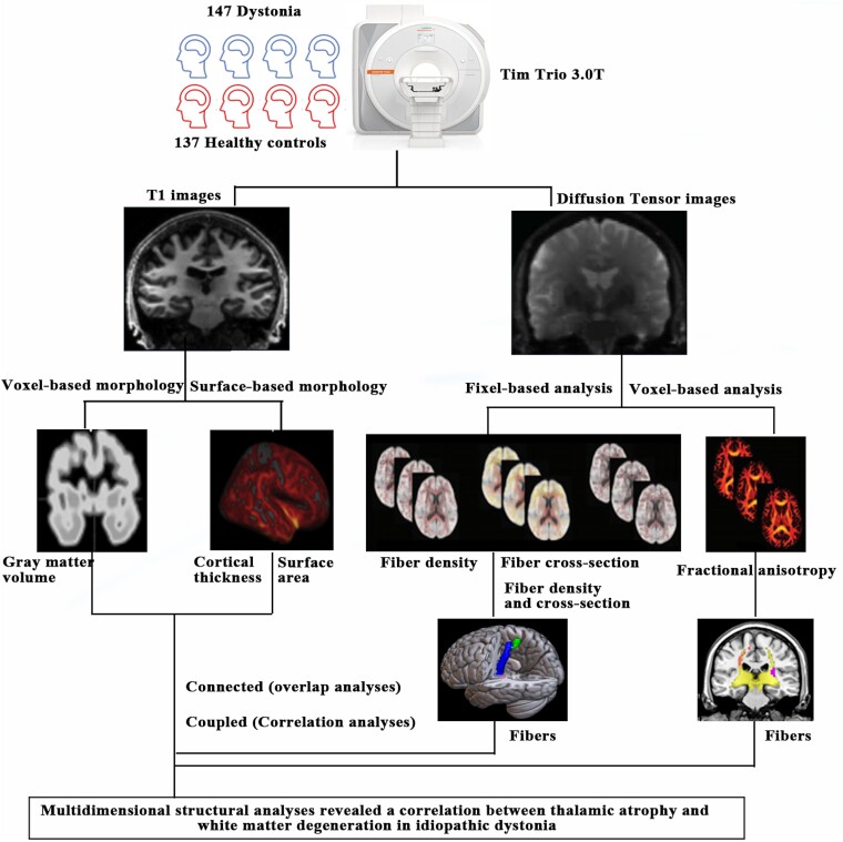
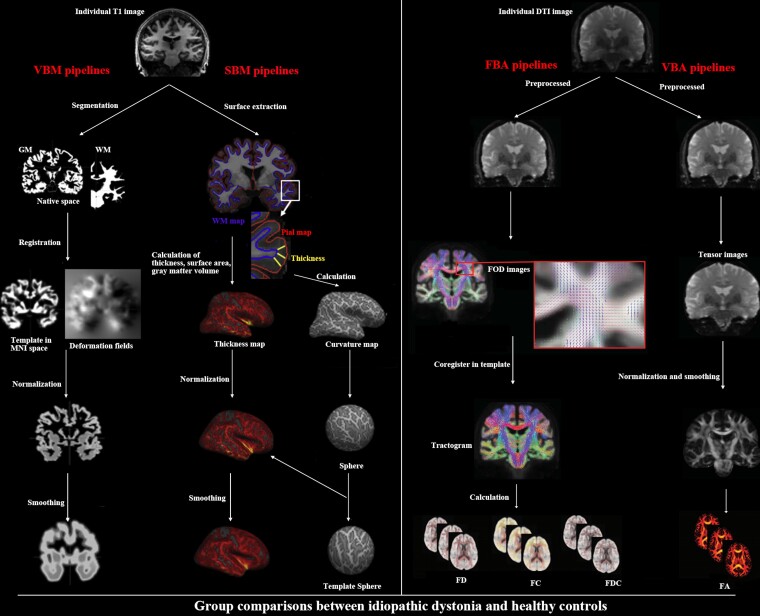
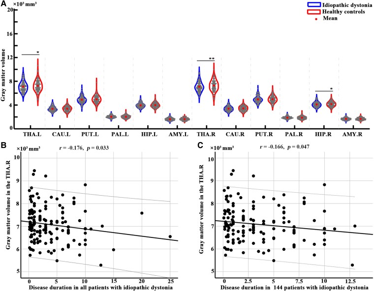
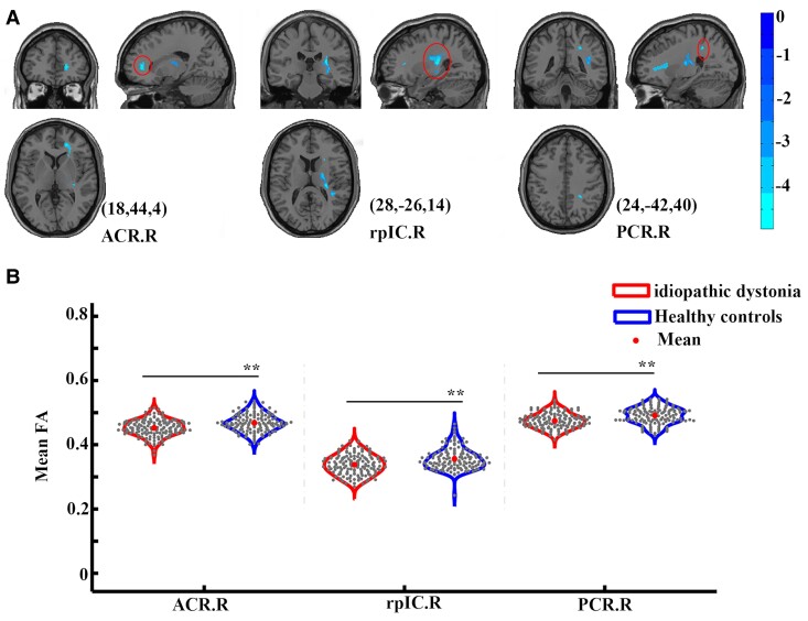
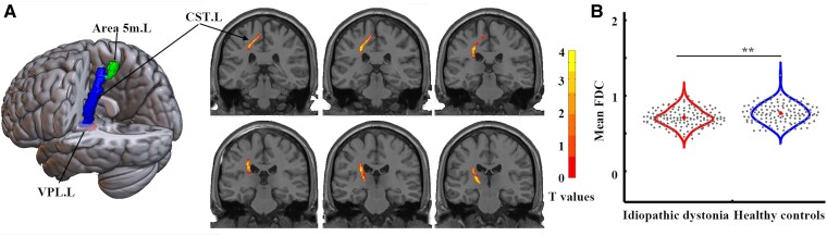
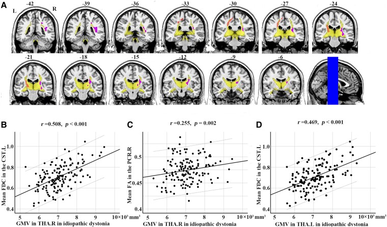
Although aberrant changes in grey and white matter are core features of idiopathic dystonia, few studies have explored the correlation between grey and white matter changes in this disease. This study aimed to investigate the coupling correlation between morphological and microstructural alterations in patients with idiopathic dystonia. Structural T1 imaging and diffusion tensor imaging were performed on a relatively large cohort of patients. Multidimensional structural analyses, including voxel-based analyses, voxel-based morphology, fixel-based analyses and surface-based morphometry, were performed to explore these structural alterations. Probabilistic tractography and correlation analyses were employed to examine these relationships. A total of 147 patients with idiopathic dystonia and 137 healthy controls were recruited in this study. There were no significant differences in the cortical morphometry between patients with idiopathic dystonia and healthy controls using voxel- and surface-based morphometry. However, the grey matter volume of the bilateral thalamus, fractional anisotropy in the right anterior corona radiata, right retrolenticular part of the internal capsule and right posterior corona radiata, and the fibre density and cross-section combined in the fibre tract connecting the left ventral posterolateral thalamic nucleus and left area 5 m, were significantly decreased in patients with idiopathic dystonia compared with those in healthy controls. Furthermore, the reduced grey matter volume in the right thalamus not only correlated with the disease duration but also with the reduced fractional anisotropy in the right posterior corona radiata and decreased the fibre density and cross-section combined in the fibre tract connecting the left ventral posterolateral thalamic nucleus and the left area 5 m in patients with idiopathic dystonia. These findings suggest that the thalamus is structurally impaired in idiopathic dystonia and that microstructural disruption in thalamocortical projections occurs secondary to thalamic atrophy.

Keywords: fixel-based analyses; idiopathic dystonia; surface-based morphometry; voxel-based analyses; voxel-based morphology.


Conflict of interest statement

The authors report no competing interests.

Figures

Graphical abstract
Graphical abstract
Figure 1
Figure 1
Pipelines of multidimensional structural analyses. Voxel-based morphology and surface-based morphology were performed for T1 images to investigate grey matter alterations, as well as the voxel-based analyses and fixel-based analyses were performed for diffusion tensor images to investigate white matter alterations. DTI, diffusion tensor imaging; FA, fractional anisotropy; FBA, fixel-based analysis; FC, fibre cross-section; FD, fibre density; FDC, fibre density and cross-section combined; FOD, fibre orientation distribution; GM, grey matter; MNI, Montreal Neurological Institute; SBM, surface-based morphometry; VBA, voxel-based analysis; VBM, voxel-based morphometry; WM, white matter.
Figure 2
Figure 2
Differences in volume in subcortical regions between 147 patients with idiopathic dystonia and 137 healthy controls. (A) The results were obtained using two-sample t-tests with age, sex and estimated total intracranial volume as covariates. The grey dots within the violin plot represent the grey matter volume values for each subject in 12 different subcortical regions, and the central dots indicate the average grey matter volume for intra-group for the corresponding regions. The left side of the each violin plot represents patients with idiopathic dystonia group, and the right side of the each violin plot represents the healthy control group.* represents P < 0.05, ** represents P < 0.05, false discovery rate corrected. (B) Spearman correlations revealed that the decreased grey matter volume in the right thalamus was significantly correlated with disease duration in patients with idiopathic dystonia. (C) The decreased grey matter volume in the right thalamus was significantly correlated with disease duration in 144 patients with idiopathic dystonia (3 patients with disease duration over 15 years were excluded). Abbreviations of subcortical regions are listed in Table 2.
Figure 3
Figure 3
Differences in white matter diffusion between 147 patients with idiopathic dystonia and 137 healthy controls. (A) Decreased FA levels in three clusters were identified between groups via whole-brain voxel-based analysis. The results were obtained using a general linear model analysis with age and sex as covariates and corrected by family-wise error permutation testing of P < 0.05. Peaks with Montreal Neurological Institute coordinates (x, y, z) were shown for each cluster. The colour bar represents the T-value. (B) Differences in FA in regions obtained from whole-brain voxel-based analysis between groups. The results were obtained using two-sample t-tests with age and sex as covariates. The grey dots within the violin plot represent the mean FA values for each subject in three different brain regions, and the central dots indicate the average FA values for intra-group for the corresponding regions. The left side of the each violin plot represents patients with idiopathic dystonia group, while the right side of the each violin plot represents the healthy control group. ** represents P < 0.05 with false discovery rate corrections. ACR.R, right anterior corona radiate; FA, fractional anisotropy; L, left; PCR.R, right posterior corona radiate; R, right; rpIC.R, right retrolenticular part of internal capsule.
Figure 4
Figure 4
Differences in fibre density and FDC between 147 patients with idiopathic dystonia and 137 healthy controls. (A) Decreased FDC in the fibre tract connecting the left ventral posterolateral thalamic nuclear and left area 5 m was identified in patients with idiopathic dystonia compared with healthy controls. The results were obtained using a general linear model analysis with age and sex as covariates and were corrected for multiple comparisons with family-wise error permutation testing of P < 0.05. The left image was shown in MRIcroGL (https://www.nitrc.org/projects/mricrogl), and the right images were shown in MRICron (https://www.nitrc.org/projects/mricron). (B) Differences in FDC in regions obtained from whole-brain analysis between groups. The results were obtained using two-sample t-tests with age and sex as covariates. The grey dots within the violin plot represent the mean FDC values for each subject in the whole brain, and the central dots indicate the average FDC values for intra-group in the whole brain. The left side of the violin plot represents patients with idiopathic dystonia group, and the right side of the violin plot represents the healthy control group. ** represents P < 0.05, false discovery rate corrected. area 5 m. L, left area 5 m; CST.L, the left corticospinal tract; FDC, fibre density and cross-section combined; L, left; R, right; VPL.L, left ventral posterolateral nucleus.
Figure 5
Figure 5
Overlap results and correlations between morphological and microstructural alterations in idiopathic dystonia. (A) Overlay of thalamocortical connections obtained from comparison results in FDC and FA between groups on the population maps of the probabilistic tractography patterns for bilateral thalamus in the healthy controls. The multi-slice views of fibres were shown with MRIcron (https://people.cas.sc.edu/rorden/mricron/). (B)-(C) The black dots of the scatter plot in figure B/C represent the relationship between the mean FDC in the CST.L or mean FA in the PCR.R and GMV in the THA.R in patients with idiopathic dystonia. Spearman correlations showed that GMV in the THA.R was significantly correlated with decreased FDC in the CST.L and FA in the PCR.R in 147 patients with idiopathic dystonia. (D) Spearman correlations showed that GMV in the THA.L was significantly correlated with decreased FDC in the CST.L in patients with idiopathic dystonia. CST.L, the left corticospinal tract; FA, fractional anisotropy; FDC, fibre density and cross-section combined; GMV, grey matter volume; L, left; PCR.R, right posterior corona radiate; R, right; THA.L, left thalamus; THA.R, right thalamus.

Similar articles

References

    1. Battistella G, Termsarasab P, Ramdhani RA, Fuertinger S, Simonyan K. Isolated focal dystonia as a disorder of large-scale functional networks. Cereb Cortex. 2017;27:1203–1215. - PMC - PubMed
    1. Scorr LM, Cho HJ, Kilic-Berkmen G, et al. . Clinical features and evolution of blepharospasm: A multicenter international cohort and systematic literature review. Dystonia. 2022;1:10359. - PMC - PubMed
    1. Marsden CD. The problem of adult-onset idiopathic torsion dystonia and other isolated dyskinesias in adult life (including blepharospasm, oromandibular dystonia, dystonic writer's cramp, and torticollis, or axial dystonia). Adv Neurol. 1976;14:259–276. - PubMed
    1. Kaji R, Bhatia K, Graybiel AM. Pathogenesis of dystonia: Is it of cerebellar or basal ganglia origin? J Neurol Neurosurg Psychiatry. 2018;89:488–492. - PMC - PubMed
    1. Lehéricy S, Tijssen MA, Vidailhet M, Kaji R, Meunier S. The anatomical basis of dystonia: Current view using neuroimaging. Mov Disord. 2013;28:944–957. - PubMed

LinkOut - more resources