Review
. 2025 Jan 28;16(7):3019-3044.
doi: 10.1039/d4sc08422e. eCollection 2025 Feb 12.

Natural biomolecules for cell-interface engineering

Affiliations
Review

Natural biomolecules for cell-interface engineering

Tong-Kai Zhang et al. Chem Sci. .

Abstract

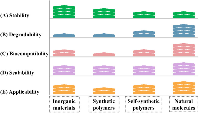
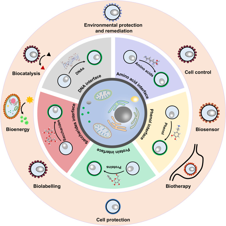
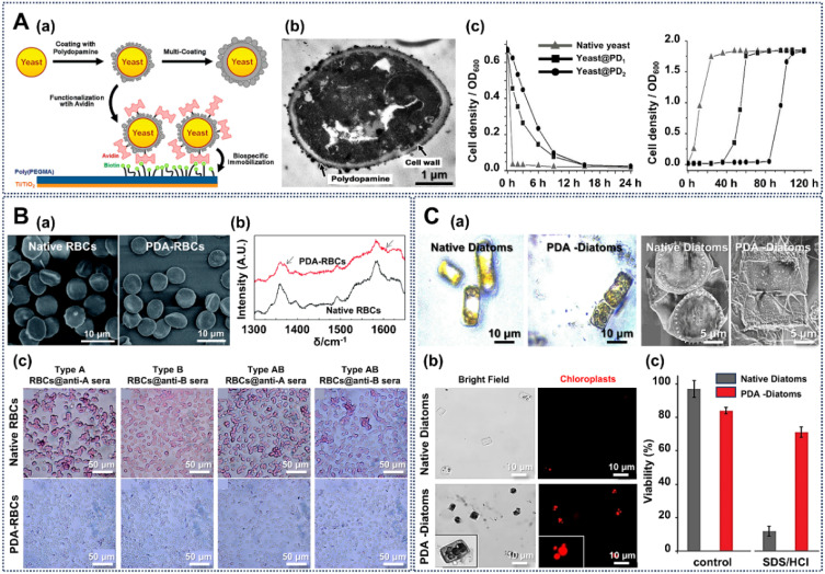
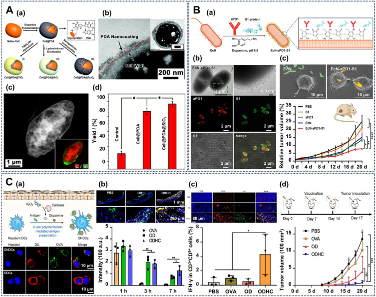
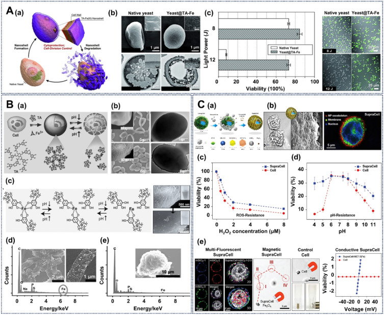
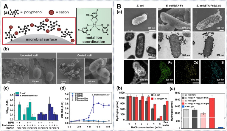
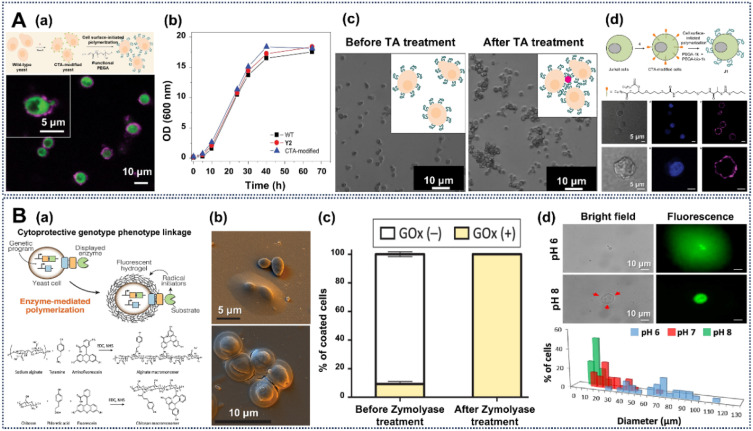
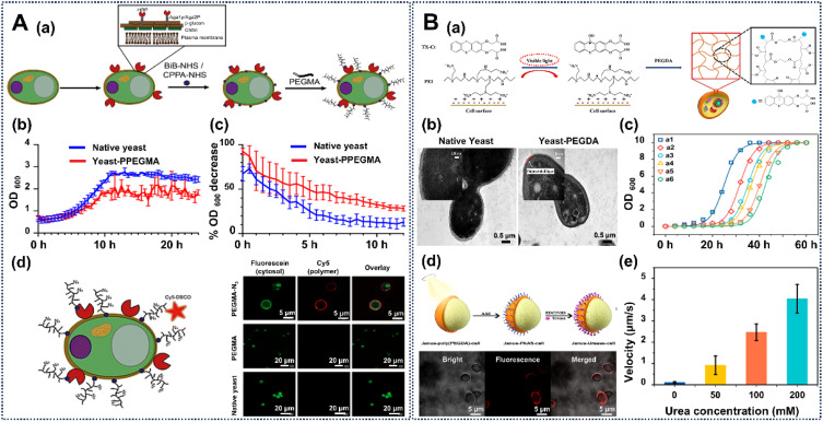
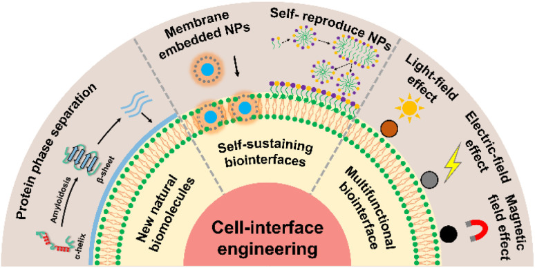
Cell-interface engineering is a way to functionalize cells through direct or indirect self-assembly of functional materials around the cells, showing an enhancement to cell functions. Among the materials used in cell-interface engineering, natural biomolecules play pivotal roles in the study of biological interfaces, given that they have good advantages such as biocompatibility and rich functional groups. In this review, we summarize and overview the development of studies of natural biomolecules that have been used in cell-biointerface engineering and then review the five main types of biomolecules used in constructing biointerfaces, namely DNA polymers, amino acids, polyphenols, proteins and polysaccharides, to show their applications in green energy, biocatalysis, cell therapy and environmental protection and remediation. Lastly, the current prospects and challenges in this area are presented with potential solutions to solve these problems, which in turn benefits the design of next-generation cell engineering.


Conflict of interest statement

The authors declare no conflict of interest.

Figures

Fig. 1
Fig. 1. Strengths and limitations of different types of material for cell-interface engineering. Each material type features inherent unique functionalities and trade-offs (A–F) in cell-interface engineering. (A) Stability represents the durability of the interface around cells, which can be evaluated from the maintenance time of the nanoshell. (B) Degradability indicates the natural degradation of the cell-surface nanoshell over a certain time, the degradation products of which can be absorbed through metabolism. (C) Biocompatibility represents the effect of the cell-surface interface on cell viability, which can be evaluated via the CCK-8 assay or a fluorescein luminescence approach. (D) Scalability indicates the potential of the cell-surface interface for further engineering. (E) Applicability indicates the application prospects of the engineering in comprehensive consideration of the above factors. Achieving the optimal outcome relies on the rational matching of material properties and living-cell functions. Furthermore, different levels of material engineering for living cells may be necessary, depending on the desired outcome.
Fig. 2
Fig. 2. A schematic showing natural biomolecular materials that are used for the creation of nanofunctionalized cells with specific nanofunctions.
Fig. 3
Fig. 3. Creation of engineered cell interfaces based on natural DNA polymers. (A) (a) Schematic illustration of cell-surface oligonucleotide-linked DNA polymer for forming stable cell–cell contacts, and the confocal laser scanning fluorescence microscope (CLSM) images of cells engineered with fluorescein-conjugated DNA assembled with cells with nonfluorescent complementary DNA strands. (b) The kinetic profiles of the DNA-based assembly process. (c) Fluorescence-activated cell-sorting separation of different Jurkat cell assemblies based on fluorescence properties, and fluorescence images of those isolated structures. (d) Multicellular structures of different sizes synthesized based on cell-surface DNA structures. Reproduced with permission from ref. . Copyright 2009, National Academy of Sciences. (B) (a) Schematic illustration of biomimetic cell wall (BCW) synthesis with DNA polymers on the cell surface. (b) Confocal fluorescence images and TEM images of the BCW template on the cell surface. (c) The relationship between the shielding enhancement and centrifugal force. (d) Staining assay of endothelial cells with an anti-CD31 antibody. Reproduced with permission from ref. . Copyright 2019, Springer Nature. (C) (a) Schematic illustration of in situ polymerization reaction with DNA polymers for cell-interface engineering and CLSM images of engineered cells. (b) The polymer density of the DNA cocoons on the MCF-7 cells through branched primers (BPs) with different concentrations. (c) The flow cytometry analysis of cell viability and the encapsulation efficiency with DNA polymers. (d) Precise handling of cells by post-editing of the DNA polymer cocoons around cells. Reproduced with permission from ref. . Copyright 2019, Springer Nature.
Fig. 4
Fig. 4. Creation of engineered cell interfaces based on natural amino acids. (A) (a) Schematic of l-cysteine-based cell-surface engineering. (b) SEM and TEM images and EDX analysis of cell@biohybrid-shell@TiO2-shell cells. (c) Post-functionalization with functional nanomaterials. (d) The desulfurizing activity of cells with different treatments. Reproduced with permission from ref. . Copyright 2014, Royal Society of Chemistry. (B) (a) Schematic of cell-surface engineering with l-cysteine-based biohybrids. (b) SEM and TEM micrographs of yeast@biohybrid interfaces. (c) Cell activity of biohybrid engineered yeast cells exposed to ultraviolet (UV) radiation. (d) Process of self-repairing biohybrid artificial interfaces during the whole process of cell division. Reproduced with permission from ref. . Copyright 2015, Royal Society of Chemistry. (C) (a) Schematic of cell-interface engineering with a bilayered interface around an S. cerevisiae cell. (b) TEM micrographs of a single yeast cell encapsulated with a bilayered interface. (c) Protection of bilayered-interface-engineered cells against UV radiation. Reproduced with permission from ref. . Copyright 2018, Royal Society of Chemistry.
Fig. 5
Fig. 5. Creation of engineered cell interfaces based on natural polydopamine (PDA). (A) (a) Schematic of cell-engineering procedure with PDA. (b) TEM images of the PDA-engineered yeast cells. (c) Growth curve of yeast with or without PDA engineering and protection against lyticase by the PDA interface around yeast cells. Reproduced with permission from ref. . Copyright 2011, American Chemical Society. (B) SEM images (a) and Raman spectra (b) of red blood cells (RBCs) without or with cell-interface engineering with PDA. (c) Antibody-mediated aggregation of human RBCs before and after PDA engineering. Reproduced with permission from ref. . Copyright 2014, Royal Society of Chemistry. (C) (a) Reflection microscopy and SEM images of native diatoms and diatoms with PDA engineering. CLSM images (b) and viability (c) of native diatoms and PDA-engineered diatoms after treatment with hot cleaning solutions. Reproduced with permission from ref. . Copyright 2022, Royal Society of Chemistry.
Fig. 6
Fig. 6. Creation of engineered cell interfaces based on natural polydopamine (PDA). (A) (a) Schematic of cell engineering with PDA. (b) TEM image showing the PDA-nanocoating-engineered cells. (c) Confocal image showing the post-engineering based on the PDA interface around cells. (d) (S)-1-Phenylethanol yield of the native cell and engineered cells. Reprinted with permission from ref. . Copyright 2017, Royal Society of Chemistry. (B) (a) Schematic of PDA-engineered EcN bacteria with further modification with proteins and antibodies. (b) TEM and confocal images of the PDA-based engineered cells. (c) SEM of engineered cells targeting tumor cells and the animal experiments on mice treated with different engineered cells. Reprinted with permission from ref. . Copyright 2023, Wiley-VCH. (C) (a) Schematic illustration of in situ PDA-mediated antigen presentation on the surface of dendritic cells (DCs, top). CLSM images of ovalbumin nanovaccine-deposited DCs (bottom). (b) Fluorescence images of subcutaneous tissues (top) and quantitative analysis of mice from different treated mice (bottom). (c) Fluorescence images of inguinal draining lymph nodes (DLNs) from mice with different treatments (top), and the percentage of IFN-γ-expressing CD8+ T cells in the DLNs (bottom). (d) Tumor growth curves of mice throughout the immunization period. Reprinted with permission from ref. . Copyright 2023, American Chemical Society.
Fig. 7
Fig. 7. Creation of engineered cell interfaces based on TA. (A) (a) Schematic of the controlled formation and degradation of TA-Feiii-interface-engineered S. cerevisiae cells. (b) FE-SEM micrograph and TEM micrograph of TA-Feiii-engineered yeast cells. (c) Protection against UV-C irradiation of the TA-Feiii-engineered interface. Reprinted with permission from ref. . Copyright 2014, Wiley-VCH. (B) (a) The acid triggered reversible engineering of a TA-Feiii interface on yeast cells. (b) SEM and TEM images of native yeast cells (top) and TA-Feiii-interface-engineered yeast cells (bottom). (c) pH-dependent reversible formation of the TA-Feiii interface. SEM images and EDS spectrum of E. coli (d) and PC12 (e) cells engineered with a TA-Feiii interface. Reprinted with permission from ref. . Copyright 2015, Wiley-VCH. (C) (a) Schematic representation of SupraCell formation via immediate, TA-assisted formation of NP exoskeletons. (b) Bright-field (left), SEM (middle) and CLSM (right) images of HeLa SupraCells based on assembly of ZIF-8 NPs via TA interparticle ligands. Viability of native HeLa cells and SupraCells-MIL-100(Fe) against ROS stimulus (c) and pH change (d). (e) Multifluorescent labeling, magnetic, and conductive properties of SupraCells with different functional nanomaterials assembled via the cell-surface TA-Feiii biointerface. Reprinted with permission from ref. . Copyright 2019, Wiley-VCH.
Fig. 8
Fig. 8. Creation of engineered cell interfaces based on TA. (A) (a) Illustration of the engineering of an MPN on microbes with FeIII ions and polyphenols. (b) SEM images of the MPN-engineered cells. (c) Enhanced survival and growth of MPN-coated E. coli and B. thetaiotaomicron. (d) The growth rates of native cells and MPN-coated cells. Reprinted with permission from ref. . Copyright 2022, American Chemical Society. (B) (a) Microscopy characterization of native E. coli, E. coli@TA–Feiii, and E. coli@TA–Feiii@CdS cells. (b) Quantitative assessment of hydrogen production by the E. coli@TA–Feiii system and control experiments within varying salinity environments. (c) Evaluation of the hydrogen production performance of the E. coli@TA–Feiii@CdS biohybrid system and control experiments under illumination. Reprinted with permission from ref. . Copyright 2023, Wiley-VCH.
Fig. 9
Fig. 9. Creation of engineered cell interfaces based on natural proteins. (A) (a) The layer-by-layer (LbL) assembly of a fibronectin-gelatin (FN-G) biointerface on a human hepatocyte carcinoma (HepG2) cell surface to protect against physical stress. (b) Growth curves and phase-contrast images of HepG2 cells with different treatments after 8 days of culture. Viability of HepG2 cells with or without protein-based engineering against centrifugation (c) and lactate dehydrogenase (d) stresses. Reprinted with permission from ref. . Copyright 2013, American Chemical Society. (B) (a) Schematic of cells functionalized with β-gal and ZIF-8. (b) CLSM images of the β-gal and ZIF-8 engineered cells. (c) Protection from the β-gal and ZIF-8 hybrid nanocoating around cells in long term survival, and lyticase and protease environments. Reprinted with permission from ref. . Copyright 2017, Wiley-VCH.
Fig. 10
Fig. 10. Creation of engineered cell interfaces based on natural proteins. (A) (a) Schematic of mammalian cells engineered with aminated and carboxylated silk protein. (b) Fluorescence microscopy shows the cell viability of the silk-engineered cells. (c) Dye reduction rate of native and engineered cells. Reprinted with permission from ref. . Copyright 2020, American Chemical Society. (B) (a) Schematic of engineered cyanobacteria cells and post-functionalization with a silicon compound. (b) CLSM and TEM images indicate the success of the introduction of a silicon hybrid coating on the cyanobacteria. (c) The cell growth of the native and engineered cells. (d) Photosynthetic activities of native and engineered cyanobacteria, measured via oxygen production. Reprinted with permission from ref. . Copyright 2021, Oxford University Press.
Fig. 11
Fig. 11. Creation of engineered cell interfaces based on polysaccharides. (A) (a) Schematic of LbL assembly of polyelectrolytes of chitosan (CHI) and carboxymethyl cellulose (CMC) on bacterial cells walls. (b) CLSM micrographs of native and engineered L. acidophilus cells. (c) Survivability of native and engineered L. acidophilus in simulated gastric and intestinal conditions. Reprinted with permission from ref. . Copyright 2011, American Chemical Society. (B) (a) Schematic of LbL assembly of chitosan and alginate on a probiotic. (b) Bright-field and SEM images of uncoated-BC (Bacillus coagulans) and LbL-(CHI/ALG)2-BC cells. (c) Effect of LbL coatings on probiotic survival against acid and bile insults. (d) Representative in vivo imaging system (IVIS) images of plain-BC and LbL-BC after 1 h oral gavage. Reprinted with permission from ref. . Copyright 2016, Wiley-VCH. (C) (a) Procedure of the fabrication of a coacervate-based engineered artificial cell wall around cells (top) and TEM and SEM images of a coacervate-coated S. cerevisiae cell (bottom). (b), E. coli-induced agglutination assay (top) and the adsorption ability of native or engineered S. cerevisiae cells toward fluorescein after different times (bottom). (c) CLSM images of the engineered cells at different budding stages. Reprinted with permission from ref. . Copyright 2018, Wiley-VCH.
Fig. 12
Fig. 12. Creation of engineered cell interfaces based on Jurkat cell-surface artificial oligosaccharides. (A) (a) Metabolic delivery of artificial azido sialic acid to Jurkat cell surfaces with Ac4ManNAz. (b) Reaction of biotinylated phosphine and azides of artificial sialic acid on cell surfaces. (c) Specificity of the Staudinger reaction based on cell-surface unnatural azido sialic acid. Reprinted with permission from ref. . Copyright 2000, American Association for the Advancement of Science. (B) (a) The Staudinger ligation and metabolic oligosaccharide engineering with Ac4ManNAz in vivo in mice. (b) Mean fluorescence intensity (MFI) of the cells treated with different azido-sugar doses. Reprinted with permission from ref. . Copyright 2004, Springer Nature. (C) (a) The schematic of the metabolic labeling with Ac4GalNAz and click chemistry with a fluorescent probe for zebrafish in vivo. (b) Identification of temporally distinct artificial glycans during zebrafish development using fluorescence labeling. Reprinted with permission from ref. . Copyright 2008, American Association for the Advancement of Science. (D) (a) Schematic of the non-natural-substrate biosynthetic strategy of incorporating chemically tagged sugars onto the cell surface through engineered GalNAc-T glycosyl transferases. (b) Fluorescence images of HepG2 cells transfected with T2 constructs. Reprinted with permission from ref. . Copyright 2020, Elsevier. (E) (a) Schematic of preparation of glycopolymer-engineered dendritic cell vaccines (G-DCV) and confocal images of DC labeling with glycopolymer-DBCO after treatment with Ac4ManNAz. (b) The adhesion behavior between G-DCV and T cells. (c) The percentage of CD3+CD8+ T cells with or without G-DCV cells. (d) Survival rates of B16-OVA tumor-bearing mice with different treatments. Reprinted with permission from ref. . Copyright 2024, Wiley-VCH.
Fig. 13
Fig. 13. Creation of engineered cell interfaces based on cell-surface synthesized polymers. (A) (a) Schematic representation of yeast cells modified with surface-initiated photoinduced electron transfer–reversible addition fragmentation chain-transfer polymerization (PET-RAFT) and CLSM images of polymer-modified yeast cells. (b) Growth curves of yeast cells with or without engineering. (c) Bright-field microscope images of engineered yeast cells before and after TA treatment. (d) The surface-initiated PET-RAFT strategy used for engineering Jurkat cells, and the CLSM images of engineered Jurkat cells. Reprinted with permission from ref. . Copyright 2017, Springer Nature. (B) (a) Schematic illustration of cell-surface enzyme-mediated polymerization used for engineering yeast cells. (b) SEM images of yeast cells with or without the cell-surface polymerization. (c) Resistance of the encapsulated yeast cells to enzymatic degradation. (d) pH-responsive cell-surface polymerization on cell surface. Reprinted with permission from ref. . Copyright 2019, American Chemical Society.
Fig. 14
Fig. 14. Creation of engineered cell interfaces based on cell-surface synthesized polymers. (A) (a) Schematic illustration of a horseradish peroxidase (HRP)-catalyzed, grafting-from method on yeast cell surfaces. Growth curve (b) and resistance to zymolyase (c) of native and PPEGMA-engineered yeast cells. (d) Schematic representation of Cy5 conjugation to PPEGMA-N3 on the cell surface, and CLSM images of Cy5-labeled yeast cells. Reprinted with permission from ref. . Copyright 2023, Royal Society of Chemistry. (B) (a) Schematic representation of visible-light-initiated radical graft polymerization of poly(PEGDA) to engineer yeast cells. (b) TEM images of native yeast and yeast-poly(PEGDA) cells. (c) Growth curves of yeast cells with different treatments. (d) Schematic illustration of the preparation process for Janus-Urease cells (top) and CLSM images (bottom) of those cells. (e) The velocity of Janus-Urease cells in urea solutions. Reprinted with permission from ref. . Copyright 2021, American Chemical Society.
Fig. 15
Fig. 15. The challenges and future research outlook of next-generation cell-interface engineering systems.
None
Tong-Kai Zhang
None
Zi-Qian Yi
None
Yao-Qi Huang
None
Wei Geng
None
Xiao-Yu Yang

Similar articles

Cited by

References

    1. Lynch M. Field M. C. Goodson H. V. Malik H. S. Pereira-Leal J. B. Roos D. S. Turkewitz A. P. Sazer S. Proc. Natl. Acad. Sci. U. S. A. 2014;111:16990–16994. doi: 10.1073/pnas.1415861111. - DOI - PMC - PubMed
    1. Arendt D. Musser J. M. Baker C. V. H. Bergman A. Cepko C. Erwin D. H. Pavlicev M. Schlosser G. Widder S. Laubichler M. D. Wagner G. P. Nat. Rev. Genet. 2016;17:744–757. doi: 10.1038/nrg.2016.127. - DOI - PubMed
    1. Guo Z. Richardson J. Kong B. Liang K. Sci. Adv. 2020;6:eaaz0330. doi: 10.1126/sciadv.aaz0330. - DOI - PMC - PubMed
    1. Sun J. Gao L. Wang L. Sun X. Talanta. 2021;234:122671. doi: 10.1016/j.talanta.2021.122671. - DOI - PubMed
    1. Nicholson W. L. Munakata N. Horneck G. Melosh H. J. Setlow P. Microbiol. Mol. Biol. Rev. 2000;64:548–572. doi: 10.1128/MMBR.64.3.548-572.2000. - DOI - PMC - PubMed

LinkOut - more resources