. 2025 Feb 25;99(2):e0167124.
doi: 10.1128/jvi.01671-24. Epub 2025 Jan 30.

A yeast-based reverse genetics system to generate HCoV-OC43 reporter viruses encoding an eighth subgenomic RNA

Affiliations

A yeast-based reverse genetics system to generate HCoV-OC43 reporter viruses encoding an eighth subgenomic RNA

Brett A Duguay et al. J Virol. .

Abstract

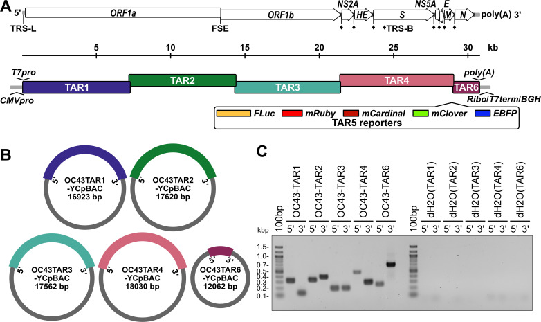
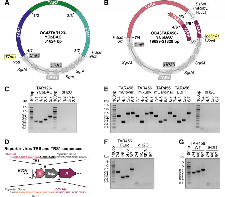
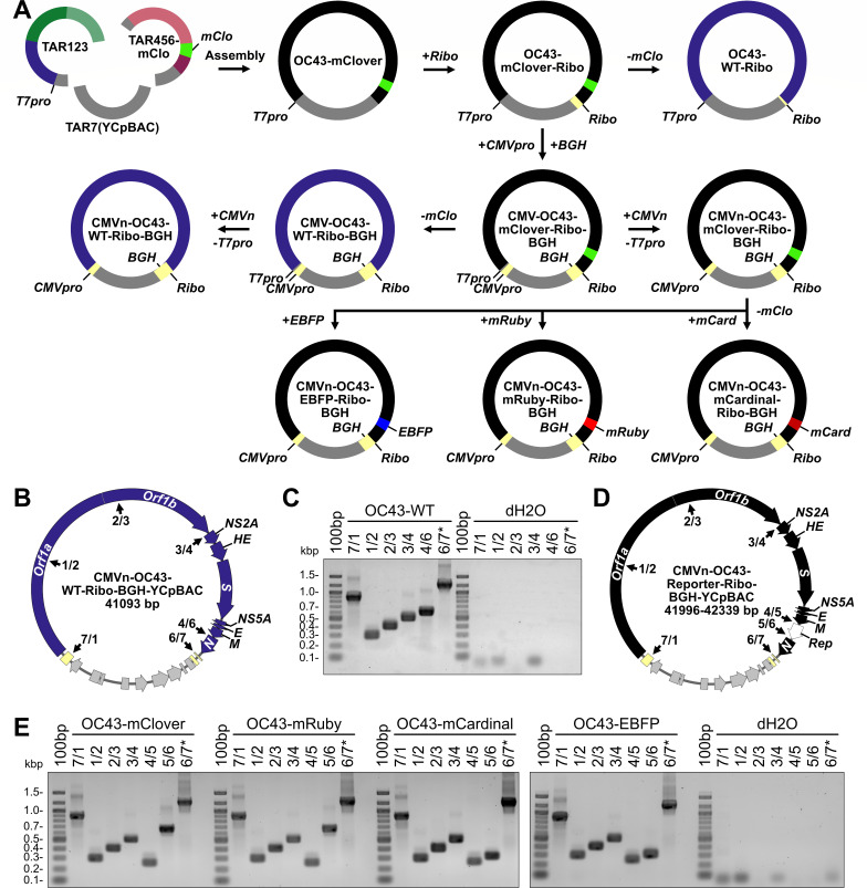
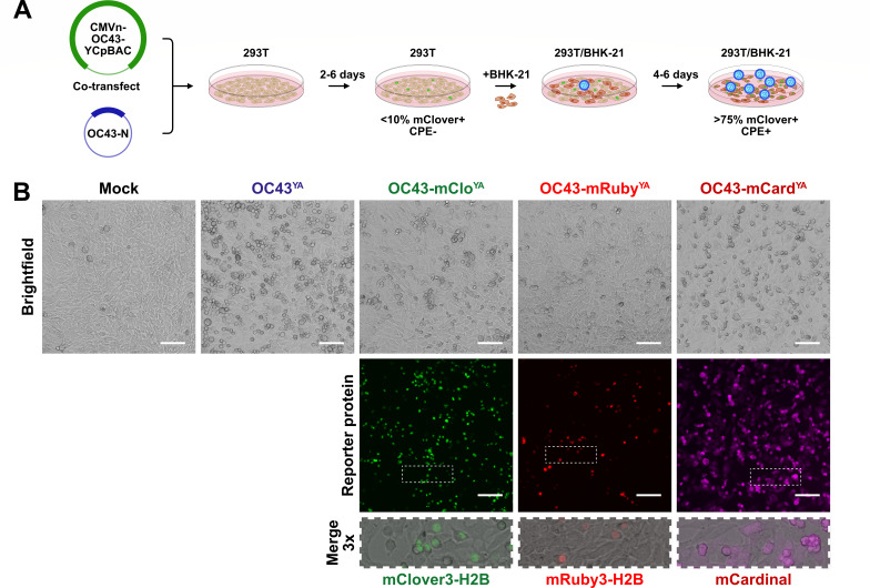
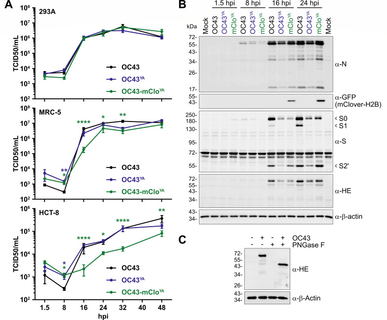
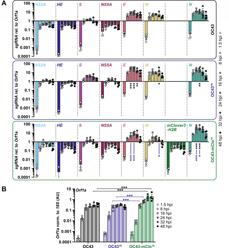
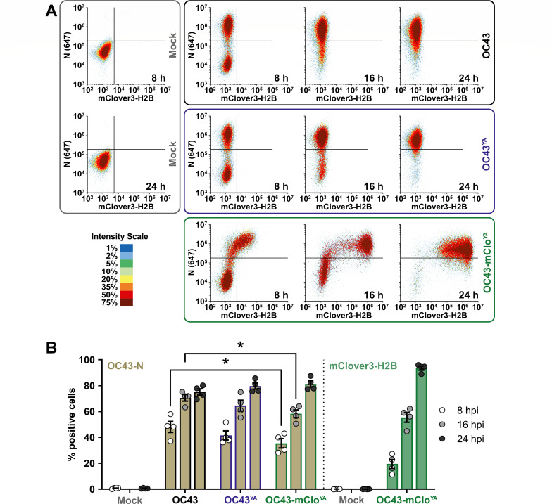
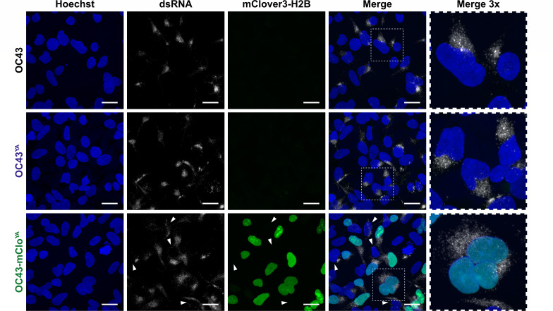
Coronaviruses have large, positive-sense single-stranded RNA genomes that challenge conventional strategies for mutagenesis. Yeast genetics has been used to manipulate large viral genomes, including those of herpesviruses and coronaviruses. This method, known as transformation-associated recombination (TAR), involves assembling complete viral genomes from dsDNA copies of viral genome fragments via homologous recombination in Saccharomyces cerevisiae. Here, we report our development of a TAR assembly and mutagenesis system for the endemic, seasonal human coronavirus (HCoV) strain OC43. HCoV-OC43 generally causes mild respiratory symptoms and is classified as a biosafety level 2 agent, making it useful for studying fundamental aspects of coronavirus biology and for comparative studies of more highly pathogenic betacoronaviruses. Following cDNA synthesis from HCoV-OC43 viral RNA, we generated five plasmids encompassing ~7.2 kb portions of the ORF1ab gene, the NS2 to M segment, or the N gene and structured to facilitate reporter gene insertions in the M-to-N intergenic region. Using these plasmids, we completed independent assemblies of yeast centromeric plasmids encoding ORF1ab, NS2a to N, as well as full-length HCoV-OC43 plasmids. A wild-type virus (OC43YA), as well as mClover3-H2B (OC43-mCloYA), mRuby3-H2B (OC43-mRubyYA), and mCardinal (OC43-mCardYA) reporter viruses, were rescued. The OC43-mCloYA reporter virus replicated comparably to an OC43 reference strain and produced the mClover3-H2B protein from a novel subgenomic RNA through insertion of an eighth body transcription regulatory sequence, preventing the need to delete or mutate viral genes. This updated HCoV-OC43 reverse genetics system will contribute to a better understanding of betacoronavirus host-pathogen interactions and can accelerate studies of novel antivirals.

Importance: Coronaviruses are ubiquitous pathogens that infect humans resulting in both mild and severe respiratory infections. Human coronavirus strain OC43 (HCoV-OC43) is one of many viruses responsible for common colds and is a useful model of more severe coronavirus infections. In this study, we describe an updated HCoV-OC43 mutagenesis system that uses yeast to capture six DNA fragments of the viral RNA genome and assemble them into full-length genomes in yeast/bacterial plasmids. The design of this system allowed for the rapid assembly and rescue of functional HCoV-OC43 viruses, including fluorescent reporter viruses with expanded genetic capacity. This updated reverse genetics system will enhance our ability to monitor viral replication, through building new reporter viruses, while also enhancing the study of betacoronavirus biology through the generation of mutant HCoV-OC43 viruses.

Keywords: HCoV-OC43; TAR; coronavirus; mutagenesis; reporter virus; reverse genetics; transformation-associated recombination; virus; yeast.


Conflict of interest statement

The authors declare no conflict of interest.

Figures

Fig 1
Fig 1
Design of HCoV-OC43 TAR plasmids for yeast assembly of viral sequences. (A) A schematic of the HCoV-OC43 genome including annotation of the leader transcription regulatory sequence (TRS-L), ribosomal frameshifting RNA element (FSE), and body TRSs (TRS-B, ♦) (top) and the orientations of the six, overlapping coronavirus TAR fragments (bottom). Inserted regulatory sequences are indicated at the 5′ (T7pro, T7 polymerase promoter; CMVpro, cytomegalovirus promoter) and 3′ (poly(A) tail; Ribo, hepatitis delta virus ribozyme; T7term, T7 terminator; BGH, bovine growth hormone polyadenylation signal) ends of the genome. Five different optional reporter genes (TAR5 reporters) are designed to be inserted between the M and N genes. (B) Illustration of the circular yeast centromeric plasmid/bacterial artificial chromosome (YCpBAC) vectors generated through homologous recombination in yeast with the corresponding HCoV-OC43 TAR sequences. HCoV-OC43 sequences are color-coded to match the diagram in panel A. YCpBAC sequences are shown in gray. (C) Plasmids containing the indicated HCoV-OC43 TAR fragments were subjected to PCR using primer sets designed to amplify the 5′ or 3′ junctions between the HCoV-OC43 TAR fragments and the YCpBAC sequences. PCR water controls (dH2O) are shown to the right of the panel. Primer pairs used for screening are listed in Table S2.
Fig 2
Fig 2
Yeast assembly of plasmids encoding HCoV-OC43 Orf1ab and NS2A-to-N. Diagrams of the OC43TAR123-YCpBAC plasmid containing HCoV-OC43 TAR fragments 1, 2, and 3 encoding Orf1ab (A) and OC43TAR456-YCpBACs containing HCoV-OC43 TAR fragments 4, 5 (indicated in white in the plasmid map), and 6 or fragments (for reporter virus assembly) or 4 and 6 (for wild-type (WT) virus assembly; inset) encoding NS2A to N +/- reporter genes (B). HCoV-OC43 sequences are color-coded to match the diagram in Fig. 1. YCpBAC sequences (TAR7) are shown in gray. Numbered junctions detected by PCR (black arrows) and restriction sites used for TAR cloning are indicated. (C) Following assembly in yeast, the TAR123-YCpBAC was subjected to PCR using primer sets designed to amplify junctions 7/1, 1/2, 2/3, and 3/7 with water controls (dH2O) to the right of the panel. (D) Arrangement of the M-N locus in reporter virus genomes with the authentic N TRS used to drive reporter gene sgRNA synthesis and an inserted copy of the N TRS (TRS*, sequence in orange) for N sgRNA synthesis indicated. (E) Following assembly in yeast, TAR456-YCpBACs were subjected to PCR using primer sets designed to amplify junctions 7/4, 4/5 (present when flanking a reporter gene), 5/6 (for fluorescence reporter genes), and 6/7 with dH2O controls to the right of the panel. (F) PCR screening of assembled TAR456-FLuc-YCpBAC with screening for junctions: 7/4, 4/5, 5F/6 (for the FLuc reporter gene), and 6/7 with dH2O controls to the right of the panel. (G) PCR screening of assembled TAR456-WT-YCpBAC with screening for junctions: 7/4, 4/6, and 6/7 with dH2O controls to the right of the panel. Primer pairs used for screening are listed in Table S2. Abbreviations: 100bp, 100 bp ladder; bp/kbp, base/kilobase pairs; CmR, chloramphenicol resistance gene; EBFP, enhanced blue fluorescent protein; FLuc, firefly luciferase; poly(A), encoded A34 sequence; Rep., reporter gene; T7pro, T7 polymerase promoter; TRS, transcription regulatory sequence; URA3, orotidine 5′-phosphate decarboxylase gene; WT, wild-type.
Fig 3
Fig 3
Yeast assembly of full-length HCoV-OC43 wild-type and reporter virus plasmids. (A) Assembly of full-length HCoV-OC43-mClover viral sequences (black) was performed by sequential assembly steps in yeast. First, restriction digested OC43TAR123-YCpBAC and OC43TAR456-mClover plasmids were assembled, followed by the sequential insertion of hepatitis delta virus ribozyme (Ribo), bovine growth hormone poly(A) signal (BGH), and CMV promoter (CMVpro) sequences (light yellow). Assembly of full-length HCoV-OC43-WT viral sequences (blue) was performed by removing the mClover3-H2B (mClo, light green) sequence from a previously assembled plasmid. Additional reporter virus plasmids (black) encoding enhanced blue fluorescent protein 2 (EBFP, blue), mRuby3-H2B (mRuby, red), or mCardinal (mCard, dark red) were assembled by recombination which simultaneously removed the mClover3-H2B sequence and replaced it with a new reporter sequence. (B) Plasmid map for the CMVn-OC43-WT-Ribo-BGH-YCpBAC used for virus rescue. Numbered junctions detected by PCR (black arrows) are indicated. (C) PCR screening of assembled DNA junctions 7/1, 1/2, 2/3, 3/4, 4/6, and 6/7 for CMVn-OC43-WT-Ribo-BGH plasmid with water controls (dH2O) to the right of the panel. (D) Plasmid map for the CMVn-OC43-Reporter-Ribo-BGH-YCpBAC used for virus rescues. Numbered junctions detected by PCR (black arrows) are indicated. The white arrow in the plasmid map indicates the location of the inserted reporter (Rep) genes. (E) PCR screening of assembled DNA junctions 7/1, 1/2, 2/3, 3/4, 4/5, 5/6, and 6/7* for CMVn-OC43-Reporter-Ribo-BGH plasmids with water controls (dH2O) to the right of the panel. Primer pairs used for screening are listed in Table S2. Abbreviations: 100bp, 100 bp ladder; bp/kbp, base/kilobase pairs; T7pro, T7 promoter sequence; WT, wild type.
Fig 4
Fig 4
Rescue of OC43YA, OC43-mCloYA, OC43-mRubyYA, and OC43-mCardYA viruses. (A) Overview of yeast-assembled OC43 rescue procedure: Assembled CMVn-OC43-YCpBAC plasmids and plasmids encoding OC43-N were co-transfected into 293T (tan) cells for 2–6 days followed by re-seeding with BHK-21 (brown) cells for 4–6 days leading to enhanced cytopathic effect (CPE), reporter protein expression (example showing mClover3-H2B-positive nuclei, green), and virus (blue) propagation. (B) Rescue of yeast-assembled viruses: After the 293T/BHK-21 co-culture, cleared culture supernatants were used to infect naïve BHK-21 cells for 24 h prior to fixation and imaging to assess CPE and reporter protein expression in the infected cell monolayers. The areas indicated with the white dashed rectangles are shown at higher magnification to show sub-cellular localization of reporter proteins. Scale bars = 100 µm.
Fig 5
Fig 5
Yeast-assembled HCoV-OC43 viruses replicate to comparable titers as wild-type HCoV-OC43 viruses and produce similar amounts of viral proteins. (A) Third passage OC43YA and OC43-mCloYA viruses were used to infect 293A cells (top), MRC-5 cells (middle), or HCT-8 (bottom) at a multiplicity of infection (MOI) for 0.1 and the supernatants were collected at the indicated times and titered using BHK-21 cells. The data are plotted as the mean ± standard error of the mean from four independent replicates. Statistically significant differences compared to OC43(ATCC) are indicated from two-way ANOVA analysis. (B) 293A cells were infected at an MOI of 0.1 with OC43 (black), OC43YA (yeast-assembled OC43; blue), or OC43-mCloYA (yeast-assembled OC43-mClover; green) viruses for the indicated times. Protein lysates were subjected to SDS-PAGE and immunoblotted with the indicated antibodies where α-GFP antibodies were used to detect mClover3-H2B. The data presented are from one of four independent experiments. (C) Lysates from 293A cells infected with OC43 (24 hpi) were incubated with or without PNGase F prior to immunoblotting with antibodies against OC43-HE or β-Actin. Abbreviations: *P ≤ 0.05; **P ≤ 0.01; ****P ≤ 0.0001; GFP, green fluorescent protein; hpi, hours post-infection; PNGase F, peptide:N-glycosidase F; S0/S1/S2′, Spike subunit 0/1/2′; TCID50, 50% tissue culture infectious dose.
Fig 6
Fig 6
gRNA/sgRNA accumulation is altered by the insertion of the mClover3-H2B reporter gene. (A) 293A cells were infected at a multiplicity of infection (MOI) of 0.1 with OC43 (black box/top panel), OC43YA (yeast-assembled OC43; blue box/middle panel), or OC43-mCloYA (yeast-assembled OC43-mClover; green box/bottom panel) viruses for the indicated times. Total RNA was reverse transcribed and used for qPCR with a common forward primer that binds in the 5′ leader sequence and reverse primers that bind downstream of the leader (Orf1a) or downstream of ORF-specific transcription regulatory sequences. All data were normalized to 18S rRNA and plotted relative to Orf1a. Data were plotted as the mean ± standard error of four independent experiments. Data points < 0.0001 are not shown. (B) Orf1a (gRNA) abundance normalized to 18S rRNA expressed as arbitrary units (AU) was plotted as the mean ± standard error of four independent experiments. The same data set was used for panels A and B. Statistical comparisons were made between matched time points by two-way ANOVA analysis. P values: *≤ 0.05, **≤ 0.01, ***≤ 0.001. Black asterisks represent differences compared to OC43 and blue asterisks represent differences between OC43YA and OC43-mCloYA.
Fig 7
Fig 7
N protein accumulation is delayed during OC43-mCloYA infection. (A) 293A cells were infected at an MOI of 0.1 with OC43 (black box/top panel), OC43YA (yeast-assembled OC43; blue box/middle panel), or OC43-mCloYA (yeast-assembled OC43-mClover; green box/bottom panel) viruses for 8, 16, or 24 hours post-infection (hpi) or mock infected for 8 or 24 hours (gray box/left panel). Cells were fixed, permeabilized, and stained with OC43-N antibodies prior to analysis by flow cytometry. Density plots of N (y-axis) vs. mClover3-H2B (x-axis) are shown from one representative experiment. (B) The data were analyzed for % positive single cells expressing OC43-N (light brown) or mClover3-H2B (light green) and plotted as the mean ± standard error of the mean from four independent experiments. Statistically significant differences in protein expression between different viruses are indicated (two-way ANOVA), *P ≤ 0.05. Abbreviations: 647, Alexa Fluor 647.
Fig 8
Fig 8
Replication compartment formation is unimpaired in cells infected with yeast-assembled HCoV-OC43 viruses. 293A cells were infected at an MOI of 0.1 with OC43, OC43YA (yeast-assembled OC43), or OC43-mCloYA (yeast-assembled OC43-mClover) viruses for 16 hours prior to fixation, permeabilization, and staining with anti-dsRNA antibodies to stain replication compartments (white cytoplasmic puncta) and Hoechst 33342 to stain cell nuclei (blue). mClover3-H2B is shown in green. White arrowheads denote dsRNA+/mClover- cells. The areas indicated with the white dashed rectangles are shown at higher magnification to show cytoplasmic dsRNA staining. All images were acquired using a confocal laser scanning microscope and are presented as maximum intensity projections from one of three independent experiments. Scale bars = 20 µm. Abbreviations: dsRNA, double-stranded RNA; H2B, histone H2B.
Fig 9
Fig 9
OC43-mCloYA is susceptible to antiviral treatment. 293A, MRC-5, or BHK-21 cells were mock infected or infected at an MOI of 0.1 with OC43-mCloYA (yeast-assembled OC43-mClover) for 1 hour prior to a medium change to 2.5% DMEM+ Pen/Strep/Gln supplemented with 0.1% DMSO or 1 µM nirmatrelvir (Nir). At 24 hours post-infection, the supernatants were collected for titering on BHK-21 cells (A). The cell monolayers were collected and fixed prior to analysis of mClover3-H2B expression by flow cytometry (B) with the average mClover3-H2B median fluorescence intensity (MFI) from mock or infected cells plotted in panel (C). Data were plotted as the mean ± standard error of three independent experiments. Statistical comparisons were made by two-way ANOVA analysis. P values: * ≤ 0.05. Abbreviations: DMSO, dimethyl sulfoxide; TCID50, 50% tissue culture infectious dose.

Similar articles

References

    1. Saberi A, Gulyaeva AA, Brubacher JL, Newmark PA, Gorbalenya AE. 2018. A planarian nidovirus expands the limits of RNA genome size. PLoS Pathog 14:e1007314. doi:10.1371/journal.ppat.1007314 - DOI - PMC - PubMed
    1. Ferron F, Sama B, Decroly E, Canard B. 2021. The enzymes for genome size increase and maintenance of large (+)RNA viruses. Trends Biochem Sci 46:866–877. doi:10.1016/j.tibs.2021.05.006 - DOI - PubMed
    1. Malone B, Urakova N, Snijder EJ, Campbell EA. 2022. Structures and functions of coronavirus replication–transcription complexes and their relevance for SARS-CoV-2 drug design. Nat Rev Mol Cell Biol 23:21–39. doi:10.1038/s41580-021-00432-z - DOI - PMC - PubMed
    1. Yan S, Zhu Q, Hohl J, Dong A, Schlick T. 2023. Evolution of coronavirus frameshifting elements: competing stem networks explain conservation and variability. Proc Natl Acad Sci U S A 120:e2221324120. doi:10.1073/pnas.2221324120 - DOI - PMC - PubMed
    1. Jahirul Islam M, Nawal Islam N, Siddik Alom M, Kabir M, Halim MA. 2023. A review on structural, non-structural, and accessory proteins of SARS-CoV-2: highlighting drug target sites. Immunobiology 228:152302. doi:10.1016/j.imbio.2022.152302 - DOI - PMC - PubMed

LinkOut - more resources