. 2025 Jan 24;53(3):gkaf039.
doi: 10.1093/nar/gkaf039.

RNA G-quadruplex structure-based PROTACs for targeted DHX36 protein degradation and gene activity modulation in mammalian cells

Affiliations

RNA G-quadruplex structure-based PROTACs for targeted DHX36 protein degradation and gene activity modulation in mammalian cells

Kun Zhang et al. Nucleic Acids Res. .

Abstract

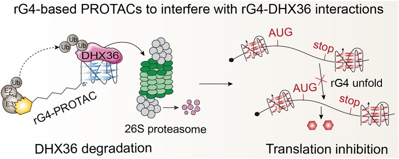
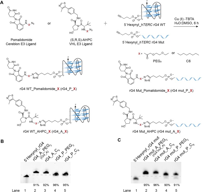
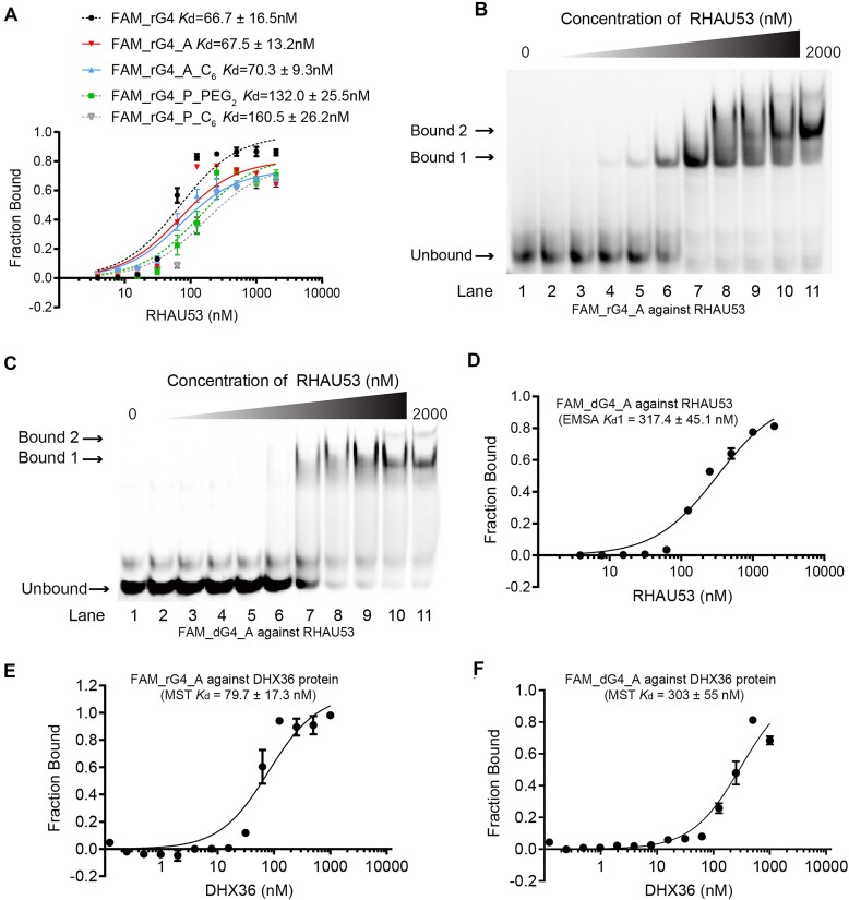
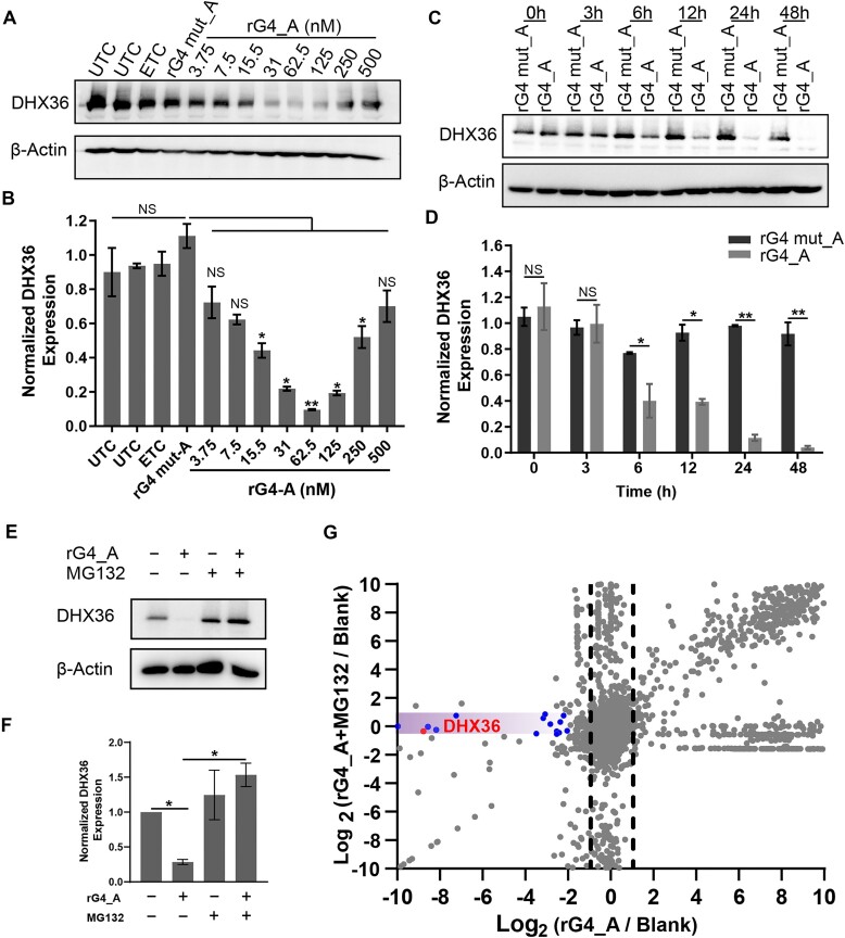
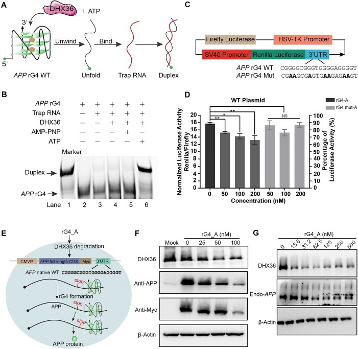
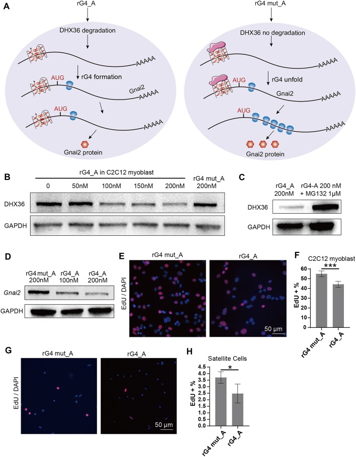
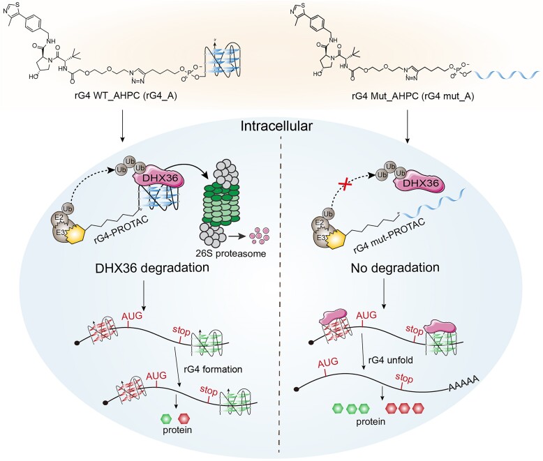
RNA G-quadruplexes (rG4s) are non-canonical secondary nucleic acid structures found in the transcriptome. They play crucial roles in gene regulation by interacting with G4-binding proteins (G4BPs) in cells. rG4-G4BP complexes have been associated with human diseases, making them important targets for drug development. Generating innovative tools to disrupt rG4-G4BP interactions will provide a unique opportunity to explore new biological mechanisms and potentially treat related diseases. Here, we have rationally designed and developed a series of rG4-based proteolytic targeting chimeras (rG4-PROTACs) aimed at degrading G4BPs, such as DHX36, a specific G4BP that regulates gene expression by binding to and unraveling rG4 structures in messenger RNAs (mRNAs). Our comprehensive data and systematic analysis reveals that rG4-PROTACs predominantly and selectively degrade DHX36 through a proteosome-dependent mechanism, which promotes the formation of the rG4 structure in mRNA, leading to the translation inhibition of rG4-containing transcripts. Notably, rG4-PROTACs inhibit rG4-mediated APP protein expression, and impact the proliferative capacity of skeletal muscle stem cells by negatively regulating Gnai2 protein expression. In summary, rG4-PROTACs provide a new avenue to understand rG4-G4BP interactions and the biological implications of dysregulated G4BPs, promoting the development of PROTACs technology based on the non-canonical structure of nucleic acids.


Conflict of interest statement

The authors declare that they have no competing financial interest.

Figures

Graphical Abstract
Graphical Abstract
Figure 1.
Figure 1.
Rational design and preparation of rG4-PROTACs by click chemistry. (A) Schematic representation of the synthesis of rG4-PROTACs between azide-modified E3 ligase ligands and alkyne-modified rG4 oligos. (S, R, S)-AHPC and Pomalidomide are E3 ligase ligand-linker conjugates. Alkyne was introduced to the 5′ end of hTERC rG4 WT or hTERC rG4 Mut sequences (Supplementary Table S1). The E3 ligase recruiter compounds were separately conjugated onto 5′Hexynyl_ hTERC rG4 WT and 5′Hexynyl_ hTERC rG4 Mut by click reaction for 6 h to generate rG4-PROTACs (rG4_A, rG4_A_C6, rG4_P_PEG2, rG4_P_C6, rG4 mut_A, rG4 mut_A_C6, rG4 mut_P_PEG2, and rG4 mut_P_C6). X: PEG2 or C6. (B) Confirmation of rG4-PROTACs conjugates. (C) Confirmation of rG4 mut-PROTACs conjugates. The click reaction products were subjected to 12% denaturing PAGE at 300 V for 30 min. All these conjugates achieved >90% of reaction yields. Dotted lines are used to distinguish bands.
Figure 2.
Figure 2.
rG4_PROTACs demonstrate stronger molecular recognition toward RHAU53 and DHX36 than dG4_PROTACs. (A) Binding curves of FAM_rG4 WT_Protacs against RHAU53 from the first shift bound (Bound 1). For 2 transitions, Kd1 was determined from the fraction of first transition band (R1) versus the concentration of RHAU53 and Kd2 was determined from the fraction of second transition band (R2) versus the concentration of RHAU53. (B) Native gel of the binding between FAM_rG4_A and RHAU53 detected by EMSA. The free rG4 band was completely shifted to bound 1 at lane 7, and bound 2 appeared at lane 9. (C) EMSA result of the binding between FAM_dG4_A and RHAU53. The free dG4 band was completely shifted to bound 1 at lane 10, and the bound 2 band appeared at lane 10. (D) Binding curves of FAM_dG4_A against RHAU53 from the first shift bound (Bound 1) and the Kd1 was calculated to be 317.4 ± 45.1 nM. (E) MST binding curve between FAM_rG4_A and DHX36 protein and the Kd was found to be 79.7 ± 17.3 nM, which verifies the binding interaction between FAM_rG4_A and DHX36. (F) MST binding curve between FAM_dG4_A and DHX36 protein and the Kd was found to be 303 ± 55 nM, which showed a relatively weaker binding interaction between FAM_dG4_A and DHX36. The data were obtained from three biological replicates with the standard deviation as an error bar.
Figure 3.
Figure 3.
rG4_PROTACs degrade DHX36 protein in a proteasome-dependent manner in different human cells. (A) rG4_A degrades DHX36 in a dose-dependent manner. HeLa cells were treated with different concentrations of rG4_A and 250 nM rG4 mut_A for 24 h, and the cells were harvested for western blotting analysis of DHX36. UTC: Untreated cells; and ETC: empty transfection control. (B) Quantitative analysis of western blot from panel (A). (C) Degradation effect of rG4_A (50 nM) and rG4 mut_A (50 nM) on DHX36 0–48 h post-transfection. (D) Quantitative analysis of western blot results at different time points. (E) MG132 blocked rG4_A induced DHX36 protein reduction. HeLa cells were treated with rG4_A (50 nM) with or without MG132 (1 μg/ml), and the cells were harvested for western blotting analysis of DHX36. (F) Quantification diagram of western blot from panel (E). (G) Proteomic analysis of rG4_A selectively degraded DHX36 through the proteasome. Hela cells were treated with 50 nM rG4_A, or rG4 mut_A plus MG132 for 24 h. Lysates were subjected to mass spec-based proteomics analysis. The volcano plot shows protein abundance (Log2) as a function of significance level. The significantly downregulated proteins through proteasome were found in the purple zone [−0.41< [Log2 (rG4_A + MG132 / Blank) ≈ 0] <0.32. Nonaxial vertical lines mark log2Fold Change >1 or <−1 significance threshold. Log2fold change was obtained from three independent experiments of Log2 (rG4_A / Blank) or Log2 (rG4_A + MG132 / Blank). *P < 0.05 and **P < 0.01. Normalized DHX36 expression was obtained from biological triplicates with the standard deviation as an error bar.
Figure 4.
Figure 4.
rG4_A negatively regulates APP gene and native protein expression in Hela cells. (A) Scheme of the functional mechanism of ATP-dependent DHX36 unwinding activity on APP 3′ UTR rG4 WT in vitro. (B) Native gel profile of DHX36 unwinding activity. Lane 1: APP rG4 WT and Trap RNA were thermally denatured and hybridized to form a duplex, which exhibits a shift band in the upper position compared with the free APP rG4 band. Lanes 2–5: negative controls in which the duplex cannot be formed in the absence of either DHX36 or ATP (lane 2, lane 3, and lane 4), or replacing ATP with AMP-PNP, a non-hydrolyzable analog of ATP (lane 5). Lane 6: after the addition of ATP, DHX36 bounds and unfolds the APP rG4 motif. The fully complementary sequence, Trap RNA, will hybridize with the unfolded rG4 motif to form a duplex. (C) Schematic illustration of dual luciferase reporter plasmids. The APP rG4 WT or APP rG4 Mut DNA sequence was inserted into the 3′ UTR of Renilla luciferase. (D) Normalized luciferase activity on APP rG4 WT plasmid. The luciferase activity of the APP rG4 WT construct group (the first black columns) was 100%, and the luciferase signals of all other groups were normalized by that of the APP rG4 WT construct group. (E) Scheme of translation inhibition of rG4_PROTACs on APP native WT plasmid. After co-transfection of the APP rG4-containing plasmid and rG4_A for 24 h, rG4 motif-mediated APP protein expression was reduced. (F) Western blot analysis of APP protein expression after treatment with different concentrations of rG4_A (0−100 nM). APP protein decreased along with DHX36 degradation. Anti-Myc tag and anti-APP antibodies were used to detect APP protein, respectively. β-Actin: internal loading control. Mock: empty transfection control. (G) Relative APP mRNA expression levels on endogenous APP expression. Western blot analysis was conducted to assess the expression of endogenous APP protein following treatment with various concentrations of rG4_A (0, 15.6, 31.2, 62.5, 125, 250, and 500 nM). The results showed that endogenous APP protein levels decreased in parallel with the degradation of DHX36. Normalized luciferase activities are obtained from biological triplicates with the standard deviation as an error bar. *P < 0.05, **P < 0.01, NS: not significant.
Figure 5.
Figure 5.
rG4_A impacts the proliferative capacity of SCs and C2C12 cells by negatively regulating Gnai2 expression. (A) Illustration of translation inhibition of rG4_PROTACs on endogenous Gnai2 gene. In C2C12 cells, rG4_A treatment induced the deletion of DHX36 protein and increased rG4 motif formation in the 5′UTR of Gani2 transcript, resulting in the Gnai2 protein expression suppression. (B) rG4_A inhibits DHX36 protein expression in a concentration-dependent manner in C2C12 cells. (C) MG132 blocked rG4_A-induced DHX36 protein loss. C2C12 cells were treated with rG4_A (200 nM) with or without MG132 (1 μg/ml), and the cells were harvested for western blotting analysis of DHX36. (D) Gnai2 protein was reduced with an increased dose of rG4_A. Quantification diagrams of the western blot are shown in Supplementary Fig. S21. Normalized protein expression was obtained from biological triplicates with the standard deviation as an error bar. (E) The effect of rG4-PROTACs on proliferative capacity was investigated by EdU assay in C2C12 cells. Representative images are shown. Scale bar = 50 μm. (F) Quantitative analysis of the relative EdU incorporation percentage from five randomly selected fields per sample of panel (E). (G) The effect of rG4-PROTACs on proliferative capacity was investigated by EdU assay in SCs. Representative images are shown. Scale bar = 50 μm. (H) Quantitative analysis of the relative EdU incorporation percentage from five randomly selected fields per sample of panel (G). The relative EdU incorporation percentage is obtained from three biological replicates with the standard deviation as an error bar. *P < 0.05 and ***P < 0.001.
Figure 6.
Figure 6.
Schematic illustration of the functional mechanism of rG4-PROTACs in regulating rG4-mediated gene translation by degradation DHX36 protein. After transfection into cells, treatment with rG4_A caused a significant DHX36 degradation, which promoted rG4 motif formation and resulted in translation inhibition. While rG4 mut_A showed no effect on DHX36 protein expression and unfolded rG4 structures to induce the translation of the downstream genes.

Similar articles

References

    1. Georgakopoulos-Soares I, Parada GE, Hemberg M Secondary structures in RNA synthesis, splicing and translation. Comput Struct Biotechnol J. 2022; 20:2871–4.10.1016/j.csbj.2022.05.041. - DOI - PMC - PubMed
    1. Gellert M, Lipsett MN, Davies DR Helix formation by guanylic acid. Proc Natl Acad Sci USA. 1962; 48:2013–8.10.1073/pnas.48.12.2013. - DOI - PMC - PubMed
    1. Sen D, Gilbert W Formation of parallel four-stranded complexes by guanine-rich motifs in DNA and its implications for meiosis. Nature. 1988; 334:364–6.10.1038/334364a0. - DOI - PubMed
    1. Davis JT G-quartets 40 years later: from 5′-GMP to molecular biology and supramolecular chemistry. Angew Chem Int Ed Engl. 2004; 43:668–98.10.1002/anie.200300589. - DOI - PubMed
    1. Huppert JL, Balasubramanian S Prevalence of quadruplexes in the human genome. Nucleic Acids Res. 2005; 33:2908–16.10.1093/nar/gki609. - DOI - PMC - PubMed

LinkOut - more resources