Review
. 2025 Jun;26(3):397-412.
doi: 10.1007/s11154-024-09939-0. Epub 2025 Jan 30.

Growth hormone-releasing hormone signaling and manifestations within the cardiovascular system

Affiliations
Review

Growth hormone-releasing hormone signaling and manifestations within the cardiovascular system

Raul A Dulce et al. Rev Endocr Metab Disord. 2025 Jun.

Abstract

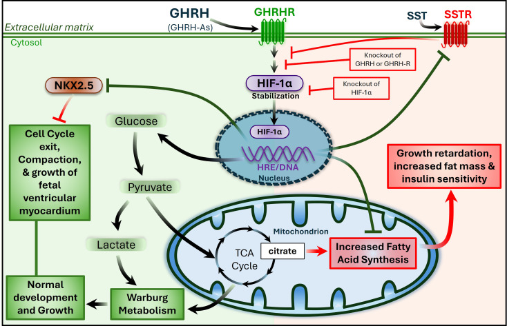
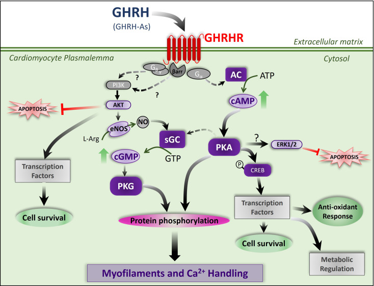
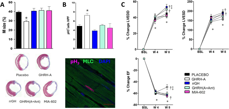
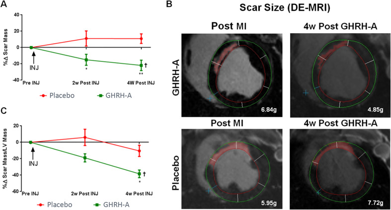
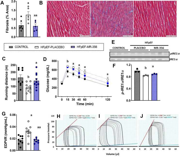
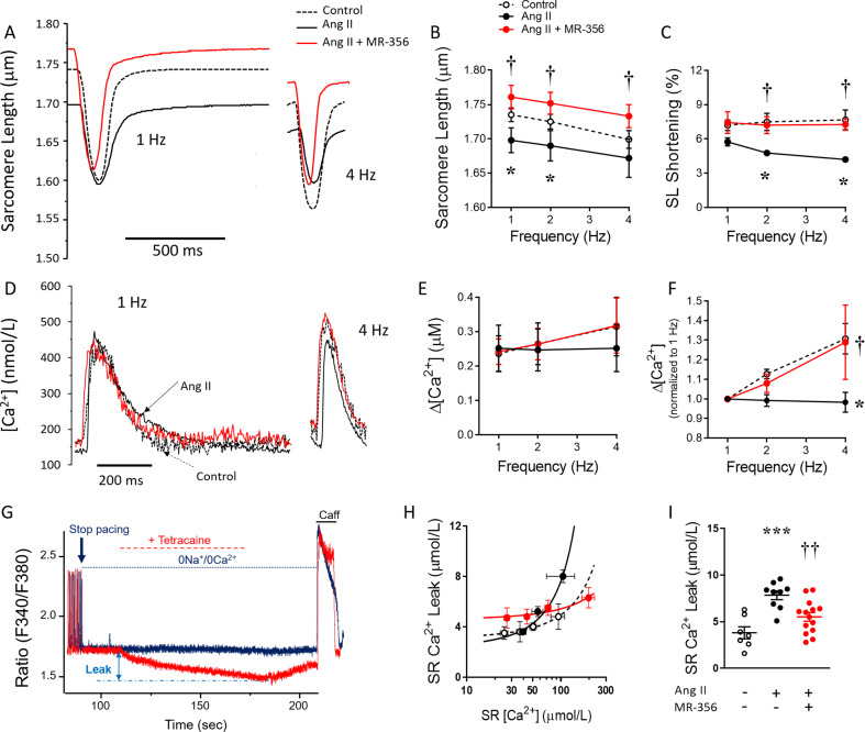
Growth hormone (GH)-releasing hormone (GHRH), a hypothalamic peptide initially characterized for its role in GH regulation, has gained increasing attention due to its GH-independent action on peripheral physiology, including that of the cardiovascular system. While its effects on the peripheral vasculature are still under investigation, GHRH and synthetic agonists have exhibited remarkable receptor-mediated cardioprotective properties in preclinical models. GHRH and its analogs enhance myocardial function by improving contractility, reducing oxidative stress, inflammation, and offsetting pathological remodeling. Studies performed in small and large animal models have demonstrated the efficacy of these compounds in diverse cardiomyopathies, suggesting their potential as promising therapeutic agents. However, the clinical translation of GHRH synthetic analogs still faces challenges related to the route of administration and potential side effects mainly associated with activation of the GH/IGF-I axis. Despite these hurdles, the compelling evidence supporting their role in cardiac repair makes GHRH analogs attractive candidates for clinical testing in the treatment of various cardiac diseases.

Keywords: Cardioprotective; Cardiovascular; GHRH receptors; GHRH-analogs; Growth hormone-releasing hormone.


Conflict of interest statement

Declarations. Competing interests: R.M.KT. and J.M.H. are listed as co-inventors on patents on GHRH analogues which were assigned to the University of Miami and Veterans Affairs Department. J.M.H. previously owned equity in Biscayne Pharmaceuticals, license of intellectual property used in this study. Biscayne Pharmaceuticals did not provide funding for this study. JMH reported having a patent for cardiac cell-based therapy. He holds equity in Vestion Inc. and maintains a professional relationship with Vestion Inc. as a consultant and member of the Board of Directors and Scientific Advisory Board. Dr. Joshua Hare is the Chief Scientific Officer, a compensated consultant and advisory board member for Longeveron, and holds equity in Longeveron. Dr. Hare is also the co-inventor of intellectual property licensed to Longeveron. The University of Miami also stands to gain royalties from the commercialization of the IP.K.E.H. holds equity in Vestion Inc. He is also the co-inventor of intellectual property licensed to Vestion Inc and Longeveron. K.E.H. is the Chief Executive Officer for KosBio P.C. and holds equity in KosBio. He is also the co-inventor of intellectual property related to GHRH/GHRHR signaling, described in this work. Longeveron LLC, Vestion Inc. and KosBio P.C. did not participate in funding this work. All other authors have declared that no conflict of interest exists.

Figures

Fig. 1
Fig. 1
Proposed Role of GHRH/GHRHR in the Metabolic Control of Heart Development. During development, cardiac cells switch between glucose and fatty acid metabolism. In NKX2.5 + cardiac progenitor cells, autocrine activation of GHRH/GHRHR stimulates oxygen-independent, cell-specific, localized activation of HIF-1α. This activation represses NKX2.5 expression, promoting a shift to aerobic glycolysis (Warburg metabolism), cell cycle exit, and thickening growth of the ventricular wall (compaction). Conversely, inactivation of GHRH/GHRHR (or HIF-1α) maintains NKX2-5 expression, redirecting glycolytic products toward fatty acid synthesis, which supports cardiac cell cycle reentry and trabecular cardiomyogenesis, at the expense of ventricular compaction. SST, somatostatin; SSTR, SST receptor; TCA, tricarboxylic acid; Adapted from Wanschel ACBA et al. 2023, BioRxiv 2023:2022.2001.2031.478572 [33].
Fig. 2
Fig. 2
Proposed mechanism of action for GHRH agonists on the modulation of calcium signaling, contractility and reparative mechanisms in myocardial cells. Activation of GHRHRs (GPCR) induces the adenylyl cyclase (AC)/cyclic AMP/protein kinase A (PKA) pathway. PKA can both phosphorylate sarcomeric and calcium handling proteins and activate soluble guanylyl cyclase (sGC). This pathway also promotes antioxidant, reparative and survival mechanisms at transcriptional level. GHRHRs signaling also promotes activation of AKT (PKB) and ERK1/2 mediate the anti-apoptotic effects of GHRH agonists (GHRH-As). Endothelial Nitric Oxide Synthase (eNOS) is proposed to be activated by GHRH. The generated nitric oxide (NO) in turn activates sGC to produce the second messenger cGMP, an activator of protein kinase G (PKG). This kinase is also able to modulate myofilament and calcium cycling. This pathway has been proposed to be activate downstream GHRHRs through activation of PI3K by the Gβγ. proteins or β-arrestins (among other possible mechanisms). Adapted from Dulce RA et al. 2022, Cardiovasc Res [18].
Fig. 3
Fig. 3
Impact of GHRH agonist administration on infarct size, cardiomyocyte proliferation and ventricular remodeling following myocardial infarction. A, Averaged percentage of infarct size (top) and representative images of hearts from every group comparing the extension of the fibrotic scar (bottom). B, Averaged abundance of mitotic cardiomyocytes (top) and a representative image of a cardiomyocyte positive for phosphorylated histone H3 (pH3) (bottom). MLC, myosin light chain. C, Depicted are changes over time in LV end-diastolic (LVEDD) (Upper Left), end-systolic (LVESD) diameters (Upper Right), and ejection fraction (EF) (Lower). As shown, the GHRH agonist (GHRH-A) offsets the increase in LVESD and LVEDD, resulting in an improvement in EF. The effect of the agonists is blocked by the receptor antagonist MR-602 (Ant) consistent with a receptor mediated effect. Treatment with rrGH does not reproduce the effects of JI-38, All values represent mean ± SEM (n = 7–10), *P < 0.05 vs. baseline (BSL), same group; †P < 0.05 vs. wk 4 (W4), same group; ‡P < 0.05 vs. all other groups at week 8 (W8), except GHRH (A + Ant). §P < 0.05 vs. all other groups at W8. Adapted from Kanashiro-Takeuchi RM et al. 2012, PNAS [14].
Fig. 4
Fig. 4
GHRH agonist treatment reduces myocardial infarct size in swine following myocardial infarction. A, Percent change of scar mass in the GHRH agonist (GHRH-A) versus placebo group at 2- and 4-weeks (w) post initiation of treatment (2-way ANOVA, between group: *P = 0.04 and **P = 0.003 vs. placebo, respectively). One-way ANOVA: †P = 0.02 for GHRH agonist group. B, Cardiac Magnetic Resonance images of hearts from an animal in each group before and 4 weeks after the initiation of treatment. C, Percent change of scar as a percentage of left ventricular mass in GHRH agonist group versus placebo group at 2- and 4-weeks post initiation of treatment (2-way ANOVA, between group: P = NS and *P = 0.02 vs. placebo, respectively). One-way ANOVA: †P = 0.0002 for GHRH agonist group. From Bagno LL et al. 2015, J Am Heart Assoc. [16]
Fig. 5
Fig. 5
Cardiac performance and hemodynamic changes in cardiometabolic HFpEF induced by 9 weeks of HFD + L-NAME. A, Quantification of interstitial fibrosis in hearts from mice: control, HFpEF treated with placebo (HFpEF-placebo) and HFpEF treated with MR-356 (HFpEF-MR-356). One-way ANOVA followed by Tukey’s test, aP < 0.05 vs. control, aaP < 0.05 vs. HFpEF-placebo, n = 4 or 6. B, Representative images of Masson-Trichrome staining from each group. C, Exercise tolerance test showed a reduced running capacity in the HFpEF-placebo mice compared with control and HFpEF-MR-356 mice (bP < 0.01 vs. control, aaP < 0.05 vs. HFpEF-placebo by unpaired t test, n = 9 or 10). D, Intraperitoneal glucose tolerance test (ipGTT) showed increased glucose levels in HFpEF-placebo mice compared with control mice in all time points after glucose injection, whereas MR-356-treated mice showed an increase in glucose levels only at 15- and 30-min time point, two-way ANOVA followed by Sidak multiple-comparisons test, aP < 0.05, bP < 0.01 vs. control, n = 9. E, Representative immunoblots of phosphorylated IRE1a (top) at Ser724 and total IRE1a (bottom). F, Western blot quantification showing reduced ratio of phospho-IRE1a to IRE1a in HFpEF-placebo mice compared with control (one-way ANOVA followed by Tukey’s test, aP < 0.05 vs. control, aaP < 0.05 vs. HFpEF-placebo, n = 6), G, Slope of end-diastolic pressure-volume relationship (EDPVR; E),: Kruskal–Wallis test followed by Dunn’s multiple-comparisons test, aP < 0.05, vs. control, aaP < 0.05 vs. HFpEF-placebo, n = 9 or 10.H–J, Representative pressure-volume (P-V) loops from control (H). Adapted from Kanashiro-Takeuchi RM et al. 2023, Am J Physiol Heart & Circ Physiol. [20]
Fig. 6
Fig. 6
Sarcomere dysfunction, depressed contractile reserve and calcium handling alterations in the Ang-II-induced model are prevented by co-treatment with the GHRH agonist MR-356. A, Representative traces of sarcomere twitches of cardiomyocytes, electric field stimulated at 1- and 4 Hz, from male CD1 control mice (N = 4, n = 30 cells) and mice that underwent either Ang-II (N = 5, n = 41 cells) or Ang-II + MR-356 treatment (N = 5, n = 38 cells). B, Resting sarcomere length (SL) in cardiomyocytes from each group at different pacing rates. C, Sarcomere shortening in response to pacing. (D) Representative traces of intracellular [Ca2+] in electric field-stimulated cardiomyocytes. (E) Δ[Ca2+] transient amplitude in response to pacing. F, Δ[Ca2+] normalized (with respect to 1 Hz) in response to pacing. G, Representative traces of intracellular [Ca2+] in cardiomyocytes illustrating the method for measuring SRCa2+ leak using tetracaine. Upper (Blue) trace corresponds to Ca2+ from a cardiomyocyte not exposed to tetracaine; lower (red) trace is the Ca2+ from the same cardiomyocyte treated with tetracaine for 70 s after pacing was stopped. H, SRCa2+ leak-load relationship in cardiomyocytes from control (N = 4 mice, n = 31 cells), Ang-II-treated (N = 5 mice, n = 38 cells) or Ang-II + MR-356-treated (N = 5 mice, n = 35 cells) CD1 mice. I, Average and individual SRCa2+ leak in cardiomyocytes from all groups at matched SRCa2+ load = 99.1 µmoL/L. From each mouse, a total of 5–9 cells were studied. *P < 0.05, ***P < 0.001 in Ang-II vs. control, and †P < 0.05, ††P < 0.01 in Ang-II + MR-356-treated vs. Ang-II; one- or two-way ANOVA as appropriate. From Dulce RA et al. 2022, Cardiovasc Res [18].

Similar articles

Cited by

References

    1. Barinaga M, Yamonoto G, Rivier C, Vale W, Evans R, Rosenfeld MG. Transcriptional regulation of growth hormone gene expression by growth hormone-releasing factor. Nature. 1983;306:84–5. - PubMed
    1. Ling N, Esch F, Bohlen P, Brazeau P, Wehrenberg WB, Guillemin R. Isolation, primary structure, and synthesis of human hypothalamic somatocrinin: growth hormone-releasing factor. Proc Natl Acad Sci U S A. 1984;81:4302–6. - PMC - PubMed
    1. Guillemin R, Brazeau P, Bohlen P, Esch F, Ling N, Wehrenberg WB. Growth hormone-releasing factor from a human pancreatic tumor that caused acromegaly. Science. 1982;218:585–7. - PubMed
    1. Rivier J, Spiess J, Thorner M, Vale W. Characterization of a growth hormone-releasing factor from a human pancreatic islet tumour. Nature. 1982;300:276–8. - PubMed
    1. Lance VA, Murphy WA, Sueiras-Diaz J, Coy DH. Super-active analogs of growth hormone-releasing factor (1–29)-amide. Biochem Biophys Res Commun. 1984;119:265–72. - PubMed

MeSH terms

Substances

LinkOut - more resources