. 2025 Jan 2;3(1):2.
doi: 10.1007/s44307-024-00053-5.

Therapeutic gene correction of HBB frameshift CD41-42 (-TCTT) deletion in human hematopoietic stem cells

Affiliations

Therapeutic gene correction of HBB frameshift CD41-42 (-TCTT) deletion in human hematopoietic stem cells

Qianyi Liu et al. Adv Biotechnol (Singap). .

Abstract

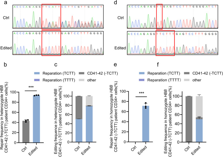
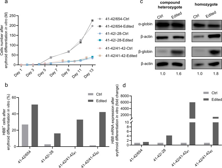
Β-thalassemia is one of the global health burdens. The CD41-42 (-TCTT) mutation at HBB is the most prevalent pathogenic mutation of β-thalassemia in both China and Southeast Asia. Previous studies focused on repairing the HBB CD41-42 (-TCTT) mutation in β-thalassemia patient-specific induced pluripotent stem cells, which were subsequently differentiated into hematopoietic stem and progenitor cells (HSPCs) for transplantation. In this study, we directly applied the CRISPR/Cas9-based gene editing therapy to correct the HBB CD41-42 (-TCTT) mutation in patient-derived HSPCs. The effective editing induced by Cas9:sgRNA ribonucleoprotein and single-stranded oligodeoxynucleotides (ssODNs) was confirmed in HUDEP-2 cell lines harboring the HBB CD41-42 (-TCTT) mutation. Further correction of heterozygote and homozygote HBB CD41-42 (-TCTT) mutations in patient-derived HSPCs resulted in a 13.4-40.8% increase in the proportion of HBB-expressing (HBB +) cells following erythroid differentiation in vitro. At 16 weeks post-xenotransplantation of the edited HSPCs into coisogenic immunodeficient mice, the reparation efficiency in engrafted bone marrow was 17.21% ± 3.66%. Multiparameter flow cytometric analysis of the engrafted bone marrow showed an increase in the percentage of HBB + cells without impairing the ability of engraftment, self-renewal, and multilineage hematopoietic repopulation of HSPCs. For the safety evaluation, 103 potential off-target sites were predicted by SITE-seq and CRISPOR, with one site displaying significant off-target editing. Since this off-target site is located in the intergenic region, it is presumed to pose minimal risk. Taken together, our study provides critical preclinical data supporting the safety and efficacy of the gene therapy approach for HBB CD41-42 (-TCTT) mutation.

Keywords: HBB CD41-42 (-TCTT); CRISPR/Cas9; Gene editing therapy; Hematopoietic stem and progenitor cells; β-thalassemia.


Conflict of interest statement

Declarations. Ethics approval and consent to participate: The human CD34 + HSPCs were obtained from mobilized peripheral blood of patients with HBB CD41-42 (-TCTT) mutation through apheresis at Sun Yat-sen Memorial Hospital, with approval from the Institutions’ Ethical Committee (No. 2020–328). All patients provided informed consent. All the animal experiments were approved by the Institutional Animal Care and Use Committee of Sun Yat-sen University (SYSU-IACUC-2020-B079) and followed the legal requirements in China and Guangdong Province. Competing interests: Hui Xu, Ying Luo, Lin Cheng, and Junbin Liang are employees of Reforgene Medicine. Author Junjiu Huang is a member of the Editorial Board for Advanced Biotechnology, and the author is not involved in the journal’s review of and decisions related to this manuscript.

Figures

Fig. 1
Fig. 1
Correcting HBB CD41-42 (-TCTT) mutation in the disease modeling cell lines. a Schematic of HBB CD41-42 (-TCTT) mutation. The exons of HBB were labeled with blue boxes. The HBB CD41-42 (-TCTT) mutation was indicated in red. The sequences of sgRNAs for gene correction were shown in the middle with the PAM sequence shown in a grey background. The sequences of ssODNs for gene correction are shown below. b Sanger sequencing chromatographs of the PCR amplicons of the wild-type HUDEP-2 and the HBB CD41-42 (-TCTT) mutant stable HUDEP-2 cell lines (HUDEP-2-CD41-42 M), with the homozygotes 4 bp loss at the HBB CD41-42 (-TCTT) mutation site. c Representative Sanger sequencing chromatographs showing the correction efficiency of HBB CD41-42 (-TCTT) mutation in the HUDEP-2-CD41-42 M cell line mediated by sgRNA_1 or sgRNA_2 with ssODNs as the template for reparation. The HBB CD41-42 (-TCTT) mutation was labeled with red boxes
Fig. 2
Fig. 2
Correcting efficiency in heterozygote or homozygote HBB CD41-42 (-TCTT) patient-derived CD34+ cells. a Representative Sanger sequencing chromatographs showing the effective gene reparation in heterozygote HBB CD41-42 (-TCTT) patient-derived CD34+ cells. The red boxes indicated the HBB CD41-42 (-TCTT) mutation sites. (b) Statistics of functional reparation frequency of HBB CD41-42 (-TCTT) mutation in heterozygote HBB CD41-42 (-TCTT) patient-derived CD34+ cells detected by Sanger sequencing and calculated by ICE analysis. (c) Statistics of particular reparation frequency of HBB CD41-42 (-TCTT) mutation in heterozygote HBB CD41-42 (-TCTT) patient-derived CD34+ cells detected by NGS. d Representative Sanger sequencing chromatographs showing the effective gene reparation in homozygote HBB CD41-42 (-TCTT) patient-derived CD34+ cells. The red boxes indicated the HBB CD41-42 (-TCTT) mutation sites. e Statistics of functional reparation frequency of HBB CD41-42 (-TCTT) mutation in homozygote HBB CD41-42 (-TCTT) patient-derived CD34+ cells detected by Sanger sequencing and calculated by ICE analysis. f Statistics of particular reparation frequency of HBB CD41-42 (-TCTT) mutation in homozygote HBB CD41-42 (-TCTT) patient-derived CD34.+ cells detected by NGS. In panel a-f, Ctrl indicated the untransfected HSPCs, while Edited indicated the HSPCs transfected with Cas9:sgRNA_1 RNP and ssODNs as templates. In panels b-c and e–f, the data represent the mean ± SD for 3 replicates using HSPCs from three independent donors. ***, P < 0.001 (by unpaired t-test)
Fig. 3
Fig. 3
Correcting HBB CD41-42 (-TCTT) mutation in patient-derived CD34 + cells induced HBB expression. a The growth curve analysis of the unedited or edited heterozygote or homozygote HBB CD41-42 (-TCTT) patient-derived CD34 + cells during erythroid differentiation. The progeny cells were counted after AO/PI staining. Samples labeled with the same genotype derived from the same donor. b The percentage of HBB + cells after erythroid differentiation (21 days) of the unedited or edited HSPCs, which is detected by flow cytometry after cell immunostaining. Samples were derived from two independent donors with heterozygote HBB CD41-42 (-TCTT) mutation and two independent donors with homozygote HBB CD41-42 (-TCTT) mutation. c The expression of α-globin and β-globin after erythroid differentiation (21 days) of the unedited or edited heterozygote (41–42/654) or homozygote (41–42/41–42#2) HBB CD41-42 (-TCTT) patient-derived CD34 + cells. β-actin served as an internal control to determine the relative expression of α-globin and β-globin. The β-globin/α-globin ratio normalized to the control is shown below. The full uncropped blots images were given in Online Resource. d HBB mRNA expression quantified by RT-qPCR after erythroid differentiation (21 days) of the unedited or edited heterozygote or homozygote HBB CD41-42 (-TCTT) patient-derived CD34 + cells. GADPH mRNA expression was used as input control. The HBB mRNA expression in the Edited group was normalized to that in the Ctrl group. Samples were derived from two independent donors with heterozygote HBB CD41-42 (-TCTT) mutation and two independent donors with homozygote HBB CD41-42 (-TCTT) mutation, which is consistent with the samples shown in panel b. Ctrl indicated the untransfected HSPCs, while Edited indicated the HSPCs transfected with Cas9:sgRNA_1 RNP and ssODNs as templates
Fig. 4
Fig. 4
Xenotransplantation of patient-derived gene-edited CD34.+ cells into NCG-X mice improved the HBB expression. a Schematic of the xenotransplantation of patient-derived CD34 + cells into NCG-X mice. An equal number of unedited (Ctrl) or edited hCD34 + cells transfected with Cas9:sgRNA_1 RNP and ssODNs as templates were transplanted into NCG-X mice, which supported human erythroid reconstitution without irradiation. The ability of human engraftment, self-renewal, erythroid differentiation, and the therapeutic benefit in improved HBB expression were analyzed 16 or 26 weeks after transplantation. b The human engraftment in peripheral blood (PB) or bone marrow (BM) of engrafted mice 16 weeks after transplantation, shown as the percentage of human (h) CD45 + cells within the population of CD45 + cells (mouse and human) detected by flow cytometry. c The self-renewal of HSPCs in the bone marrow of engrafted mice 16 weeks after transplantation, shown as the percentage of hCD34 + cells within the population of hCD45 + cells detected by flow cytometry. d The erythroid differentiation of HSPCs in the bone marrow of engrafted mice 16 weeks after transplantation, shown as the percentage of hCD235a + cells detected by flow cytometry. e The percentage of HBB + cells within the population of hCD235a + cells in the bone marrow of engrafted mice 16 weeks after transplantation, which was detected by flow cytometry. f Statistics of the functional reparation frequency of HBB CD41-42 (-TCTT) mutation in the input HSPCs and the cells derived from the bone marrow of engrafted mice 16 weeks after transplantation, which was detected by NGS. g Representative DNA profiles of the input HSPCs and the cells derived from the bone marrow of engrafted mice 16 weeks after transplantation, which was detected by NGS. The sequence of sgRNA_1 was shown at the top with the PAM sequence shown in grey background and the HBB CD41-42 (-TCTT) mutation shown in red. The bordered sequences indicated the substitutions. The red boxes indicated the insertions. The short line indicated the deletions. The vertical dotted line indicated the predicted cleavage position. In panel b-g, Ctrl indicated the mice transplanted with untransfected HSPCs, while Edited indicated the mice transplanted with HSPCs transfected with Cas9:sgRNA_1 RNP and ssODNs as templates. In panel b-f, the data represent the mean ± SD for 2 to 4 engrafted mice transplanted by unedited or edited homozygote HBB CD41-42 (-TCTT) patient-derived CD34 + cells. Each dot represented a single recipient mouse. ***, P < 0.001, **, P < 0.01 (by unpaired t test)
Fig. 5
Fig. 5
Off-target analysis of HBB CD41-42 (-TCTT) patient-derived CD34+ cells corrected by CRISPR/Cas9. a Overview of the top 20 candidate off-target sites for Cas9:sgRNA_1 RNP predicted by SITE-seq analysis and CRISPOR. The sequence of on-target sites and PAM was shown at the top. Dots represented matches to the on-target site, while the colored nucleotides represented mismatches. b The indel frequency at the 103 candidate off-target sites in both patient-derived HSPCs edited in vitro and the engrafted cells gathered from mice bone marrow 8 weeks or 16 weeks after xenotransplantation, which was detected by amplicon deep sequencing. A highly complementary site (chr3_23029106_T1P69, OT001) with definite off-target editing efficiency was shown. c The specific indel frequency of the on-target site and OT001 detected by amplicon deep sequencing. The Ctrl indicated the untransfected HSPCs. The In vitro indicated the patient-derived HSPCs edited in vitro. The In vivo indicated the engrafted cells gathered from mice bone marrow 8 weeks or 16 weeks after xenotransplantation. The data represent the mean ± SD for 3 to 9 replications. Each dot represented independent donor HSPCs or a single recipient mouse. ***, P < 0.001, *, P < 0.05 (by unpaired t-test). d Representative Sanger sequencing chromatographs showing the definite off-target editing efficiency at OT001 in the edited homozygote HBB CD41-42 (-TCTT) patient-derived CD34+ cells. Ctrl indicated the untransfected HSPCs, while Edited indicated the HSPCs transfected with Cas9:sgRNA_1 RNP and ssODNs as templates

Similar articles

References

    1. Adachi K, Konitzer P, Pang J, Reddy KS, Surrey S. Amino Acids Responsible for Decreased 2, 3-Biphosphosphoglycerate Binding to Fetal Hemoglobin. Blood. 1997;90(8):2916–20. 10.1182/blood.V90.8.2916. - PubMed
    1. Allen, D., Kalter, N., Rosenberg, M., & Hendel, A. (2023). Homology-Directed-Repair-Based Genome Editing in HSPCs for the Treatment of Inborn Errors of Immunity and Blood Disorders. Pharmaceutics, 15(5). 10.3390/pharmaceutics15051329 - PMC - PubMed
    1. Andreani, M., Testi, M., Gaziev, J., Condello, R., Bontadini, A., Tazzari, P. L., . . . Lucarelli, G. (2011). Quantitatively different red cell/nucleated cell chimerism in patients with long-term, persistent hematopoietic mixed chimerism after bone marrow transplantation for thalassemia major or sickle cell disease. Haematologica, 96(1), 128–133. 10.3324/haematol.2010.031013 - PMC - PubMed
    1. Azhagiri MKK, Babu P, Venkatesan V, Thangavel S. Homology-directed gene-editing approaches for hematopoietic stem and progenitor cell gene therapy. Stem Cell Res Ther. 2021;12(1):500. 10.1186/s13287-021-02565-6. - PMC - PubMed
    1. Cameron, P., Fuller, C. K., Donohoue, P. D., Jones, B. N., Thompson, M. S., Carter, M. M., . . . May, A. P. (2017). Mapping the genomic landscape of CRISPR-Cas9 cleavage. Nat Methods, 14(6), 600–606. 10.1038/nmeth.4284 - PubMed

LinkOut - more resources