. 2025 Feb 4;122(5):e2418613122.
doi: 10.1073/pnas.2418613122. Epub 2025 Jan 30.

Activation of the conserved Hippo kinases by inflammasome-triggered proteolytic cleavage controls programmed cell death in macrophages

Affiliations

Activation of the conserved Hippo kinases by inflammasome-triggered proteolytic cleavage controls programmed cell death in macrophages

Yu-Ting Su et al. Proc Natl Acad Sci U S A. .

Abstract

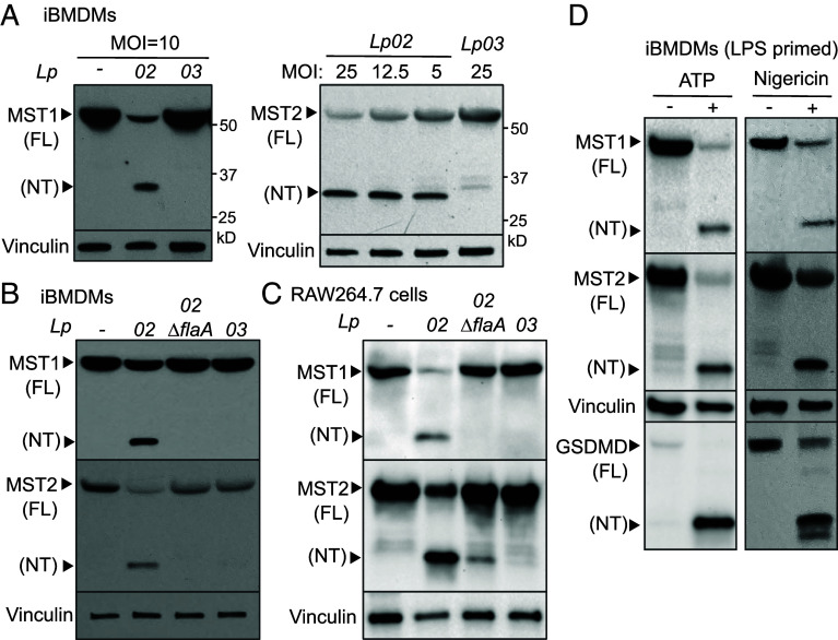
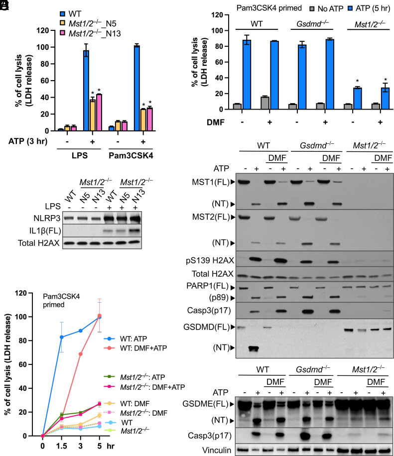
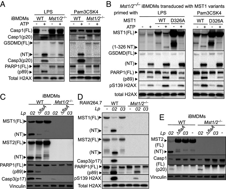
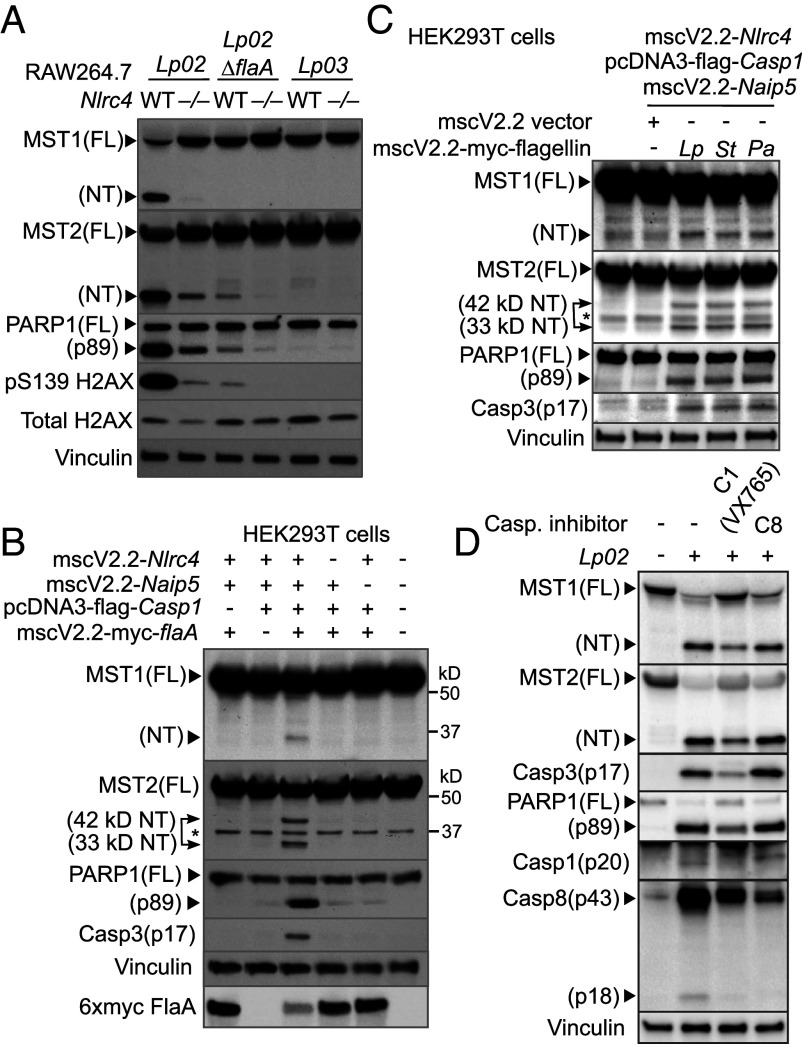
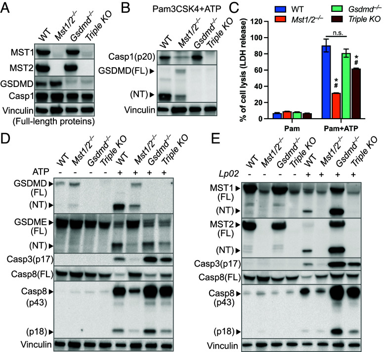
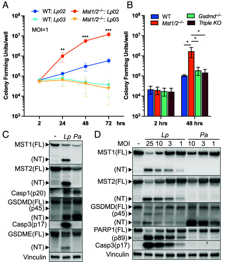
The mammalian Hippo kinases, MST1 and MST2, regulate organ development and suppress tumor formation by balancing cell proliferation and death. In macrophages, inflammasomes detect molecular patterns from invading pathogens or damaged host cells and trigger programmed cell death. In addition to lytic pyroptosis, the signatures associated with apoptosis are induced by inflammasome activation, but how the inflammasomes coordinate different cell death processes remains unclear. Here, we identify the crucial role of MST1/2 in inflammasome-triggered cell death. Macrophages proteolytically convert full-length MST1/2 into the MST1/2 N-terminal fragments (MST1/2-NT) when the NLRC4 inflammasome detects flagellin from the pathogenic bacterium, Legionella pneumophila. Activation of the NLRP3 inflammasome by the damage-associated molecular pattern, extracellular ATP, also produces MST1/2-NT. Caspase-1, the protease activated by these inflammasomes, directly cleaves MST1/2, and blockage of caspase-1 inhibits MST1/2-NT production in macrophages challenged with L. pneumophila. Importantly, MST1/2-NT production is critical for macrophages to trigger a set of death processes associated with apoptosis upon inflammasome activation and knocking out Mst1/2 causes dysregulated gasdermin protein cleavage for pyroptotic death. Furthermore, macrophages lacking MST1/2 have increased susceptibility to virulent L. pneumophila, revealing that the Hippo kinases are important restriction factors against the pathogen. These findings demonstrate that proteolytic cleavage of MST1/2 induced by inflammatory stimuli is an immune pathway to regulate programmed cell death in macrophages and uncover a unique link between the tumor-suppressive Hippo kinases and the inflammasomes in innate immunity.

Keywords: GSDME; STK3; STK4; pattern recognition receptors; type IV secretion.


Conflict of interest statement

Competing interests statement:The authors declare no competing interest.

Figures

Fig. 1.
Fig. 1.
Inflammasome activation triggers cleavage of the Hippo kinases. (A) iBMDMs were challenged with the indicated Lp strains for 3 h. Levels of MST1, MST2, and vinculin (as internal controls) in cell lysate were determined by immunoblotting. MOI, multiplicity of infection. (B and C) Macrophages were challenged with the Lp strains for 3 h at MOI =10 for iBMDMs or MOI = 20 for RAW264.7 cells. (D) iBMDMs were primed with LPS for 3 h, followed by ATP stimulation for 3 h or nigericin for 4 h. Levels of MST1, MST2, GSDMD cleavage (as an indicator of NLRP3 activation) and vinculin (as internal controls) were detected by immunoblotting.
Fig. 2.
Fig. 2.
The Hippo kinases control lytic cell death in macrophages treated with ATP. (A) LPS- or Pam3CSK4-primed iBMDMs were stimulated with ATP for 3 h. LDH activity within the conditioned media was determined using a colorimetric cytotoxicity assay and converted into percentages of the positive controls (iBMDMs lysed by Triton X-100). Data were presented as mean ± SD of technical triplicates and representative of at least three independent experiments. *P < 0.001, Student’s t test, two tailed; unpaired, compared to WT+ATP. (B) Protein levels of NLRP3 and full-length (FL) IL1β in cell lysate of iBMDMs primed with or without LPS were detected by immunoblotting. Histone H2AX served as an internal control. (C) Time course of ATP-triggered cell lysis in Pam3CSK4-primed iBMDMs with/without the presence of DMF was determined by the LDH release assay described in A. (D) Cell lysis induced by 5-h ATP stimulation in Pam3CSK4-primed iBMDMs treated with/without DMF was measured by the LDH release assay as described in A. *P < 0.001, Student’s t test, two tailed; unpaired, compared to WT iBMDMs receiving the same treatment. (E and F) Levels of the indicated proteins in cell lysates from the experiments in D were determined by immunoblotting. Total H2AX and vinculin served as internal controls.
Fig. 3.
Fig. 3.
The Hippo kinases are critical for various death processes in macrophages upon ATP stimulation and Lp challenge. (A) LPS- or Pam3CSK4-primed iBMDMs were stimulated with ATP for 3 h. (B) Mst1/2−/− iBMDMs were transduced to express WT MST1 or the MST1D326A variant. The transduced iBMDMs were primed with LPS or Pam3CSK4 and stimulated with ATP for 3 h. (C) iBMDMs were challenged with the Lp strains at MOI = 20 for 3 h. (D) RAW264.7 macrophages were challenged with the Lp strains at MOI = 25 for 4.5 h. (E) iBMDMs were challenged with the Lp strains at MOI = 10 for 3 h. Levels and processing of indicated proteins in the cell lysates were analyzed by immunoblotting. Histone H2AX and vinculin served as internal controls.
Fig. 4.
Fig. 4.
Processing of the Hippo kinases is a downstream effect of the NLRC4 inflammasome. (A) WT or Nlrc4−/− RAW264.7 macrophages were challenged with the indicated Lp strains at MOI = 20 for 4.5 h. Levels and processing of the indicated proteins were determined by immunoblotting. (B) Cell lysates from HEK293T cells producing mouse NLRC4, NAIP5, caspase-1, and 6xmyc-tagged Lp flagellin FlaA were collected 24 h after transfection. Levels and processing of the indicated proteins were determined by immunoblotting. *nonspecific, cross-reacting band. An additional 42 kDa MST2-NT was observed in the cells with activated NAIP5/NLRC4 inflammasomes. (C) Mouse NAIP5/NLRC4 inflammasome was reconstituted in HEK293T cells with cotransfection of the flagellin genes encoded by different pathogens using transient transfection as described in B. (D) WT iBMDMs were pretreated with the caspase-1 inhibitor VX765 (100 μM) or caspase-8 inhibitor (200 μM) 1 h prior to challenge of the Lp02 strain (MOI = 20, 3 h). Levels and processing of the indicated proteins were analyzed by immunoblotting. Vinculin served as an internal control.
Fig. 5.
Fig. 5.
The Hippo kinases affect activation of caspases in wildtype macrophages in response to ATP and Lp challenge. (A) Presence or absence of the indicated proteins in the gene knockout iBMDMs was determined by immunoblotting. (B) Pam3CSK4-primed iBMDMs were treated with ATP for 5 h. Activation of caspase-1 and GSDMD cleavage in the cell lysates were analyzed by immunoblotting. (C) Pam3CSK4-primed WT and the gene knockout iBMDMs were stimulated with ATP for 5 h. Cell lysis was determined by the LDH release assay as described in Fig. 2A. Data were presented as mean ± SD of technical triplicates and representative of at least three independent experiments. *P < 0.05 compared to WT+ATP, #P < 0.05 compared to Gsdmd−/−+ATP. Student’s t test, two tailed; unpaired. (D) Cell lysates of the Pam3CSK4-primed iBMDMs after 5 h ATP stimulation were analyzed by immunoblotting. (E) Cell lysates of the iBMDMs challenged with Lp02 (MOI = 20, 3 h) were analyzed by immunoblotting. Vinculin served as an internal control.
Fig. 6.
Fig. 6.
The Hippo kinases are key determinants of host defense against virulent Lp. (A) WT or Mst1/2−/− iBMDMs were challenged with the Lp strains at MOI = 1. iBMDMs were washed at 2 h post infection to remove free Lp. At the indicated time points, iBMDMs were lysed with digitonin to release intracellular Lp. Lp colony-forming units (CFUs) were enumerated by plating assays. Data are the mean ± SD of six biological replicates (N = 6). **P < 0.01, ***P < 0.001 (Student’s t test, two tailed, unpaired, compared to WT: Lp02 at the same time points). (B) Intracellular replication of the virulent Lp02 strain in WT and the gene knockout iBMDMs was determined as described in A. Data are the mean ± SD of four biological replicates (N = 4). *P < 0.05, Student’s t test, two tailed, unpaired. (C and D) WT iBMDMs were challenged with the virulent Lp02 strain or P. aeruginosa at MOI = 10 in C or at various MOIs in D for 3 h. Levels of the indicated proteins in the cell lysates were determined by immunoblotting. Vinculin served as an internal control.

References

    1. Ketelut-Carneiro N., Fitzgerald K. A., Apoptosis, pyroptosis, and necroptosis—Oh My! The many ways a cell can die. J. Mol. Biol. 434, 167378 (2022). - PubMed
    1. Lamkanfi M., Dixit V. M., Inflammasomes and their roles in health and disease. Annu. Rev. Cell Dev. Biol. 28, 137–161 (2012). - PubMed
    1. de Zoete M. R., Palm N. W., Zhu S., Flavell R. A., Inflammasomes. Cold Spring Harb. Perspect. Biol. 6, a016287 (2014). - PMC - PubMed
    1. Ren T., Zamboni D. S., Roy C. R., Dietrich W. F., Vance R. E., Flagellin-deficient Legionella mutants evade Caspase-1- and Naip5-mediated macrophage immunity. PLoS Pathog. 2, e18 (2006). - PMC - PubMed
    1. Molofsky A. B., et al. , Cytosolic recognition of flagellin by mouse macrophages restricts Legionella pneumophila infection. J. Exp. Med. 203, 1093–1104 (2006). - PMC - PubMed

MeSH terms

LinkOut - more resources