. 2025 Mar 10;11(3):1847-1856.
doi: 10.1021/acsbiomaterials.4c02175. Epub 2025 Jan 30.

Biomimetic Silk Nanoparticle Manufacture: Calcium Ion-Mediated Assembly

Affiliations

Biomimetic Silk Nanoparticle Manufacture: Calcium Ion-Mediated Assembly

Napaporn Roamcharern et al. ACS Biomater Sci Eng. .

Abstract

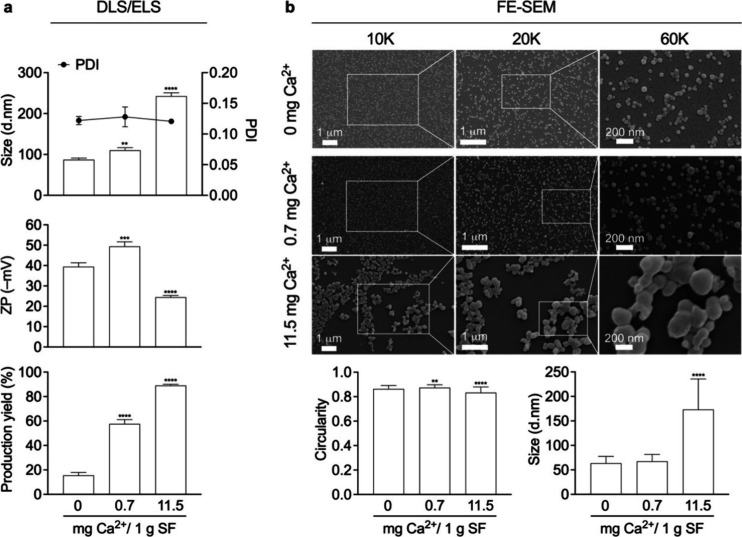
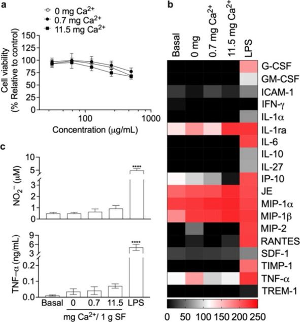
Silk has emerged as an interesting candidate among protein-based nanocarriers due to its favorable properties, including biocompatibility and a broad spectrum of processing options to tune particle critical quality attributes. The silk protein conformation during storage in the middle silk gland of the silkworm is modulated by various factors, including the most abundant metallic ion, calcium ion (Ca2+). Here, we report spiking of liquid silk with calcium ions to modulate the silk nanoparticle size. Conformational and structural analyses of silk demonstrated Ca2+-induced silk assemblies that resulted in a liquid crystalline-like state, with the subsequent generation of β-sheet-enriched silk nanoparticles. Thioflavin T studies demonstrated that Ca2+ effectively induces self-assembly and conformation changes that also increased model drug loading. Ca2+ incorporation in the biopolymer feed significantly increased the nanoparticle production yield from 16 to 89%, while simultaneously enabling Ca2+ concentration-dependent particle-size tuning with a narrow polydispersity index and altered zeta potential. The resulting silk nanoparticles displayed high biocompatibility in macrophages with baseline levels of cytotoxicity and cellular inflammation. Our strategy for manufacturing biomimetic silk nanoparticles enabled overall tuning of particle size and improved yields─features that are critical for particle-based nanomedicines.

Keywords: Bombyx mori; antisolvent precipitation; desolvation; metal ion; nanomedicine; silk fibroin.


Conflict of interest statement

The authors declare no competing financial interest.

Figures

Figure 1
Figure 1
Structural and conformational analyses of silk solutions and nanoparticles. (a) Structural changes of aqueous silk fibroin (SF) in the presence of Ca2+ as assessed by thioflavin T assays (ThT); isopropanol (IPA) (n = 3), (b) 1D 1H NMR, and (c) and 2D [1H, 15N] HSQC NMR spectra in the NH region (n = 1). The 1D 1H NMR spectra are shown at the left of the panel; difference spectra (red and blue) compared with control spectrum (black) are shown at the right of panel; all NMR data: black, control; red, with 0.7 mg Ca2+ (7% increase); blue, with 11.5 mg Ca2+ (17.5% increase). The difference spectra are adjusted with the vertical scale multiplied by a factor of 4. FTIR secondary structure analyses of control silk films with low (silk I) and high (silk II) β-sheet content and silk nanoparticles with (d) the normalized amide I absorbance spectra, (e) secondary structure content, and (f) and correlation coefficient. The percentage of β-sheet content was shown as a summation of β-sheet antiparallel amyloid, β-sheet native, and β-sheet intermolecular structures (n = 3). The second-derivative amide I spectrum of an air-dried silk film (amorphous silk denoted silk I) was used as a reference for the correlation coefficient (R) calculations (mean ± SD, n = 3). (g) The overlay of model drug-loaded silk nanoparticle concentration corresponds to size distribution measured by the nanoparticle-tracking analysis (NTA). The light scatter data are shown as black lines, and the fluorescence data are shown as green lines for 1 mg/mL samples. The percentage is obtained by calculating the area under the curve of the fluorescence data relative to the light scatter data, showing the percentages of model drug-loaded nanoparticles. (h) The fluorescence intensity of the payload detected by nanoparticle tracking. One-way ANOVA and Dunnett’s multiple comparisons test were used for statistical analysis, p < 0.05 (*), p < 0.01 (**), p < 0.001 (***), and p < 0.0001 (****). (i) Schematic of the proposed Ca2+-enhanced silk self-assembly process. Ca2+ increased the yield and enabled particle size tuning, ultimately resulting in β-sheet-rich nanoparticles following antisolvent precipitation. Abbreviations: thioflavin T assays (ThT); isopropanol (IPA); liquid silk fibroin (SF); solid silk nanoparticle (SNP).
Figure 2
Figure 2
Silk nanoparticle characteristics. (a) Analysis of size (dynamic light scattering, DLS; polydispersity index, PDI), zeta potential (ZP) (electrophoretic light scattering, ELS), and production yield of silk nanoparticles (n = 3). (b) Field emission scanning electron microscopy (FE-SEM) images were reported at 10, 20, and 60K magnifications. 400–500 particles and 100–150 particles derived from at least three different regions of interest were used to perform size and circularity calculations, respectively. Metal ion ratios were expressed as mg per 1 g of silk fibroin (SF). One-way ANOVA and Dunnett’s multiple comparison test were used for statistical analysis; p < 0.05 (*), p < 0.01 (**), p < 0.001 (***), and p < 0.0001 (****). PDI: polydispersity index.
Figure 3
Figure 3
Impact of silk nanoparticles on macrophages. (a) Cell viability and inflammatory responses, (b) inflammatory cytokine expression level (scale bar pixel intensity), and (c) endogenous nitrite (NO2) and TNF-α levels in response to the silk nanoparticles. Macrophages treated with complete media supplemented with 200 ng/mL lipopolysaccharide (LPS) served as an inflammation-positive control. SF: Silk fibroin. One-way ANOVA and Dunnett’s multiple comparisons test were used for statistical analysis, p < 0.05 (*), p < 0.01 (**), p < 0.001 (***), and p < 0.0001 (****) (n = 3).

References

    1. Zheng M.; Wang X.; Chen Y.; Yue O.; Bai Z.; Cui B.; Jiang H.; Liu X. A review of recent progress on collagen-based biomaterials (Adv. Healthcare Mater. 16/2023). Adv. Healthcare Mater. 2023, 12 (16), 237008010.1002/adhm.202370080. - DOI - PubMed
    1. Saeedi M.; Vahidi O.; Moghbeli M.; Ahmadi S.; Asadnia M.; Akhavan O.; Seidi F.; Rabiee M.; Saeb M. R.; Webster T. J.; Verma R. S.; Sharifi E.; Zarrabi A.; Rabiee N. Customizing nano-chitosan for sustainable drug delivery. J. Controlled Release 2022, 350, 175–192. 10.1016/j.jconrel.2022.07.038. - DOI - PubMed
    1. Eichhorn S. J.; Etale A.; Wang J.; Berglund L. A.; Li Y.; Cai Y.; Chen C.; Cranston E. D.; Johns M. A.; Fang Z.; Li G.; Hu L.; Khandelwal M.; Lee K.-Y.; Oksman K.; Pinitsoontorn S.; Quero F.; Sebastian A.; Titirici M. M.; Xu Z.; Vignolini S.; frka-Petesic B. Current international research into cellulose as a functional nanomaterial for advanced applications. J. Mater. Sci. 2022, 57 (10), 5697–5767. 10.1007/s10853-022-06903-8. - DOI
    1. Thurber A. E.; Omenetto F. G.; Kaplan D. L. In vivo bioresponses to silk proteins. Biomaterials. 2015, 71, 145–157. 10.1016/j.biomaterials.2015.08.039. - DOI - PMC - PubMed
    1. Nguyen T. P.; Nguyen Q. V.; Nguyen V.-H.; Le T.-H.; Huynh V. Q. N.; Vo D.-V. N.; Trinh Q. T.; Kim S. Y.; Le Q. V. Silk fibroin-based biomaterials for biomedical applications: A review. Polymers. 2019, 11 (12), 1933.10.3390/polym11121933. - DOI - PMC - PubMed