. 2025 Apr 22;9(8):1940-1951.
doi: 10.1182/bloodadvances.2025015842.

A model of zymogen factor XII: insights into protease activation

Affiliations

A model of zymogen factor XII: insights into protease activation

Aleksandr Shamanaev et al. Blood Adv. .

Abstract

In plasma, the zymogens factor XII (FXII) and prekallikrein reciprocally convert each other to the proteases FXIIa and plasma kallikrein (PKa). PKa cleaves high-molecular-weight kininogen (HK) to release bradykinin, which contributes to regulation of blood vessel tone and permeability. Plasma FXII is normally in a "closed" conformation that limits activation by PKa. When FXII binds to a surface during contact activation it assumes an "open" conformation that increases the rate of activation by PKa. Mutations in FXII that disrupt the closed conformation have been identified in patients with conditions associated with excessive bradykinin formation. Using FXII structures from the AlphaFold database, we generated models for the closed form of human FXII that we tested with site-directed mutagenesis. The models predict multiple interactions between the fibronectin type 2 (FN2), kringle, and catalytic domains involving highly conserved amino acids that restrict access to the FXII activation cleavage sites. Based on the model, we expressed FXII with single-amino acid substitutions and studied their effects on FXII activation by PKa. Replacements for Arg36 in the FN2 domain; Glu225, Asp253, or Trp268 in the kringle domain; or Lys346 near the activation cleavage site were activated >10-fold faster by PKa than wild-type FXII. Adding these proteins to plasma resulted in rapid HK cleavage due to markedly enhanced reciprocal activation with prekallikrein. The results support a model that explains the behavior of FXII in solution. Conformational changes involving the identified amino acids likely occur when FXII binds to a surface to facilitate activation.


Conflict of interest statement

Conflict-of-interest disclosure: D.G. receives consultant fees from Anthos, Bayer, Bristol Myers Squibb, and Kanssen with an interest in inhibition of contact activation and the kallikrein-kinin system for therapeutic purposes. The remaining authors declare no competing financial interests.

Figures

None
Graphical abstract
Figure 1.
Figure 1.
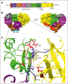
A model for human FXII. (A) Schematic diagram of human FXII showing the positions of the heavy chain N-terminal peptide (N-term, orange-red), fibronectin type 2 (FN2, purple), epidermal growth factor 1 (EGF1, brown), fibronectin type 1 (FN1, magenta), EGF2 (dark gray), and kringle (KNG, light green) domains, the proline-rich region (PRR, dark green), the activation loop (light gray) and the catalytic domain (CD, yellow). The position of an anion-binding site (ABS) between FN2 and EGF1 is indicated in light blue, the activation cleavage site at Arg353 is indicated by a blue arrow, and the active site serine residue (Ser544) is indicated by the black bar. (B) AF-adjusted prediction (supplemental Figure 3C) for full-length human FXII shown as molecular surface representations. The 2 images shown are rotated 180° relative to each other. The color scheme is identical to that in panel A. Positions for the cleavage sites at Arg343 and Arg353 are indicated in dark blue. Most of the activation loop (Cys340-Arg353) is buried between the KNG and CD. (C) Predicted intramolecular interactions between the FXII FN2 domain (purple), KNG domain (light green), activation loop (light gray), and CD (yellow) are shown as cartoon and stick diagrams. Important basic amino acids are shown in dark blue, acidic amino acids in red, and tryptophan residues in olive. Positions of Arg343 and Arg353 are indicated in dark blue on the peptide backbone of the activation loop. The positions of Leu266 and Val359 on the peptide backbone are indicated. Hydrogen bonds are shown as dashed light blue lines, and cation-π interactions as dashed orange lines.
Figure 2.
Figure 2.
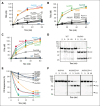
FXII activation. (A-C) Activation of FXII by (A-B) PKa or (C) FXIa. One hundred nanomolar FXII-WT, FXII lacking most of its heavy chain region (ΔFXII), or FXII variants with single–amino acid substitutions were incubated with PKa (12.5 nM) or FXIa (2.5 nM) at 37°C. For panels A-C, at indicated times, aliquots were removed and tested for FXIIa generation by chromogenic assay. Results represent averages ± 1 standard deviation (SD) for at least 3 experiments. (D) FXII cleavage by PKa. One micromolar FXII-WT (left) or the variant FXII-Ala346 (right), was incubated with 125 nM PKa. At indicated time, samples were removed into reducing sample buffer and size fractionated by SDS-PAGE. Positions of molecular mass standards are shown on the left. Shown on the right are positions of standards for the zymogen FXII band (Z) and the heavy chain (HC) and light chain (LC) of FXIIa. (E) Densitometric quantification of FXII Z band disappearance in reactions with PKa identical to those shown in panel D. The curves show changes in Z band intensity as a percent of the intensity at 0 minutes. Each point represents the average for 2 experiments. (F) FXII-Ala544 (left), FXII-Ala268, Ala544 (middle), and FXII-Ala502, Ala544 (right), 100 nM each, was incubated with 12.5 nM PKa at 37°C. At indicated times, samples were removed into reducing SDS sample buffer, size fractionated by SDS-PAGE, and transferred to nitrocellulose membranes. Immunoblotting was with an HRP-conjugated anti-FXII IgG. Positions of molecular mass standards are shown on the left. Positions of standards for the Z band, and the HC, and LC of FXIIa are indicated at the right of each image. Shown are representative blots for experiments that were run in duplicate.
Figure 3.
Figure 3.
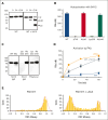
The FXII KNG domain and protein conformation. (A) FXII-WT (400 nM) was incubated with vehicle (left panel) or an equimolar concentration of IgG 5A12 (right panel). At indicated times, samples were removed into reducing SDS sample buffer, size fractionated by SDS-PAGE, and transferred to nitrocellulose membranes. Immunoblotting was with an HRP-conjugated anti-FXII IgG. Positions of molecular mass standards are shown on the left. Positions of standards for the Z band, and the HC, and LC of FXIIa are indicated at the right of each image. (B) FXII-WT, ΔFXII, FXII-Ala, FXII-Lys253, and FXII-Ala346 (400 nM) were incubated overnight (O/N) with an equimolar concentration of IgG 5A12. FXIIa generation was assessed by chromogenic substrate cleavage. (C) FXII-WT (WT), FXIIa-WT (XIIa), FXII-Lys253, and FXII-Ala346 (200 ng) were size fractionated by SDS-PAGE, transferred to nitrocellulose membrane, and immunoblotted with IgG 5A12 (left and middle) or polyclonal anti-FXII IgG (right). (D) One hundred nanomolar FXII-WT (WT), ΔFXII, or FXII-WT mixed with 100 nM IgG 5A12 (WT + 5A12) was incubated with PKa (12.5 nM) at 37°C. At indicated times, aliquots were removed and tested for FXIIa generation by chromogenic assay. (E) smFRET efficiency histograms of FXII S544A in the absence (left panel) or presence (right panel) of 100 mM εACA. Representative graphs are shown. Each experiment was done in triplicate. Gaussian distribution is indicated by red dashed lines. The number of each population is indicated at the left of each image.
Figure 4.
Figure 4.
FXII binding to PK. (A) Standard curve of different concentrations (1-120 μg/mL) of WT FXII or FXIIa binding to plate-immobilized PK. Results are shown as optical density values, measured as described in “Methods.” (B) Binding of WT FXII and FXIIa, and FXII variants (30 μg/mL) to plate-immobilized PK. Values are shown as a percent of the signal for FXII-WT (assigned a value of 100%) ± 1 SD. (C) Plasma FXII or FXIIa (10 μg) was incubated O/N with anti-PK IgG beads alone or with plasma PK (10 μg) at 4°C. (D) Plasma or recombinant FXII (10 μg) were incubated O/N with anti-PK IgG beads and 10 μg PK lacking an active site serine (PK-Ala559) at 4°C. For panels C and D. After incubation, the beads were washed, samples were eluted with nonreducing SDS sample buffer, size fractionated by SDS-PAGE, and either (C) stained with Coomassie blue or (D) transferred to nitrocellulose membranes and probed with anti–FXII HRP-conjugated IgG. Positions of molecular mass standards are shown on the left. Positions of standards for PK and FXII/FXIIa are indicated at the right of each image. Shown are representative gels/blots for experiments that were run in duplicate.
Figure 5.
Figure 5.
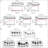
Activation of the KKS in human plasma. (A) Human FXII-deficient plasma was supplemented with 400 nM FXII-WT and incubated in the presence (top row) or absence (bottom row) of kaolin (2.5 mg/mL). Kaolin will induce contact activation in plasma. (B) Human FXII-deficient plasma was supplemented with 400 nM FXII-Ala36 (top row) or FXII-Ala346 (bottom row). For all experiments, FXII-supplemented plasma was incubated at 37°C. Samples were removed at the indicated times into nonreducing sample buffer, size fractionated by SDS-PAGE, and transferred to nitrocellulose membranes. Membranes were developed with antibodies to FXII (left), PK (middle), or FXI (right). Positions of molecular mass standards in kilodaltons are shown to the left of each panel. Positions for free FXII and FXIIa (XII[a]), free PK and PKa (PK[a]), and FXI and FXIa; and FXIIa, PKa, and FXIa in complex with plasma protease inhibitors (XIIa + INH, PKa + INH, and XIa + INH) are shown to the right of each panel. Note that free FXIa migrates more slowly than free FXI in panel A (right column). Shown are representative blots for experiments that were run in duplicate.
Figure 6.
Figure 6.
HK cleavage in human plasma and in mice. (A) Human FXII-deficient plasma was supplemented with FXII-WT, ΔFXII, FXII-Ala36, FXII-Lys253, and FXII-Ala346 (140 nM) and incubated at 37°C. At indicated times, samples were removed into non-reducing sample buffer, size fractionated by SDS-PAGE, transferred to nitrocellulose membranes, and probed with goat anti-human HK IgG. Positions of markers for uncleaved HK (white arrow) and cleaved HKa forms (black arrows) are indicated to the right of each image. Western blots underwent densitometry scanning to generate the curves shown below each western blot. Curves show the disappearance of HK (blue), appearance of HKa intermediate (single cleavage, gray), and appearance of the final form of HKa (2 cleavages, red). Percentile values were assigned to each band based on comparison to the density of the HK band at time 0 (assigned a value of 100%). Data are averages of 2 experiments. (B) FXII-deficient mice received intravenous infusions of FXII-WT, ΔFXII, FXII-Ala36, FXII-Lys253, or FXII-Ala346 to achieve estimated plasma concentrations of 40 nM. Shown are nonreducing western blots of plasma collected 0, 15, or 30 minutes, or ∼16 hours (O/N) after infusion. Blots were developed with anti-murine HK IgG. Positions of markers for HK (white arrows) and cleaved HKa forms (black arrows) are indicated to the right of each image. Positions of molecular mass markers are shown on the left. Shown are representative blots for experiments that were run in duplicate.

Similar articles

Cited by

References

    1. de Maat S, Maas C. FXII: form determines function. J Thromb Haemost. 2016;14(8):1498–1506. - PubMed
    1. Maas C, Renné T. Coagulation factor XII in thrombosis and inflammation. Blood. 2018;131(17):1903–1909. - PubMed
    1. Shamanaev A, Litvak M, Ivanov I, et al. Factor XII structure-function relationships. Semin Thromb Hemost. 2024;50(7):937–952. - PMC - PubMed
    1. Schmaier AH. The contact activation and kallikrein/kinin systems: pathophysiologic and physiologic activities. J Thromb Haemost. 2016;14(1):28–39. - PubMed
    1. Long AT, Kenne E, Jung R, Fuchs TA, Renné T. Contact system revisited: an interface between inflammation, coagulation, and innate immunity. J Thromb Haemost. 2016;14(3):427–437. - PubMed