Review
. 2025 Feb 13;68(3):2219-2237.
doi: 10.1021/acs.jmedchem.4c02342. Epub 2025 Jan 30.

Exploring the Ascorbate Requirement of the 2-Oxoglutarate-Dependent Dioxygenases

Affiliations
Review

Exploring the Ascorbate Requirement of the 2-Oxoglutarate-Dependent Dioxygenases

Carlos C Smith-Díaz et al. J Med Chem. .

Abstract

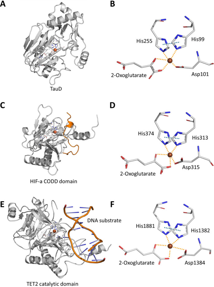
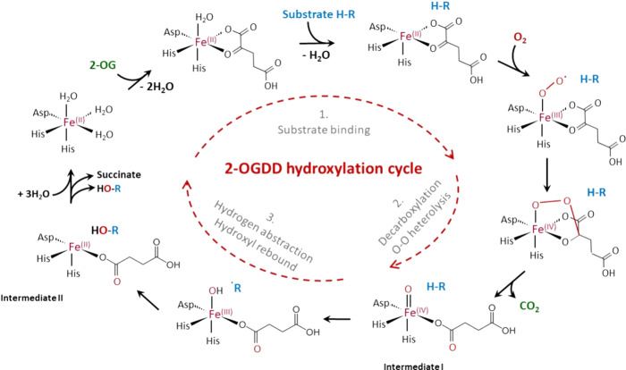
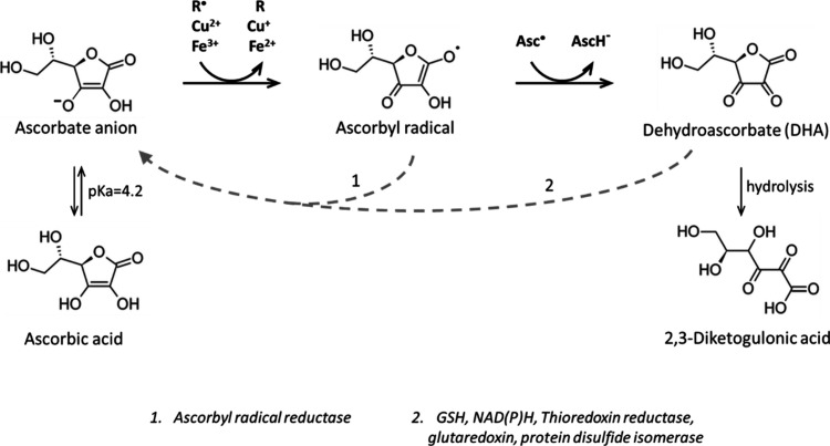
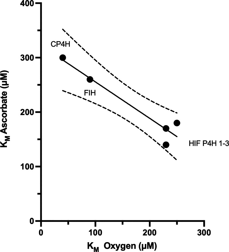
In humans, the 2-oxoglutarate-dependent dioxygenases (2-OGDDs) catalyze hydroxylation reactions involved in cell metabolism, the biosynthesis of small molecules, DNA and RNA demethylation, the hypoxic response and the formation of collagen. The reaction is catalyzed by a highly oxidizing ferryl-oxo species produced when the active site non-heme iron engages molecular oxygen. Enzyme activity is specifically stimulated by l-ascorbic acid (ascorbate, vitamin C), an effect not well mimicked by other reducing agents. In this perspective article we discuss the reliance of the 2-OGDDs on ascorbate availability. We draw upon findings from studies with different 2-OGDDs to piece together a comprehensive theory for the specific role of ascorbate in supporting enzyme activity. Our discussion centers on the capacity for ascorbate to act as an efficient radical scavenger and its propensity to reduce and chelate transition metals. In addition, we consider the evidence supporting stereospecific binding of ascorbate in the enzyme active site.


Conflict of interest statement

The authors declare no competing financial interest.

Figures

Figure 1
Figure 1
A comparison of the crystal structures of TauD, PHD2 and TET2. The crystal structures for the 2-OGDD enzymes (A,B) TauD [E. coli, PDB reference 1os7(19)], (C,D) PHD2 (Homo sapiens, PBD reference 5L9B(20)) and (E,F) TET2 [Homo sapiens, PDB reference 4nm6(21)] are depicted with some computational transformations (notably the transformation of N-oxalylglycine to 2-OG in TET2, Mn to Fe in PHD2). The TauD structure depicts one monomer from the oligomeric crystal structure and PHD2 one monomer from the dimeric crystal structure. These three enzymes are characterized by a conserved iron and 2-OG binding site. While the proteins are of vastly different size, have different substrates, and in the case of TauD, are from different organisms, they share a characteristic triad of amino acids (2 His and 1 Asp) which coordinate iron at the active site. Another key feature is the conserved 2-OG binding site, where 2-OG coordinates to iron in a bidentate manner with stabilizing interactions from a nearby arginine residue (not shown).
Figure 2
Figure 2
2-OGDD hydroxylation consensus catalytic cycle. Human enzymes in the 2-OGDD family are proposed to employ the following hydroxylation mechanism. Step 1. In its ground state, enzyme bound Fe at the active site lies in an octahedral geometry, coordinating to three amino acid residues and three water molecules. Binding of the 2-OG substrate displaces two water molecules, generating the 2-OG-bound ligand configuration frequently observed in crystallographic structures. This process is followed by the binding of the principal substrate and the displacement of another water, generating a vacant binding site for molecular oxygen at the Fe center. Step 2. The resulting Fe(III)-superoxo species primes the enzyme for the decarboxylation of 2-OG, releasing CO2 and creating a Fe(IV) ferryl intermediate with bound succinate. Step 3. The highly reactive ferryl intermediate hydroxylates the principal substrate via a two-step hydrogen abstraction/hydroxyl rebound mechanism.
Figure 3
Figure 3
Reactions of ascorbate in mammalian systems. Ascorbate is a highly efficient reducing agent for biological free radicals and oxidized transition metals. Sequential oxidation of the ascorbate anion by single electron steps generates the ascorbyl radical and DHA. Both products can be recycled intracellularly but DHA is unstable at neutral pH and readily undergoes irreversible hydrolysis to generate inactive breakdown products.
Figure 4
Figure 4
Relationship between the 2-OGDD Km values for oxygen and ascorbate. This relationship was constructed from published data available for the collagen and HIF hydroxylases.,, The values are as follows: CP4H1: Ascorbate Km 300 μM, Oxygen Km 40 μM. FIH: Ascorbate Km 260 μM, Oxygen Km 90 μM. HIF P4H 1–3: Ascorbate Km 140–180 μM, Oxygen Km 230–250 μM.
Figure 5
Figure 5
Impact of ascorbate on the inhibition of the HIF hydroxylases by iron chelation, Co(II) and Ni(II) exposure, competitive 2-OG inhibitor DMOG and hypoxia. HIF-1α protein accumulates when the regulatory HIF hydroxylases are inhibited. The Western blots shown demonstrate HIF-1α stabilization in ascorbate-deficient compared with ascorbate-loaded Jurkat cells (500 μM ascorbate overnight). Ascorbate completely prevented hydroxylase inhibition following induction with (A) the Fe chelator DFO, (B) Co(II) and (C) Ni(II). In contrast, ascorbate only partially blocked the effects of (D) DMOG, the competitive inhibitor of 2-OG and (E) O2 deprivation. The data shown are representative of 2–3 independent experiments with Jurkat cells, showing HIF-1α band accumulation as a marker for HIF hydroxylase inhibition and β-actin as a loading control. The Figure has been adapted from previously published results.
Figure 6
Figure 6
Ascorbate binding site within the TET homologue CMD1. The active site of TET homologue CMD1 (PDB code 7cy8) shows that iron is bound by a characteristic triad of amino acids (2 His and 1 Asp). Ascorbate also directly coordinates to the enzyme bound iron, with the 5-methylcytosine substrate sitting in a nearby pocket.
Figure 7
Figure 7
Uptake of ascorbate in humans and distribution to the tissues. Humans are dependent on dietary sources of ascorbate, mostly from fresh fruit and vegetables in which it is not destroyed by cooking. Following uptake in the bowel, ascorbate is transported via the circulation into tissues around the body. Plasma levels are maintained below 100 μM by filtration and reabsorption through the kidneys. Accumulation in tissues is controlled by active transport via the SVCTs, and tissue saturation levels vary across a wide range, as shown. The highest levels are recorded in those tissues that are sites of 2-OGDD activity or Cu enzyme-dependent hormone synthesis (marked with an asterisk).
Figure 8
Figure 8
Modeling penetration of ascorbate from plasma into the tissues and accompanying cellular uptake. This schematic illustrates the pharmacokinetic modeling of the relationship between ascorbate diffusion from the circulation, penetration into the tissues, and cellular uptake by tissue cells. The central circle represents a blood vessel with 10, 50, or 100 μM ascorbate, with a heat map showing the corresponding diffusion distance and cellular uptake. The black bars indicate 100 μm: intervessel distance in normal tissues is 50 μm. The image shows that plasma saturation levels of ∼100 μM ascorbate are required to ensure intracellular ascorbate levels can reach the mM range required to support 2-OGDD activity. Average levels in the human population are regularly reported to be ∼50 μM which, according to this model, would compromise ascorbate delivery at short distances form the blood vessels, even in normal tissues. Figure drawn to summarize data from study modeling ascorbate penetration through multicellular layers.

Similar articles

Cited by

References

    1. Brodie B. B.; Axelrod J.; Shore P. A.; Udenfriend S. Ascorbic acid in aromatic hydroxylation. II. Products formed by reaction of substrates with ascorbic acid, ferrous ion and oxygen. J. Biol. Chem. 1954, 208, 741–750. 10.1016/S0021-9258(18)65599-1. - DOI - PubMed
    1. Hutton J. J. Jr.; Trappel A. L.; Udenfriend S. Requirements for alpha-ketoglutarate, ferrous ion and ascorbate by collagen proline hydroxylase. Biochem. Biophys. Res. Commun. 1966, 24 (2), 179–84. 10.1016/0006-291X(66)90716-9. - DOI - PubMed
    1. Myllylä R.; Kuutti-Savolainen E. R.; Kivirikko K. I. The role of ascorbate in the prolyl hydroxylase reaction. Biochem. Biophys. Res. Commun. 1978, 83 (2), 441–8. 10.1016/0006-291X(78)91010-0. - DOI - PubMed
    1. Myllylä R.; Majamaa K.; Gunzler V.; Hanauske-Abel H. M.; Kivirikko K. I. Ascorbate is consumed stoichiometrically in the uncoupled reactions catalyzed by prolyl 4-hydroxylase and lysyl hydroxylase. J. Biol. Chem. 1984, 259 (9), 5403–5. 10.1016/S0021-9258(18)91023-9. - DOI - PubMed
    1. Myllylä R.; Tuderman L.; Kivirikko K. I. Mechanism of the Prolyl Hydroxylase Reaction. 2. Kinetic Analysis of the Reaction Sequence. Eur. J. Biochem. 1977, 80, 349–357. 10.1111/j.1432-1033.1977.tb11889.x. - DOI - PubMed