Review
. 2025 Jan 30;24(2):39.
doi: 10.1007/s12311-025-01790-6.

Cerebellar Development and the Burden of Prematurity

Affiliations
Review

Cerebellar Development and the Burden of Prematurity

Tobias Muehlbacher et al. Cerebellum. .

Abstract

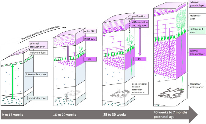
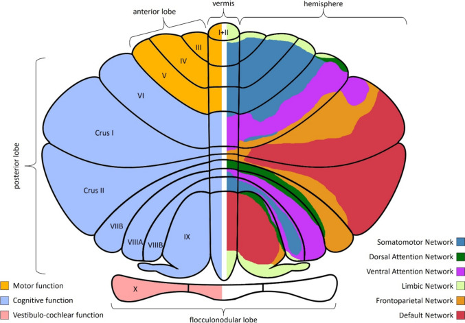
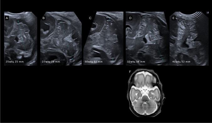
The role of the cerebellum in the neurodevelopmental outcomes of preterm infants has often been neglected. However, accumulating evidence indicates that normal cerebellar development is disrupted by prematurity-associated complications causing cerebellar injury and by prematurity itself. This hampers not only the normal development of motor skills and gait, but also cognitive, language, and behavioral development, collectively referred to as "developmental cognitive affective syndrome." In this comprehensive narrative review, we provide the results of an extensive literature search in PubMed and Embase to summarize recent evidence on altered cerebellar development in premature infants, focusing on neuroimaging findings, its causative factors and its impact on long-term neurodevelopmental outcomes.

Keywords: Cerebellar disease; Cerebellum; Neonate; Preterm infant.


Conflict of interest statement

Declarations. Competing Interest: TM receives funding from Anna-Mueller-Grocholski Foundation for the prospective CSI NeO Study. SJS received funding from the Raynor Foundation for the Raynor Cerebellar Project. No funding has been received for this review. The authors have nothing further to disclose.

Figures

Fig. 1
Fig. 1
Development of the cerebellar cortex. Two proliferative zones are present in the developing cerebellum. The first is the dorsolateral subventricular zone of the rhombic lips from where excitatory granule precursor cells migrate from the 5th week of gestation and proliferate tangentially to form the external granular layer (EGL) above the developing molecular layer. The second is the ventricular zone from where gamma-aminobutyric acid (GABA)ergic inhibitory cells including the Purkinje cells (PC) originate and migrate through the intermediate zone to form the PC layer and inhibitory interneurons within the molecular layer as well as the cerebellar nuclei within the cerebellar white matter [9, 15]. At week 16, the PC layer is still organized in multiple layers before becoming the characteristic PC monolayer at 28 weeks. The EGL is a transient developmental layer reaching a peak thickness with six to nine cell layers around 25 weeks of gestation. The outer cell layers of the EGL (adjacent to the cerebrospinal fluid) proliferate under the secretion of Sonic Hedge Hog (Shh) from the growing dendritic tree of the PC, while GCP in the inner EGL start to differentiate and migrate through the molecular layer. During the migration into the initially paucicellular internal granular layer (IGL), the T-shaped axons grow perpendicular as so-called parallel fibers forming en-passant synapses with the dendritic tree of the PC. With the huge numbers of migrating GCP and their parallel fibers, the molecular layer and IGL grow from the neonatal period and during the first year of life, while the EGL gradually fades and vanishes during this period. Illustration inspired by Volpe [9].
Fig. 2
Fig. 2
Illustration of cerebellar functional topography. Left part of the illustration: The anterior lobe (orange) consists of lobules I-V and has primarily motor function. The posterior lobe (light-blue) is the largest part of the cerebellum consisting of lobules VI to XI and is involved in cognitive function. The oldest part of the cerebellum is the floccularnodular lobe (lobule X) with direct connections to the vestibular nuclei. The right part of the illustration is a schematic of large different cerebral-cerebellar networks. Lobules III-VI and VIIIB are part of the somatomotor networks, while the rest of the cerebellum is connected with association cortices for higher cognitive function. Intriguingly, there seems to be no connection to the primary visual cortex networks [2].
Fig. 3
Fig. 3
Mature cerebellar cortex and circuitry. The cerebellar cortex contains of three layers: the deepest cell-dense granular layer, the second mono-cell layer contains Purkinje cells (PC) and Bergmann glia cells and the molecular layer containing only few inhibitory interneurons, e.g. stellate cells (SC) and basket cells (BC) [9, 15]. Information from the cerebral cortex is relayed in pontine nuclei and reach excitatory granule cells (GC) and unipolar brush cells (UBC) as well as inhibitory Golgi-cells (GoC) via mossy fibers. Further afferent signaling from the spinal cord and vestibular system arrive via climbing fibers coming from the inferior olive nucleus, which interact directly with the dendritic tree of the PC. The latter are arranged strictly in a sagittal plane. Perpendicular to these in a coronal plane are the T-shaped axons (parallel fibers) of the GC which form “en-passant” synapses with the dendritic trees of multiple PC [15]. The only efferent signals from the cerebellar cortex are transferred via the axons of the PC to the cerebellar nuclei in the cerebellar white matter (from lateral to ventral the dentate, emboliform, globose and fastigial nuclei), which relay the information mainly via the thalamus to the cerebral cortex or to lesser extend to red nucleus or other structures in the brainstem [15, 20]. Afferent fibers from the cerebral cortex are relayed in the pontine nuclei (mossy fibers) and arrive in the contralateral cerebellum via the middle cerebellar peduncle [17, 20]. The inferior cerebellar peduncle contains afferent projections from the inferior olive nucleus (climbing fibers) and both afferent and efferent fibers from and to the spinal cord and vestibular system [16, 20].
Fig. 4
Fig. 4
Unimpaired cerebellar growth in a preterm infant of 25 weeks gestational age. Serial coronal ultrasound scans (A-E) through the mastoid fontanel show a normal development with proceeding foliation representing the rapid increase in cerebellar surface. (F) T2 MRI scan at postmenstrual age of 33 weeks results in same transverse cerebellar diameter compared to corresponding ultrasound scan (D)
Fig. 5
Fig. 5
Cerebellar hypoplasia of prematurity: Preterm infant of 25 4/7 weeks gestation age without supra- or infratentorial hemorrhage. Upper row shows serial transnuchal ultrasound scans, the lower row corresponding axial scans through the mastoid fontanel at day of life (DOL) 7 and 27, approximately postmenstrual age (PMA) 36 weeks and term equivalent age (TEA). A normal transverse cerebellar diameter (TCD) of 25-50th percentile according to Imamoglu et al. [29] was present at first scan with one week. Even without recognizable supra- or infratentorial hemorrhage in serial ultrasound scans a mild cerebellar hypoplasia developed during the unremarkable neonatal course with a TCD < 3rd percentile at PMA 36 weeks and TEA
Fig. 6
Fig. 6
Limited cerebellar hemorrhage (CBH) left posterior-lateral hemisphere in a preterm infant of 29 weeks gestational age. A Coronal ultrasound scan through the mastoid fontanel, B corresponding transnuchal ultrasound scan on the fourth day of life shows the limited CBH (< 1/3 of the left hemisphere). C On the follow-up scan at postmenstrual age (PMA) of 34 5/7 weeks through the left mastoid fontanel, an asymmetrical appearance with smaller left hemisphere can be identified. D T2 weighted MRI scan at PMA 36 6/7 weeks and susceptibility weighted imaging (SWI) confirm the asymmetry with a hypoplastic left hemisphere and residual hemosiderin deposition after the CBH
Fig. 7
Fig. 7
Massive cerebellar hemorrhage (CBH) of the right hemisphere in a preterm infant of 27 weeks gestational age. A Coronal scan via the anterior fontanel shows an increased echogenicity (CBH, arrow) in the right cerebellar hemisphere, which can be also seen in (B) the right paramedian sagittal scan (arrow). C the left cerebellar hemisphere appears normal (arrow). D Better visualization of the extend of the massive CBH (> 1/3 of the right hemisphere, arrowheads) via ultrasound scan through the mastoid fontanel with midline shift of the vermis and IVth ventricle. E Axial T2 MRI scan and paramedian sagittal T2 scans, F corresponding to (B) and (G) to (C), and susceptibility weighted imaging (H) proving the massive CBH of the right hemisphere
Fig. 8
Fig. 8
Impaired cerebellar development after high-grade intraventricular hemorrhage in a preterm infant of 25 weeks gestational age. A Coronal scan shows IVH III° right side with posthemorrhagic ventricular dilatation. B Sagittal midline scan shows an enlarged IIIrd ventricle and prominent IVth ventricle and blood clots within the posterior fossa around the cerebellar vermis and in the cisterna magna. C coronal scan through the mastoid fontanel shows even more pronounced the infratentorial blood clots in the cerebrospinal fluid around the otherwise unaffected cerebellum. D coronal scan through the mastoid fontanel at term equivalent age with severe cerebellar hypoplasia, TCD 40.6 mm. (E + F) MRI SWI proves hemosiderin deposition mainly in right lateral ventricle and IVth ventricle, especially the lateral apertures. No parenchymal hemorrhagic infarction in the cerebrum or cerebellum could be identified. G T2w scan confirms the symmetric cerebellar hypoplasia seen in US scan
Fig. 9
Fig. 9
Crossed cerebro-cerebellar diaschisis. Preterm infant 24 weeks gestational age with IVH III and left-sided periventricular hemorrhagic infarction. At postmenstrual age of 38 weeks exists an asymmetry with a hypoplastic contralateral right cerebellar hemisphere (A) in ultrasound via mastoid fontanel and (B) transnuchal ultrasound scan. C Posterior coronal scan shows the extent of the periventricular hemorrhagic infarction with porencephaly. (D + E) T2 weighted MRI scans are also with hypoplasia of the contralateral cerebellar hemisphere and without signs of a direct cerebellar lesion. (F) With no susceptibility artefacts in SWI within the cerebellum or on the cerebellar surface, a crossed cerebro-cerebellar diaschisis is proven (for more information see text)

Similar articles

References

    1. Sathyanesan A, Zhou J, Scafidi J, Heck DH, Sillitoe RV, Gallo V. Emerging connections between cerebellar development, behaviour and complex brain disorders. Nat Rev Neurosci. 2019;20(5):298–313. 10.1038/s41583-019-0152-2. - PMC - PubMed
    1. Buckner RL, Krienen FM, Castellanos A, Diaz JC, Yeo BT. The organization of the human cerebellum estimated by intrinsic functional connectivity. J Neurophysiol. 2011;106(5):2322–45. 10.1152/jn.00339.2011. - PMC - PubMed
    1. Stoodley CJ, Schmahmann JD. Functional topography of the human cerebellum. Handb Clin Neurol. 2018;154:59–70. 10.1016/B978-0-444-63956-1.00004-7. - PubMed
    1. Schmahmann JD. The cerebellum and cognition. Neurosci Lett. 2019;688:62–75. 10.1016/j.neulet.2018.07.005. - PubMed
    1. Glickstein M, Strata P, Voogd J. Cerebellum: history. Neuroscience. 2009;162(3):549–59. 10.1016/j.neuroscience.2009.02.054. - PubMed

MeSH terms

LinkOut - more resources