. 2025 Jan 31;16(1):1200.
doi: 10.1038/s41467-025-56419-3.

Rhesus Cytomegalovirus-encoded Fcγ-binding glycoproteins facilitate viral evasion from IgG-mediated humoral immunity

Affiliations

Rhesus Cytomegalovirus-encoded Fcγ-binding glycoproteins facilitate viral evasion from IgG-mediated humoral immunity

Claire E Otero et al. Nat Commun. .

Abstract

Human cytomegalovirus (HCMV) encodes four viral Fc-gamma receptors (vFcγRs) that counteract antibody-mediated activation in vitro, but their role in infection and pathogenesis is unknown. To examine their in vivo function in an animal model evolutionarily closely related to humans, we identified and characterized Rh05, Rh152/151 and Rh173 as the complete set of vFcγRs encoded by rhesus CMV (RhCMV). Each one of these proteins displays functional similarities to their prospective HCMV orthologs with respect to antagonizing host FcγR activation in vitro. When RhCMV-naïve male rhesus macaques were infected with vFcγR-deleted RhCMV, peak plasma DNAemia levels and anti-RhCMV antibody responses were comparable to wildtype infections of both male and female animals. However, the duration of plasma DNAemia was significantly shortened in immunocompetent, but not in CD4 + T cell-depleted animals. Since vFcγRs were not required for superinfection of rhesus macaques, we conclude that these proteins can prolong lytic replication during primary infection by evading virus-specific adaptive immune responses, particularly antibodies.


Conflict of interest statement

Competing interests: S.R.P. has served as a consultant to Merck, Moderna, Pfizer, GSK and Dynavax and has led sponsored programs with Moderna and Merck. P.K. has received funding from Biotest AG. None of these activities have impacted this work. The remaining authors declare no competing interests.

Figures

Fig. 1
Fig. 1. Rh152/Rh151 binds IgG and antagonizes host FcγR activation by blocking receptor engagement.
A 293 T cells were transfected with a pIRES-eGFP plasmid expressing the indicated UL119/118 or Rh152/Rh151 constructs or human CD99. Cells were probed for binding of PE-conjugated rhesusized IgG1 by flow cytometry with or without permeabilization. Polycistronic GFP expression was used to gate on transfected cells. The cartoon illustrates the orientation and configuration of the Rh152/Rh151 constructs in the cellular membrane. Extracellular and cytoplasmic termini of the proteins are indicated, and the modifications to the YXXΦ sorting motifs are highlighted in green. Created in BioRender. Kolb, P. (2024) https://BioRender.com/w36j646. B HeLa cells recombinantly expressing the RhCD4 target antigen and the indicated vFcγRs in equimolar amounts from a T2A-linked fusion protein were incubated with graded amounts of RhCD4-specific rhesusized IgG1 and tested for human CD16 activation using a cell-based reporter assay. Symbols show area under curve (AUC) values from independent experiments normalized to activation in the presence of a non-Fcγ binding glycoprotein control (human CD99). Bars show the mean of independent experiments. C 293 T cells stably expressing human CD20 were transfected with the indicated vFcγRs or human CD99, incubated with the anti-CD20 antibody Rituximab, and probed for rhesus or human CD16 binding via flow cytometry. The experimental setup is shown in detail on the left. Symbols show the mean fluorescence intensity (MFI) of independent experiments normalized to CD20 expression following transfection. Bars show the mean of independent experiments. Source data are provided as a Source Data file.
Fig. 2
Fig. 2. Rh173 binds IgG and antagonizes FcγR activation.
A 293 T cells transfected with pIRES-eGFP plasmids providing a Tapasin signal peptide and an N-terminal HA-tag encoding either an established vFcγR, an RhCMV RL11 family member or the human CD99 protein as a negative control were probed for binding of PE-conjugated rhesusized IgG1 with or without permeabilization via flow cytometry. The presented histograms show a full panel of vFcγRs and a CD99 control to illustrate IgG binding ranges. The bar graphs focus on RL11 family members without known IgG binding. Symbols show the mean fluorescence intensity (MFI) of independent experiments for IgG-binding (left panel) or MFI of protein expression using the N-terminal HA-tag (right panel), both normalized to UL119/118 signals. Bars show the mean of independent experiments. Empty gray bars represent RL11 family members that lack a predicted signal peptide and/or transmembrane domain in their original sequence. B HeLa cells were transfected with plasmid pIRES-eGFP expressing a T2A-linked fusion protein of the RhCD4 target antigen, and the indicated Rh173 sequence variants from published RhCMV sequences. The transfectants were incubated with graded amounts of RhCD4-specific rhesusized IgG1 and tested for human CD16 activation using a cell-based FcγR activation reporter assay. Equal transfection efficiency was monitored via polycistronic GFP expression. A non-Fcγ binding glycoprotein control (CD99) served as control. Symbols show mean area under curve (AUC) values of independent experiments performed in technical replicates. Bars show the mean of independent experiments. Source data are provided as a Source Data file.
Fig. 3
Fig. 3. RhCMV vFcγRs are early-late gene products that are non-essential for growth in vitro and locate to sub-cellular vesicular structures in infected cells.
A Graphical overview of FL-RhCMV-derived recombinants, showing vFcγRs either deleted (highlighted in red) or fused to HA or V5 epitope tags (blue/red). Deleted vFcγRs are annotated in red. B vFcγR-encoding or neighboring gene mRNA expression was assessed by RT-PCR. Rhesus fibroblasts (RFs) infected with the triple vFcγRs deletion mutant (ΔΔΔ) at MOI = 5 were harvested at 36 hpi. cDNA was produced by reverse transcription, and PCRs were performed using primers targeting vFcγR-encoding or neighboring ORFs. Control samples without RT, RhCMV IE1, and GAPDH were included as controls. C Multistep growth curves on RFs compared replication kinetics of vFcγR-deleted (ΔΔΔ) and parental FL-RhCMV. RFs infected at MOI = 0.01, with viral titers measured as FFU/ml at indicated times. Data represent means ± SD (n = 3). D RFs and rhesus retinal pigment epithelial cells (RPEs) were infected with serial dilutions of FL-RhCMV, ΔΔΔ, or a pentameric complex (PC)-deleted mutant. Infection rates were assessed by IFA for pp65b, expressed as relative ratios of infected epithelial cells to fibroblasts. Symbols show mean values of technical replicates. Bars show means of independent experiments. E vFcγRs expression kinetics were monitored in RFs infected with FL-RhCMV or ΔΔΔ (MOI = 5). Total mRNA harvested at multiple time points was analyzed by qRT-PCR. Viral transcripts were normalized to GAPDH. Rh156 (UL123, IE1), Rh189 (US11), and Rh137 (UL99) served as kinetic controls for IE, E, and L classes. F Co-localization of vFcγRs was analyzed by infecting RFs with FL-RhCMV/Rh05-HA/Rh173-V5 (MOI = 1). Cells were stained for HA, Rh152/151, and V5. Representative single-cell views. Similar results were obtained in four independent experiments. Uncropped images and analysis at 48 hpi are shown in Supplementary Fig. S5. Secondary antibody control is shown in Supplementary Fig. 4. G Subcellular localization of vFcγRs was assessed using organelle markers (ER, Golgi, early endosome, lysosome, autophagosome). RFs infected with FL-RhCMV/Rh05-HA/Rh173-V5 showed specific co-localization patterns. Uncropped images, uninfected cells are shown in Fig. S6. An analysis at 48 hpi is shown in Fig. S7. Similar results were obtained in four independent experiments. H Virions of FL-RhCMV, ΔΔΔ, or FL-RhCMV/Rh05-HA/Rh173-V5 were purified. Immunoblots confirmed Rh05, Rh152/151, and Rh173 expression, with pp65 and IE serving as controls. Source data are provided as a Source Data file.
Fig. 4
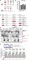
Fig. 4. RhCMV vFcγRs mirror HCMV vFcγR function and efficiently antagonize host FcγR activation in vitro.
A Graphical overview of HCMV vFcγRs and their RhCMV orthologs. Created in BioRender. Kolb, P. (2024) https://BioRender.com/f19l834. B Transfected HeLa cells expressing both the rhesus CD4 target antigen and the indicated vFcγRs as T2A-linked fusion protein from a pIRES-e-GFP plasmid were incubated with graded amounts of a rhesus CD4-specific rhesusized IgG1 antibody and tested for human CD16 activation using a cell-based reporter assay. Equal transfection was monitored via polycistronic GFP expression. Empty vector (e.v.) transfection and expression of a non-Fcγ binding glycoprotein (CD99) served as controls. Symbols show mean area under curve (AUC) values of independent experiments. Bars show the mean of independent experiments. C Overview of the RhCMV 68-1 based recombinants. To create RhCMV 68-1 R, we repaired a premature termination codon in Rh152/151. Single and triple deletion mutants of the vFcγRs were based on the repaired 68-1 strain. All deletions are indicated by highlighting the corresponding ORFs in red. D Telomerized rhesus fibroblasts (tRF) were infected with RhCMV 68-1 or 68-1 R or 68-1 R derived single vFcγR deletion mutants at an MOI of 5 for 72 h and then labeled with [35S]-Met/Cys for 2 h. Whole-cell lysates were prepared, and precipitations of IgG binding molecules were performed using ProteinG bound rhesus-CD4 specific rhesusized IgG1. Lysates from each condition were split and either left untreated or digested with either EndoH or PNGaseF. All samples were analyzed via gradient SDS-PAGE and subsequent autoradiography. One of two independent experiments is presented here. E RFs were infected with RhCMV 68-1, 68-1 R, or single vFcγR deletion mutants at an MOI of 2 for 48 h and then incubated with a 1:10 dilution of a pooled sera from eight CMV seropositive RM. The samples were assessed for human or rhesus FcγR activation using a cell-based reporter assay. The resulting mIL-2 ELISA OD values were normalized to the mean activation on the vFcγR deleted virus for each subgroup. vFcγRs expressed by viruses used for infection are indicated below. FL-RhCMV infection is indicated in pink, and complete vFcγR deletion is indicated in green. * = mock-infected cells. Symbols show independent experiments performed in technical replicates. Bars show the mean of independent experiments. Source data are provided as a Source Data file.
Fig. 5
Fig. 5. RhCMV encoded vFcγRs antagonize the clearance of viral genomes from plasma in vivo.
A Four RhCMV-seronegative male RM were inoculated with an FL-RhCMVΔΔΔ. Three RhCMV-seronegative male RM and 12 RhCMV-seronegative female RM were inoculated with FL-RhCMV or FL-RhCMV plus RhCMV UCD52 respectively. DNA was isolated form plasma samples, and viral genome copy numbers were determined by qPCR using primer/probe sets targeting exon 1 of the IE locus. Copy numbers are reported as mean +/− SD via standard curve interpolation. B Kaplan-Meyer survival analysis showing time to control of plasma DNAemia (first-time point with genome copy numbers below the detection limit (1 copy/well). The median time to control was 31.5 days post-infection in animals infected with vFcγR-deleted RhCMV compared to 59.5 days in animals infected with FL-RhCMV (p = 0.031). C IgG binding to whole FL-RhCMV virions, RhCMV gB, and RhCMV pentamer was measured by ELISA and is shown as mean +/− SD. D ADCP is reported as the percentage of live THP-1 cells containing fluorescently labeled FL-RhCMV after incubation with plasma. ADCC is reported as the percentage of live rhesus CD16-expressing NK92 cells expressing the degranulation marker CD107a following co-incubation with FL-RhCMV infected fibroblasts and plasma samples. All responses are shown as mean +/− SD and demonstrate comparable magnitude and kinetics between animals infected with FL-RhCMV (n = 3) versus vFcγR-deleted RhCMVΔΔΔ (n = 4). E Kinetics of viral genome copy numbers in the plasma of CD4 + T cell-depleted RhCMV-naïve (seronegative) animals infected with vFcγR-deleted RhCMV (red) versus FL-RhCMV (black). Averages from immunocompetent RhCMV-seronegative RM infection in (A) are shown for comparison (gray). F Two RhCMV-seropositive RMs were inoculated with 5 × 106 PFU of vFcγR-deleted RhCMVΔΔΔ carrying an SIV-5’pol transgene as an immunological marker replacing the Rh13.1 ORF. The onset of SIV 5’Pol-specific CD4+ and CD8 + T cell responses and the boosting of RhCMV-specific T cell responses were measured in peripheral blood by intracellular cytokine staining (ICS) for IFN-γ and TNF-α using either a pool of 15mer peptides overlapping by 11 amino acids (AA) corresponding to SIV-5’pol or RhCMV lysates. The frequency of IFN-γ + and/or TNF-α + memory T cells is shown for each individual animal, and each indicated time point post-inoculation. Gating Strategies in Fig. S10. Source data are provided as a Source Data file.

Update of

References

    1. Davison, A. J. et al. The order Herpesvirales. Arch. Virol.154, 171–177 (2009). - PMC - PubMed
    1. Taher, H. et al. In vitro and in vivo characterization of a recombinant rhesus cytomegalovirus containing a complete genome. PLoS Pathog.16, e1008666 (2020). - PMC - PubMed
    1. Forte, E., Zhang, Z., Thorp, E. B. & Hummel, M. Cytomegalovirus latency and reactivation: An intricate interplay with the host immune response. Front. Cell. Infect. Microbiol.10, 130 (2020). - PMC - PubMed
    1. Sylwester, A. W. et al. Broadly targeted human cytomegalovirus-specific CD4+ and CD8+ T cells dominate the memory compartments of exposed subjects. J. Exp. Med.202, 673–685 (2005). - PMC - PubMed
    1. Takenaka, K. et al. Cytomegalovirus reactivation after sllogeneic hematopoietic stem cell transplantation is associated with a reduced risk of relapse in patients with acute myeloid Leukemia who survived to day 100 after transplantation: The Japan society for hematopoietic cell transplantation transplantation-related complication working group. Biol. Blood Marrow Transpl.21, 2008–2016 (2015). - PubMed

MeSH terms

Grants and funding