. 2025 Jan;22(1):10-25.
doi: 10.30773/pi.2024.0186. Epub 2025 Jan 15.

Memory Decline and Aberration of Synaptic Proteins in X-Linked Moesin Knockout Male Mice

Affiliations

Memory Decline and Aberration of Synaptic Proteins in X-Linked Moesin Knockout Male Mice

Hua Cai et al. Psychiatry Investig. 2025 Jan.

Abstract

Objective: This study aims to investigate may moesin deficiency resulted in neurodevelopmental abnormalities caused by negative impact on synaptic signaling ultimately leading to synaptic structure and plasticity.

Methods: Behavioral assessments measured neurodevelopment (surface righting, negative geotaxis, cliff avoidance), anxiety (open field test, elevated plus maze test), and memory (passive avoidance test, Y-maze test) in moesin-knockout mice (KO) compared to wild-type mice (WT). Whole exome sequencing (WES) of brain (KO vs. WT) and analysis of synaptic proteins were performed to determine the disruption of signal pathways downstream of moesin. Risperidone, a therapeutic agent, was utilized to reverse the neurodevelopmental aberrance in moesin KO.

Results: Moesin-KO pups exhibited decrease in the surface righting ability on postnatal day 7 (p<0.05) and increase in time spent in the closed arms (p<0.01), showing increased anxiety-like behavior. WES revealed mutations in pathway aberration in neuron projection, actin filament-based processes, and neuronal migration in KO. Decreased cell viability (p<0.001) and expression of soluble NSF adapter protein 25 (SNAP25) (p<0.001) and postsynaptic density protein 95 (PSD95) (p<0.01) was observed in days in vitro 7 neurons. Downregulation of synaptic proteins, and altered phosphorylation levels of Synapsin I, mammalian uncoordinated 18 (MUNC18), extracellular signal-regulated kinase (ERK), and cAMP response element-binding protein (CREB) was observed in KO cortex and hippocampus. Risperidone reversed the memory impairment in the passive avoidance test and the spontaneous alternation percentage in the Y maze test. Risperidone also restored the reduced expression of PSD95 (p<0.01) and the phosphorylation of Synapsin at Ser605 (p<0.05) and Ser549 (p<0.001) in the cortex of moesin-KO.

Conclusion: Moesin deficiency leads to neurodevelopmental delay and memory decline, which may be caused through altered regulation in synaptic proteins and function.

Keywords: Memory; Moesin; Neurodevelopment; PSD95; Risperidone; Synapsin.


Conflict of interest statement

Conflicts of Interest

The authors have no potential conflicts of interest to disclose.

Figures

Figure 1.
Figure 1.
Schematic representation of the experimental design and procedures. A: Exon 2 of the moesin gene on chromosome X was deleted to generate moesin-KO. Neuronal cells were obtained from the cortex of WT or moesin-KO on embryonic day 15. B: Neurodevelopmental testing was performed at postnatal day 7 for hemizygous male KO and homozygous female KO. The open field test was used to assess anxiety-related behavior in male hemizygous KO at the age of 2 months, and the elevated plus maze test was used to assess anxiety-related at the age of 5 months. C: Mice were intraperitoneally injected with saline or risperidone for 2 weeks (5 days per week). After a 1-week washout period, behavioral tests for memory function (passive avoidance test and Y-maze test) were performed at 8 weeks (2 months) of age. D: Exome sequencing and sperm analysis were conducted at 3 months of age. KO, knockout mice; WT, wild-type mice.
Figure 2.
Figure 2.
Moesin-KO show abnormal behaviors. We investigated whether moesin-KO exhibit symptoms of developmental disorders. An open field test to measure anxiety status (A and B), and an elevated plus-maze test was performed on male mice 5 months of age (C). All data were analyzed using the t-test and are presented as the mean±standard error of the mean of six determinations. *p<0.05; **p<0.01. KO, knockout mice; WT, wild-type mice.
Figure 3.
Figure 3.
Exome sequencing results in moesin-KO compared to reference genes. A: Number of altered bases per chromosome. This figure shows a bar graph depicting the distribution of altered bases across different chromosomes in the moesin knockout condition. B: Counts of different types of altered bases. This result provides a breakdown of the alterations by type, including substitutions, insertions, and deletions. C: SNV class of altered bases. This graph categorizes the SNVs identified in the exome sequencing into their respective classes based on their genomic impact. KO, knockout mice; SNV, single nucleotide variant.
Figure 4.
Figure 4.
GO-term analysis of genes altered in exome sequencing results. A: Top 20 most significantly altered GO-terms. This panel displays the top 20 GO-terms with the highest degree of change, with neuron projection development, actin filament-based process, and regulation of plasma membrane bounded cell projection organization ranking among the highest. B: Network analysis of GO-terms. This panel illustrates the network relationships between the identified GO-terms, showing how these biological processes are interconnected. GO, gene ontology.
Figure 5.
Figure 5.
Moesin-KO neurons decreased cell viability and the expression of neuronal cell markers. Relative expression levels of GFAP, MAP2, Sox2, DCX, SNAP25, PSD95, and moesin are displayed. At 7 days (A) and 14 days (B) after seeding cortical neurons, molecular changes in neuronal cell markers were examined by immunoblotting. All data were analyzed by t-test and are presented as the mean±SEM of three replicates. The bar is 100 μm. *p<0.05; **p<0.01; ***p<0.001. KO, knockout mice; WT, wild-type mice.
Figure 6.
Figure 6.
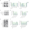
Moesin-KO decreased the expression of synaptic proteins and phosphorylation of signaling proteins in the adult brain. A and B: The expression or activation level of synaptic vesicle proteins such as Syntaxin 1A, Synaptophysin, CDK5, and radixin were confirmed by immunoblotting in the cortex and hippocampus to examine the downstream target of moesin. Relative expression levels of each protein are presented in bar graphs. C and D: The expression and phosphorylation level of synapsin I were examined in the cortex and hippocampus of moesin-KO and presented in bar graphs. E and F: The activation levels of signaling molecules such as ERK and CREB were checked with phosphorylationspecific antibodies. The expression and phosphorylation levels of MUNC18 and PSD95 were examined in the cortex, and hippocampus of moesin-KO, and their relative expression is presented in graphs. All data were analyzed by t-test and are presented as the mean±SEM of three replicates. **p<0.01; ***p<0.001. KO, knockout mice; WT, wild-type mice; CDK5, Cyclin dependent kinase 5; ERK, extracellular signal-regulated kinase; CREB, cAMP response element-binding protein; SEM, standard error of the mean.
Figure 7.
Figure 7.
Risperidone reversed behavioral and molecular deficits in moesin-KO. Risperidone was administered for 2 weeks and validated by behavioral experiments at 2 months of age. A: Risperidone reversed memory impairment in moesin-KO in the passive avoidance test and (B) total entry number (left panel) and spontaneous alternation percentage (right panel) in the Y maze. C: Risperidone induced the expression of PSD95, the phosphorylation of synapsin at Ser605 and Ser549, and ERK phosphorylation in the cortex of moesin-KO, and the data are displayed in the graph. All data were analyzed by one-way ANOVA and are presented as the mean±SEM. *p<0.05; **p<0.01; ***p<0.001. KO, knockout mice; WT, wild-type mice; ERK, extracellular signal-regulated kinase; SEM, standard error of the mean; Ris, risperidone.

Similar articles

References

    1. Sato N, Funayama N, Nagafuchi A, Yonemura S, Tsukita S, Tsukita S. A gene family consisting of ezrin, radixin and moesin. Its specific localization at actin filament/plasma membrane association sites. J Cell Sci. 1992;103(Pt 1):131–143. - PubMed
    1. Fehon RG, McClatchey AI, Bretscher A. Organizing the cell cortex: the role of ERM proteins. Nat Rev Mol Cell Biol. 2010;11:276–287. - PMC - PubMed
    1. Okamoto K, Bosch M, Hayashi Y. The roles of CaMKII and F-actin in the structural plasticity of dendritic spines: a potential molecular identity of a synaptic tag? Physiology (Bethesda) 2009;24:357–366. - PubMed
    1. Bosch M, Castro J, Saneyoshi T, Matsuno H, Sur M, Hayashi Y. Structural and molecular remodeling of dendritic spine substructures during long-term potentiation. Neuron. 2014;82:444–459. - PMC - PubMed
    1. Solinet S, Mahmud K, Stewman SF, Ben El Kadhi K, Decelle B, Talje L, et al. The actin-binding ERM protein moesin binds to and stabilizes microtubules at the cell cortex. J Cell Biol. 2013;202:251–260. - PMC - PubMed

LinkOut - more resources