. 2025 Feb 1;18(2):DMM052047.
doi: 10.1242/dmm.052047. Epub 2025 Mar 3.

The behavioural consequences of dystrophinopathy

Affiliations

The behavioural consequences of dystrophinopathy

Minou A T Verhaeg et al. Dis Model Mech. .

Abstract

Duchenne muscular dystrophy is a severe neuromuscular disorder, caused by mutations in the DMD gene. Normally, the DMD gene gives rise to many dystrophin isoforms, of which multiple are expressed in the brain. The location of the mutation determines the number of dystrophin isoforms affected, and the absence thereof leads to behavioral and cognitive impairments. Even though behavioral studies have thoroughly investigated the effects of the loss of Dp427, and to a lesser extent of Dp140, in mice, direct comparisons between models lacking multiple dystrophin isoforms are sparse. Furthermore, a behavioral characterization of the DMD-null mouse, which lacks all dystrophin isoforms, has never been undertaken. Using a wide variety of behavioral tests, we directly compared impairments between mdx5cv, mdx52 and DMD-null mice. We confirmed the role of Dp427 in emotional reactivity. We did not find any added effects of loss of Dp140 on fear, but showed the involvement of Dp140 in spontaneous behavior, specifically in habituation and activity changes due to light/dark switches. Lastly, our results indicate that Dp71/Dp40 play an important role in many behavioral domains, including anxiety and spontaneous behavior.

Keywords: Mdx52 and DMD-null; Mdx5cv; Anxiety; Cognition; Dystrophin; Learning; Social interaction; Spontaneous behavior.


Conflict of interest statement

Competing interests The authors declare no competing or financial interests.

Figures

Fig. 1.
Fig. 1.
Study overview. Mice were included at 8 weeks of age and underwent a variety of behavioral tests for 8 weeks. DL, dark-light box; 3C, three-chamber social interaction; NOR, novel object recognition; OP, object placement; MWM, Morris water maze.
Fig. 2.
Fig. 2.
Anxiety in the dark-light box and open field. Mdx5cv (n=16), mdx52 (n=15), DMD-null (n=16) and wild-type (WT) (n=33). (A) In the dark-light box, mdx5cv, mdx52 and DMD-null mice all visited the light compartment less often than WTs (P=0.009, P<0.001 and P<0.001, respectively). DMD-null mice visited the light compartment less often than mdx5cv mice (P=0.021). (B) Mdx5cv, mdx52 and DMD-null mice all spent less time in the light compartment than WTs (all P<0.001). DMD-null mice spent less time in the light compartment than mdx5cv and mdx52 mice (P=0.043 and P=0.008, respectively). (C) Visits to the light department were on average shorter for mdx5cv mice than for WTs (P=0.022). (D) In the open field test, all DMD models stayed closer to the walls than WTs (all P<0.001). (E) Mdx5cv, mdx52 and DMD-null mice spent less time in the inner zone of the box than WTs (P=0.035, P<0.001 and P<0.001, respectively). (F) Mdx52 and DMD-null mice walked less distance in the inner zone of the box than WT mice (both P<0.001). (G) DMD-null mice walked slower than WT, mdx5cv and mdx52 mice (all P<0.001). (H) DMD-null mice spent less time moving than WT, mdx5cv and mdx52 mice (all P<0.001). Mdx52 mice moved less than WTs (P=0.017). *P<0.05, **P<0.01, ***P<0.001 (we refer the reader to Table S2 for an overview of the statistical tests performed).
Fig. 3.
Fig. 3.
Unconditioned fear response after a short restraint. Mdx5cv (n=16), mdx52 (n=15), DMD-null (n=16) and WT (n=33). (A) In all DMD models, walking velocity was decreased compared to that in WTs (all P<0.001). DMD-null mice showed a further decrease compared to mdx5cv mice (P=0.012). (B) Freezing time was increased in all DMD models (all P<0.001). DMD-null mice spent more time frozen than mdx5cv mice (P=0.019). (C) Freezing time per time bin of 60 s. All DMD strains show a different pattern over time compared to WTs (P<0.001), but no differences were found between the DMD strains. *P<0.05, ***P<0.001 (Mann–Whitney test).
Fig. 4.
Fig. 4.
Spatial learning and memory in the Barnes maze and Morris water maze. (A) Schematic of the Barnes maze protocol. Mdx5cv (n=16), mdx52 (n=15), DMD-null (n=16) and WT (n=33). Created in BioRender by Verhaeg, M. (2025). https://BioRender.com/p79/689. This figure was sublicensed under CC-BY 4.0 terms. (B) Distance traveled to reach the platform during the acquisition learning. No differences were found between groups. (C) All groups traveled approximately equal distances to reach the platform zone during the probe trial. (D) No significant differences were found between groups in the relative distance traveled in the target quadrant. All groups traveled more distance in this quadrant compared to chance level (0.25, indicated by the dotted line) (all P<0.001). (E) DMD-null mice showed increased distance traveled to the new platform location during reversal learning compared to WTs (P=0.015). (F) No differences were found between groups in the distance traveled to reach the new platform zone during the reversal probe trial. (G) DMD-null mice showed a higher relative distance traveled in the new target quadrant compared to WTs (P=0.033). All groups performed above chance level (all P<0.001, indicated by the dotted line). (H,I) No differences were found between groups in distance traveled until reaching the old platform location during the reversal learning (H) or the reversal probe (I). (J) No differences were found between groups in the relative distance traveled in the old target quadrant. None of the groups differed from chance level. (K) Schematic of the Morris water maze protocol. Mdx5cv (n=16), mdx52 (n=13), DMD-null (n=11) and WT (n=33). Created in BioRender by Verhaeg, M. (2025). https://BioRender.com/p79/689. This figure was sublicensed under CC-BY 4.0 terms. (L) No differences were found between groups in the distance they needed to swim to find the platform. (M) No differences were found in the distance swam until reaching the platform during the probe trial. (N) No differences were found between groups in the relative distance swam in the target quadrant. Only the performances of WT and mdx5cv mice differed from chance level, indicated by the dotted line at 0.25 (P<0.001 and P=0.002, respectively). (O) Mdx52 and DMD-null mice spent less time in the target quadrant than mdx5cv (P=0.005 and P=0.009, respectively) and WT (P=0.012 and P=0.022, respectively) mice. (P-R) No difference was found in the time spent in any of the other quadrants. *P<0.05, **P<0.01, ***P<0.001 (we refer the reader to Table S2 for an overview of the statistical tests performed).
Fig. 5.
Fig. 5.
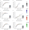
Serial reversal learning with food reward in PhenoTyper cages. Mdx5cv (n=9-10), mdx52 (n=14), DMD-null (n=10-15) and WT (n=32-33). (A) Discrimination learning (left target). Both mdx5cv and mdx52 mice showed steeper learning curves than those of WTs (P<0.001 and P=0.011, respectively) and DMD-null mice (P<0.001 and P=0.018, respectively). (B) Reversal day 1 (right target). Mdx5cv mice showed decreased learning compared to WTs (P=0.017). (C) Reversal day 2 (left target). Mdx52 mice showed increased performance compared to WTs and mdx5cv mice (P<0.001 and P=0.013, respectively). (D) Reversal day 3 (middle target). Mdx52 mice showed increased learning compared to WTs (P=0.018). (E) Reversal day 4 (right target). No differences were found between groups. (F) Reversal day 5 (left target). No differences were found between groups. All graphs were cut off at n=3 for visual purposes. Cartoons were created in BioRender by Verhaeg, M. (2025). https://BioRender.com/e60i623. This figure was sublicensed under CC-BY 4.0 terms.
Fig. 6.
Fig. 6.
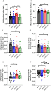
Spontaneous behavior in the PhenoTyper cages: activity-based behavior. Mdx5cv (n=14), mdx52 (n=14), DMD-null (n=16) and WT (n=33). (A) Mdx52 mice showed a higher habituation index during the dark phase compared to WT, mdx5cv and DMD-null mice (P<0.001, P=0.025 and P<0.001, respectively). (B) Mean activity duration during the dark phase was lower in DMD-null mice than in WT and mdx5cv mice (P=0.002 and P<0.001, respectively). (C) In DMD-null mice, activity in anticipation of the dark phase was less changed than in WT mice (P=0.004). (D) DMD-null mice showed less change in activity in response to the start of the dark phase than WTs (P=0.023). (E) Mdx52 mice showed a higher increase in activity in anticipation of the start of the light phase than WTs (P=0.032). (F) Mdx52 mice showed a lower decrease in activity change in response to the start of the light phase than WT and mdx5cv mice (P=0.037 and P<0.001, respectively). DMD-null mice also showed less of a decrease in activity than mdx5cv mice (P<0.001). *P<0.05, **P<0.01, ***P<0.001 (one-way ANOVA with post-hoc Tukey).
Fig. 7.
Fig. 7.
Spontaneous behavior in the PhenoTyper cages: sheltering behavior, movement and arrest. Mdx5cv (n=14), mdx52 (n=14), DMD-null (n=16) and WT (n=33). (A) The long shelter visit threshold was increased in mdx5cv mice compared to that in WT and DMD-null mice (P<0.001 and P=0.007, respectively). (B) Both mdx52 and DMD-null mice showed a lower cumulative duration of shelter visits above the long shelter visit threshold compared to that in WTs (P<0.001 and P=0.009, respectively). (C) The long movement threshold was lower in DMD-null mice than in WT, mdx5v and mdx52 mice (P<0.001, P<0.001 and P=0.027, respectively). (D) DMD-null mice had a lower duration of arrests above the threshold than WT and mdx5cv mice (P=0.014 and P=0.009, respectively). *P<0.05, **P<0.01, ***P<0.001 (we refer the reader to Table S2 for an overview of the statistical tests performed).

References

    1. Alexander, M., Gasperini, M., Tsai, P., Gibbs, D., Spinazzola, J., Marshall, J., Feyder, M., Pletcher, M., Chekler, E. P. and Morris, C. (2016). Reversal of neurobehavioral social deficits in dystrophic mice using inhibitors of phosphodiesterases PDE5A and PDE9A. Transl. Psychiatry 6, e901-e901. 10.1038/tp.2016.174 - DOI - PMC - PubMed
    1. Araki, E., Nakamura, K., Nakao, K., Kameya, S., Kobayashi, O., Nonaka, I., Kobayashi, T. and Katsuki, M. (1997). Targeted disruption of exon 52 in the mouse dystrophin gene induced muscle degeneration similar to that observed in Duchenne muscular dystrophy. Biochem. Biophy. Res. Commun. 238, 492-497. 10.1006/bbrc.1997.7328 - DOI - PubMed
    1. Bagdatlioglu, E., Porcari, P., Greally, E., Blamire, A. M. and Straub, V. W. (2020). Cognitive impairment appears progressive in the mdx mouse. Neuromuscul. Disord. 30, 368-388. 10.1016/j.nmd.2020.02.018 - DOI - PMC - PubMed
    1. Banihani, R., Smile, S., Yoon, G., Dupuis, A., Mosleh, M., Snider, A. and McAdam, L. (2015). Cognitive and neurobehavioral profile in boys with Duchenne muscular dystrophy. J. Child Neurol. 30, 1472-1482. 10.1177/0883073815570154 - DOI - PubMed
    1. Barboni, M. T. S., Liber, A. M. P., Joachimsthaler, A., Saoudi, A., Goyenvalle, A., Rendon, A., Roger, J. E., Ventura, D. F., Kremers, J. and Vaillend, C. (2021). Altered visual processing in the mdx52 mouse model of Duchenne muscular dystrophy. Neurobiol. Dis. 152, 105288. 10.1016/j.nbd.2021.105288 - DOI - PubMed

LinkOut - more resources