

Case Reports
doi: 10.1016/j.case.2024.09.006.
eCollection 2024 Dec.
Transesophageal Echocardiography Guidance during Transcatheter Mitral Valve Edge-to-Edge Repair for Severe Mitral Regurgitation Post-Mitral Annuloplasty: A Case Series
Affiliations
- PMID: 39885887
- PMCID: PMC11775905
- DOI: 10.1016/j.case.2024.09.006
Item in Clipboard
Case Reports
Transesophageal Echocardiography Guidance during Transcatheter Mitral Valve Edge-to-Edge Repair for Severe Mitral Regurgitation Post-Mitral Annuloplasty: A Case Series
CASE (Phila).
.
No abstract available
Keywords: Mitral annuloplasty; Mitral regurgitation; Transcatheter edge-to-edge repair; Transesophageal echocardiography.
Figures


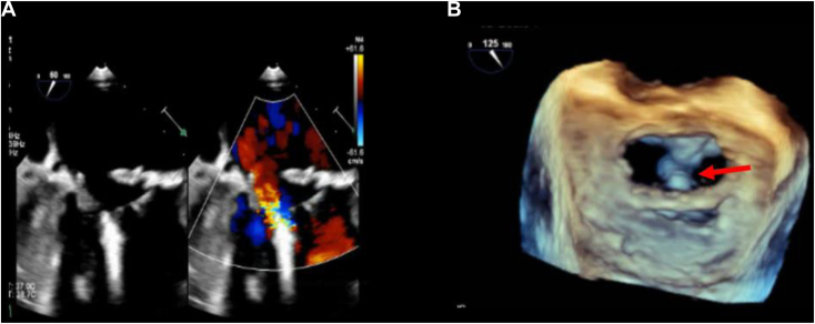
(A) Two-dimensional TEE, midesophageal commissural (63°) systolic view without (left) and with (right) color-flow Doppler, demonstrates functional disease of the P2 scallop, which was tethered with severe MR originating from poor coaptation between the A2 and P2 scallops and flowing through the posterior dehiscence of the annuloplasty band. The P2 leaflet length is 9 mm (dashed yellow line). (B) Three-dimensional TEE, volume-rendered reconstruction diastolic display, en face perspective from the left atrium, demonstrates a flexible band (Cosgrove Edwards no. 28) with posterior dehiscence (arrow).

(A) Three-dimensional TEE, midesophageal long-axis MPR of the MV in a diastolic phase, demonstrates the MVOA of 4.77 cm2 (yellow dashed outlined area). (B) Three-dimensional TEE with color-flow Doppler, MPR of the MV in a systolic phase, demonstrates the MV VCA3D; the blue line transects the MR in 2 orthogonal, long-axis planes, which creates the short-axis en face display of the VCA3D of 0.29 cm2 (red outlined area).
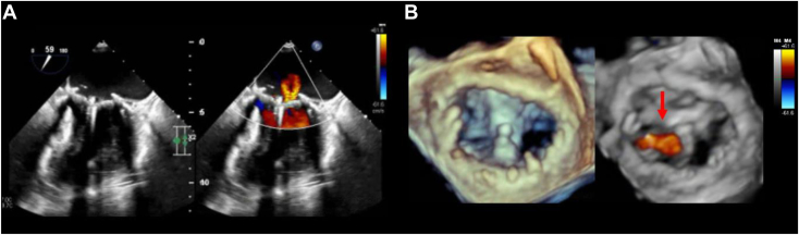
(A) Two-dimensional TEE, midesophageal commissural (60°) systolic view without (left) and with (right) color-flow Doppler, demonstrates mild residual MR after device deployment. (B) Three-dimensional TEE, volume-rendered reconstruction diastolic display, en face perspective from the left atrium, demonstrates the device between the A2 and P2 scallops (arrow) creating a double orifice MV opening.

(A) Three-dimensional TEE, midesophageal long-axis MPRs of the MV in a diastolic phase, demonstrates the total MVOA of 1.89 cm2 (2 yellow dashed outlined areas). (B) Three-dimensional TEE with color-flow Doppler, MPR of the MV in a systolic phase, demonstrates the MV VCA3D (blue line transects the MR in 2 orthogonal, long-axis planes, which creates the short-axis en face display of the VCA3D totaling 0.10 cm2 (2 tiny yellow outlined areas).

(A) Two-dimensional TEE, midesophageal long-axis (135o) systolic view without (left) and with (right) color-flow Doppler, demonstrates a flail P2 scallop (arrow) with severe, anteriorly directed MR. (B) Three-dimensional TEE, volume-rendered reconstruction display, en face systolic view from the perspective of the left atrium without (left) and with (right) color-flow Doppler, demonstrates a rigid annuloplasty ring (no. 34 Carpentier Edwards) with flail P2 scallop (arrow) and severe MR. (C) Two-dimensional TEE, midesophageal long-axis (123o) diastolic view, demonstrates P2 leaflet length of 15 mm (dashed yellow line).

(A) Three-dimensional TEE, midesophageal long-axis MPRs of the MV, in a diastolic phase, demonstrates the total MVOA of 3.82 cm2 (yellow dashed outlined area). (B) Three-dimensional TEE with color-flow Doppler, MPR of the MV in a systolic phase, demonstrates the MV VCA3D; the blue line transects the MR in 2 orthogonal, long-axis planes, which creates the short axis en face display of the VCA3D of 0.8 cm2 (red outlined area).
(A) Two-dimensional TEE, midesophageal commissural (59°) systolic view, without (left) and with (right) color-flow Doppler, demonstrates mild residual MR. (B) Three-dimensional TEE, volume-rendered reconstruction systolic display without (left) and with (right) color-flow Doppler, en face perspective from the left atrium, demonstrates the device (MitraClip XTW) between the A2 and P2 scallops with mild residual MR lateral to the device (arrow).

(A) Three-dimensional TEE, midesophageal long-axis MPRs of the MV in a diastolic phase, demonstrates the total MVOA of 2.68 cm2 (2 yellow dashed outlined areas). (B) Three-dimensional TEE with color-flow Doppler, MPR of the MV, in a systolic phase, demonstrates the MV VCA3D (blue line transects the MR in 2 orthogonal, long-axis planes, which creates the short-axis en face display of the VCA3D 0.29 cm2 (red outlined area).

(A) Three-dimensional TEE, volume-rendered reconstruction display, en face perspective from the left atrium, systolic view without (left) and with (right) color-flow Doppler, demonstrates a flexible annuloplasty ring (no. 29 Simulus) with a tethered P2 scallop (arrow) with severe, posteriorly directed MR. (B) Two-dimensional TEE, midesophageal multiplane (90o and 150o) systolic views with color-flow Doppler, identified the maximal PISA radius (arrow). (C) Two-dimensional TEE, midesophageal multiplane (90o and 150o) diastolic views, demonstrates P2 leaflet length (7 mm) in the long-axis view (150o).

(A) Three-dimensional TEE, midesophageal long-axis MPR of the MV in a diastolic phase, demonstrates the MVOA of 4.29 cm2 (yellow dashed outlined area). (B) Three-dimensional TEE with color-flow Doppler, MPR of the MV in a systolic phase, demonstrates the MV VCA3D; the blue line transects the MR in 2 orthogonal, long-axis planes, which creates the short axis en face display of the VCA3D 0.61 cm2 (red outlined area).
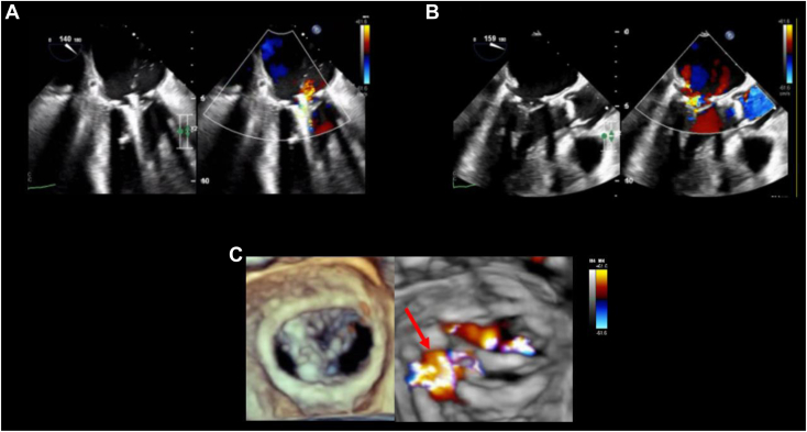
(A) Two-dimensional TEE, midesophageal long-axis (140o) systolic view, without (left) and with (right) color-flow Doppler, demonstrates the device (MitraClip NTW) grasping the A2 and P2 scallops while avoiding the neochord to the A2 scallop. (B) Two-dimensional TEE, midesophageal long-axis (159o) systolic view, without (left) and with (right) color-flow Doppler, demonstrates the released device between the A2 and P2 scallops with mild residual MR. (C) Three-dimensional TEE, volume-rendered reconstruction display, en face perspective from the left atrium, diastolic view without (left) and systolic view with (right) color-flow Doppler, demonstrates the device on the lateral side of A2/P2 with mild MR primarily lateral to the device (arrow).

(A) Three-dimensional TEE, midesophageal long-axis MPRs of the MV, in a diastolic phase, demonstrates the total MVOA of 2.78 cm2 (2 yellow dashed outlined areas). (B) Three-dimensional TEE with color-flow Doppler, MPR of the MV in a systolic phase, demonstrates the MV VCA3D (blue line transects the MR in 2 orthogonal, long-axis planes, which creates the short-axis en face display of the VCA3D 0.39 cm2 (red outlined area).
References
-
- Otto C.M., Nishimura R.A., Bonow R.O., et al. 2020 ACC/AHA guideline for the management of patients with valvular heart disease: a report of the American College of Cardiology/American Heart Association Joint Committee on Clinical Practice Guidelines. Circulation. 2021;143:e72–e227. - PubMed
-
- Mehaffey H.J., Hawkins R.B., Schubert S., et al. Contemporary outcomes in reoperative mitral valve surgery. Heart. 2018;104:652–656. - PubMed
-
- Petrus A.H., Dekkers O.M., Tops L.F., et al. Impact of recurrent mitral regurgitation after mitral valve repair for functional mitral regurgitation: long-term analysis of competing outcomes. Eur Heart J. 2019;40:2206–2219. - PubMed
-
- Kim J.H., Lee S.H., Joo H.C., et al. Effect of recurrent mitral regurgitation after mitral repair in patients with degenerative mitral regurgitation. Circ J. 2017;82:93–101. - PubMed
Publication types
LinkOut - more resources
Full Text Sources
