

Aberrant right renal perfusion from right internal mammary artery
- PMID: 39886218
- PMCID: PMC11780940
- DOI: 10.1016/j.jvscit.2024.101713
Aberrant right renal perfusion from right internal mammary artery
Abstract
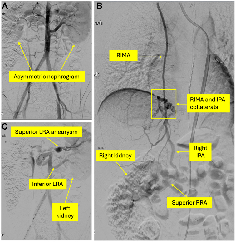
Renal artery (RA) anomaly is common and may have significant clinical implications. We present a case of a 29-year-old man in whom the upper two-thirds of the right kidney were supplied by the right internal mammary artery and collateral network. Additionally, the superior left RA had proximal stenosis with a distal aneurysm. Renin sampling confirmed renovascular hypertension. He successfully underwent aorta-to-superior right RA bypass and primary repair of superior left RA aneurysm. This case adds to existing knowledge of RA anomalies, and underscores the importance of comprehensive evaluations for alternate renal blood supply for effective surgical management of renovascular hypertension.
Keywords: Aberrant renal artery; Alport syndrome; Fibromuscular dysplasia; Renal artery aneurysm; Renovascular hypertension.
© 2024 The Authors.
Conflict of interest statement
None.
Figures


References
-
- Cocheteux B., Mounier-Vehier C., Gaxotte Y., McFadden E.P., Francke J.P., Beregi J.P. Rare variations in renal anatomy and blood supply: CT appearances and embryological background. A pictorial essay. Eur Radiol. 2001;11:779–786. - PubMed
-
- Leckie A., Tao M.J., Narayanasamy S., Khalili K., Schieda N., Krishna S. The renal vasculature: what the radiologist needs to know. Radiographics. 2021;41:1531–1548. - PubMed
-
- Satyapal K.S., Haffejee A.A., Singh B., Ramsaroop L., Robbs J.V., Kalideen J.M. Additional renal arteries: incidence and morphometry. Surg Radiol Anat. 2001;23:33–38. - PubMed
Publication types
LinkOut - more resources
Full Text Sources
