. 2025 Jan 22:2025:6587305.
doi: 10.1155/mi/6587305. eCollection 2025.

Trapa natans L. Extract Attenuates Inflammation and Oxidative Damage in Cisplatin-Induced Cardiotoxicity in Rats by Promoting M2 Macrophage Polarization

Affiliations

Trapa natans L. Extract Attenuates Inflammation and Oxidative Damage in Cisplatin-Induced Cardiotoxicity in Rats by Promoting M2 Macrophage Polarization

Vesna Matovic et al. Mediators Inflamm. .

Abstract

Background: Trapa natans L. fruits and leaf extracts have a broad range of immunomodulatory, anti-inflammatory, and antioxidant effects; however, their effects on cardiac protection have not been investigated. Objective: The study aims to test the biological activity of Trapa natans L. extract (TNE) in cisplatin (CDDP)-induced cardiotoxicity. Methods: Wistar albino rats received a single dose of CDDP intraperitoneally and TNE ones per day for 2 weeks orally. Cardiac inflammation, necrosis, and fibrosis were determined by histological and immunohistochemical analyses. Cytokines in rat sera and cardiac tissue were detected by enzyme-linked immunosorbent assay (ELISA) and quantitative real-time (qRT)-PCR. Rat macrophages cultured in the presence of TNE for 48 h were harvested for flow cytometry, while supernatants were collected for cytokine and reactive oxygen species (ROS) measurement. Results: Application of TNE significantly attenuated CDDP induced cardiotoxicity as demonstrated by biochemical and histopathological analysis. Administration of TNE once daily for 14 days decreased level of proinflammatory (TNF-α, IFN-γ, and IL-6) and prooxidative parameters (NO2, O2, and H2O2), while increased level of immunosuppressive IL-10 and antioxidative glutathione (GSH), catalase (CAT) and uperoxide dismutase (SOD) in the systemic circulation. TNE treatment resulted in attenuated heart inflammation and fibrosis accompanied with the reduced infiltration of macrophages and reduced expression of proinflammatory and profibrotic genes in heart tissue of CDDP-treated animals. In vitro lipopolysaccharide (LPS)-stimulated macrophages cultured in the presence of TNE adopted immunosuppressive phenotype characterized by decreased production of proinflammatory cytokines and prooxidative mediators. Conclusion: Our study provides the evidence that TNE ameliorates cisplatin-induced cardiotoxicity in rats by reducing inflammation and oxidative stress via promoting M2 macrophage polarization.

Keywords: Trapa natans L. extract; cardiotoxicity; cisplatin.


Conflict of interest statement

The authors declare no conflicts of interest.

Figures

Figure 1
Figure 1
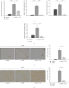
T. natans L. extract ameliorates cisplatin-induced cardiotoxicity. (A) Experimental design. Rats were injected with saline, single dose of CDDP (7 mg/kg/w, i.p.), or CDDP (7 mg/kg/w, i.p.), and TNE (100 mg/kg/w, per os ones per day for 2 weeks). The sera for CK/LDH measurement and hearts for histological analysis were collected 14 days after treatment. (B) Fold change of CK and LDH levels was significantly lower in CDDP + TNE-treated rats compared to rats that received CDDP only. (C) H&E staining of heart tissue sections of CDDP + TNE-treated animals revealed reduced myocytes degeneration, inflammatory cells infiltration, hemorrhage, and vascular congestion compared to CDDP-only treated rats. (D) Sirius red staining of heart tissues obtained from CDDP-treated rats revealed extensive collagen deposition. Stained area of fibrous dense tissue was reduced in CDDP + TNE-treated animals and absent in control animals (magnification x40). Data presented as mean ± SD; n = 5 rats per groups. p < 0.05. CDDP, cis-diamminedichloroplatinum [II]; TNE, Trapa natans L. extract.
Figure 2
Figure 2
T. natans L. extract attenuates inflammation and oxidative damage in CDDP-treated rats. (A) The concentration of proinflammatory TNF-α, IFN-γ, and IL-6 were significantly decreased and (B) level of anti-inflammatory IL-10 was significantly increased in serum of CDDP + TNE-treated animals compared to rats that received CDDP only. (C) Levels of prooxidative parameters (NO2ˉ, O2ˉ, and H2O2) were significantly lower, while (D) levels of antioxidative parameters (GSH, CAT, and SOD) were significantly higher in systemic circulation of CDDP + TNE-treated animals compared to rats that received CDDP only. Data presented as mean ± SD; n = 5 rats per groups. p < 0.05, ∗∗p < 0.01, ∗∗∗p < 0.001. CDDP, cis-diamminedichloroplatinum [II]; TNE, Trapa natans L. extract.
Figure 3
Figure 3
Application of T. natans L. extract reduces the presence of macrophages and inflammatory cytokines in the hearts of CDDP-treated rats. TNE decreased relative expression of (A) proinflammatory cytokine genes (TNF-α, IL-1β, and IL-6) as well as (B) profibrogenic marker TGF-β in the heart tissue of CDDP-treated rats. Representative immunohistochemical images and number of (C) CD68+ macrophages and (D) iNOS+ cells per field in the paraffin-embedded heart tissue sections (×20). Data presented as mean ± SD; n = 5 rats per groups. p < 0.05, ∗∗p < 0.01, ∗∗∗p < 0.001. CDDP, cis-diamminedichloroplatinum [II]; TNE, Trapa natans L. extract.
Figure 4
Figure 4
TNE suppress reactive oxygen species and proinflammatory cytokine responses of macrophages. (A) Schematic diagram describing design of in vitro experiments. In vitro LPS-stimulated peritoneal macrophages isolated from healthy rats were cultured for 48 h with or without TNE. Phenotype of peritoneal macrophages, as well as level of cytokines and prooxidative parameters in supernatants, was analyzed. (B) The significantly lower amounts of IL-6 and higher amount of IL-10 were noticed in supernatants of in vitro LPS-stimulated peritoneal macrophages cultured with TNE. Difference in the concentration of TNF-α and IFN-γ in the supernatant did not reach statistical difference. (C) Flow cytometry revealed significantly higher percentage of IL-10-producing-, and lower percentage of IL-4-producing LPS-activated CD11b+ macrophages cultured in the presence of TNE, when compared to LPS-stimulated macrophages that were cultured without TNE. (D) TNE-induced significant decrease in concentration of prooxidative parameters O2 and NO in the supernatant of LPS-stimulated CD11b+ macrophages. p < 0.05, ∗∗p < 0.01, ∗∗∗p < 0.001. TNE, Trapa natans L. extract.
Figure 5
Figure 5
Proposed mechanism of TNE-based attenuation of cisplatin-induced cardiotoxicity. After cisplatin-induced cardiac injury, application of TNE induces polarization of proinflammatory M1 macrophages to the anti-inflammatory and proregenerative M2 phenotype characterized by increased production of protective IL-10 and antioxidative GSH, CAT, and SOD, resulting with attenuated inflammation, oxidative damage, and fibrosis in the heart. CAT, catalase; GSH, glutathione; SOD, superoxide dismutase.

References

    1. Dasari S., Tchounwou P. B. Cisplatin in Cancer Therapy: Molecular Mechanisms of Action. European Journal of Pharmacology . 2014;740:364–378. doi: 10.1016/j.ejphar.2014.07.025. - DOI - PMC - PubMed
    1. Xia J., Hu J. N., Zhang R. B., et al. Icariin Exhibits Protective Effects on Cisplatin-Induced Cardiotoxicity via ROS-Mediated Oxidative Stress Injury in Vivo and in Vitro. Phytomedicine . 2022;104 doi: 10.1016/j.phymed.2022.154331.154331 - DOI - PubMed
    1. Qi Y. J., Ying Y., Zou J., et al. Kaempferol Attenuated Cisplatin-Induced Cardiac Injury via Inhibiting STING/NF-Kappa B-Mediated Inflammation. American Journal of Translational Research . 2020;12:8007–8018. - PMC - PubMed
    1. Csapo M., Lazar L. Chemotherapy-Induced Cardiotoxicity: Pathophysiology and Prevention. Medicine and Pharmacy Reports . 2014;87(3):135–142. doi: 10.15386/cjmed-339. - DOI - PMC - PubMed
    1. Pop R. M., Popolo A., Trifa A. P., Stanciu L. A. Phytochemicals in Cardiovascular and Respiratory Diseases: Evidence in Oxidative Stress and Inflammation. Oxidative Medicine and Cellular Longevity . 2018;2018(1):11. doi: 10.1155/2018/1603872.1603872 - DOI - PMC - PubMed

Supplementary concepts

LinkOut - more resources