. 2025 Mar;31(3):74.
doi: 10.3892/mmr.2025.13439. Epub 2025 Jan 31.

Triptolide reverses cis‑diamminedichloroplatinum resistance in esophageal squamous cell carcinoma by suppressing glycolysis and causing mitochondrial malfunction

Affiliations

Triptolide reverses cis‑diamminedichloroplatinum resistance in esophageal squamous cell carcinoma by suppressing glycolysis and causing mitochondrial malfunction

Kuiyuan Liu et al. Mol Med Rep. 2025 Mar.

Abstract

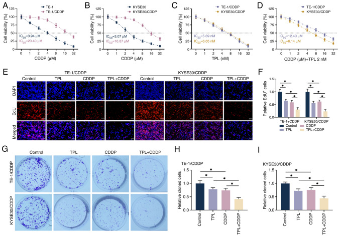
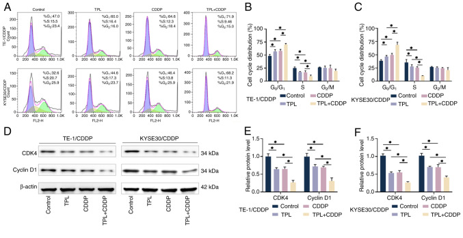
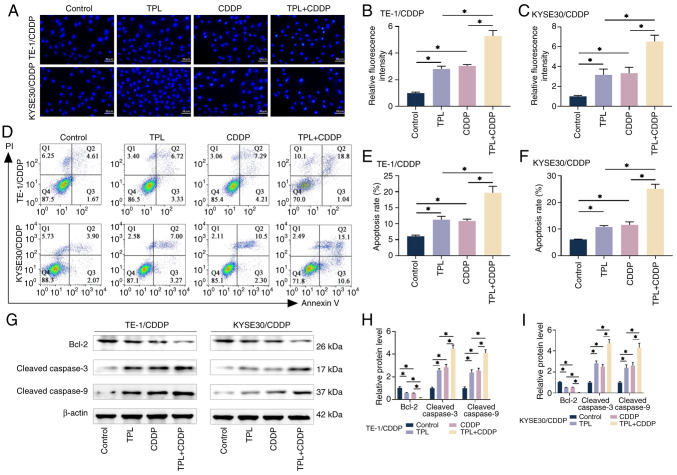
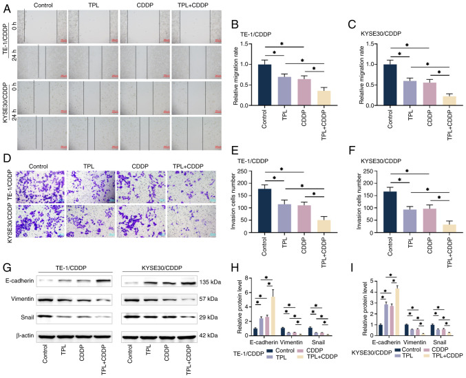
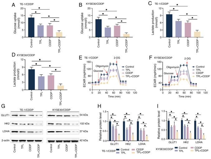
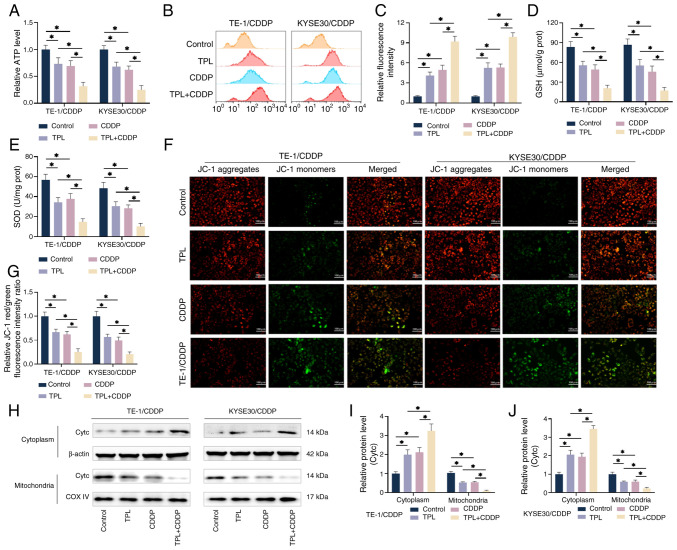
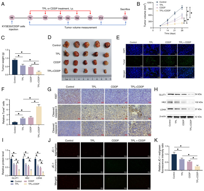
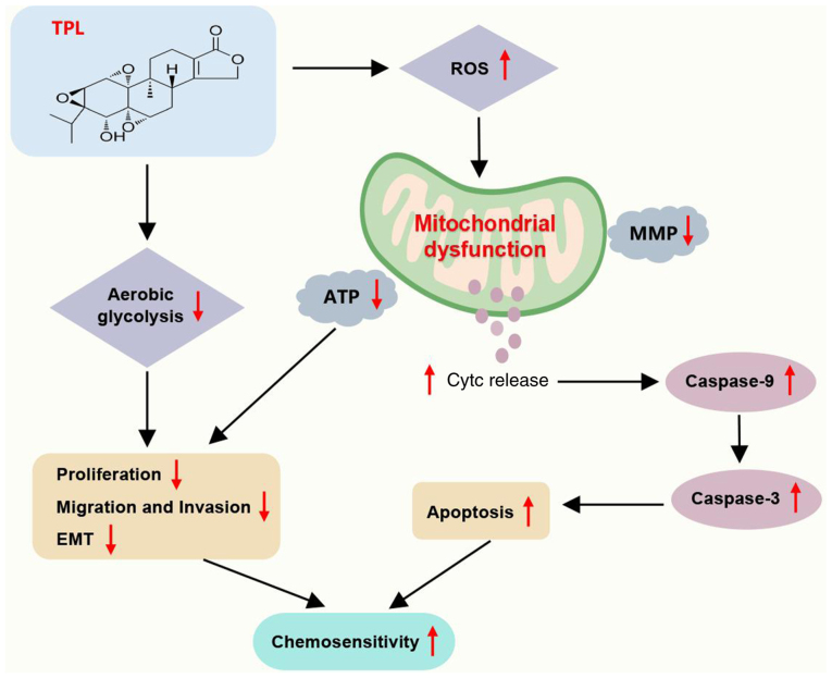
The present study investigated the sensitization mechanism of triptolide (TPL) in esophageal squamous cell carcinoma (ESCC) resistant to cis‑diamminedichloroplatinum (CDDP). CDDP‑resistant TE‑1/CDDP and KYSE30/CDDP cells were created using an incremental drug concentration approach. TPL and CDDP treatment conditions were screened based on the Cell Counting Kit‑8 cell viability assay and cell proliferation was detected using 5‑ethynyl‑2'‑deoxyuridine and clone formation assays. Flow cytometry combined with Hoechst 33258 staining was used to assess cell cycle progression and apoptosis. Scratch healing assay, Transwell assay and western blotting were used to investigate the malignant behaviors of the cells. Changes in cellular glycolysis were investigated by measuring glucose uptake, lactate production and the levels of related regulatory factors. Changes in mitochondrial function were examined by detecting ATP and reactive oxygen species levels, as well as mitochondrial membrane potential and cytochrome c release. Furthermore, a nude mouse subcutaneous graft tumor model assay was used to assess the in vivo effect of TPL. In vitro dosages of TPL and CDDP were tested at 2 nM and 4 µM, respectively. Notably, TPL decreased the proliferation, migration, invasion and epithelial‑mesenchymal transition of CDDP‑resistant ESCC cells, increased their apoptosis and significantly suppressed tumor growth in a nude mouse model of ESCC. TPL was shown to have a strong CDDP‑sensitizing effect in vitro and in vivo and its mechanism may involve inhibiting anaerobic glycolysis and causing mitochondrial energy metabolism impairment to induce apoptosis. In conclusion, TPL may be considered a potential CDDP sensitizer with substantial clinical implications for ESCC therapy.

Keywords: cis‑diamminedichloroplatinum‑resistant; esophageal squamous cell carcinoma; glycolysis; mitochondrial dysfunction; triptolide.


Conflict of interest statement

The authors declare that they have no competing interests.

Figures

Figure 1.
Figure 1.
TPL increases the proliferative effect of CDDP on anti-CDDP-resistant ESCC cells. (A and B) The cells viability was measured with CCK-8. (C) The effects of various TPL concentrations on the proliferative viability of TE-1/CDDP and KYSE30/CDDP cells were detected using CCK-8. (D) 2 nM TPL was administered concurrently with CDDP treatment of TE-1/CDDP, KYSE30/CDDP cells and viability was determined by CCK-8. (E and F) EdU detection of proliferation in TE-1/CDDP and KYSE30/CDDP cells (magnification, ×20; scale bar 100 µm). (G-I) Clone formation assay to determine the formation levels of TE-1/CDDP and KYSE30/CDDP cells. *P<0.05. TPL, triptolide; CDDP, cis-diamminedichloro-platinum; ESCC, esophageal squamous cell carcinoma; CCK, cell counting kit; TE-1/CDDP, cisplatin-resistant TE-1 cells; KYSE30/CDDP, cisplatin-resistant KYSE30 cells; IC50, median inhibition concentration; DAPI, diamidino-phenyl-indole; EdU, 5-ethynyl-2′-deoxyuridine.
Figure 2.
Figure 2.
TPL enhances CDDP-promoted CDDP-resistant ESCC cell cycle blockade. (A-C) Cell cycle arrest in TE-1/CDDP and KYSE30/CDDP cells was observed using a flow cytometric assay under various treatment conditions. (D-F) Differences in the level of cell cycle proteins in TE-1/CDDP and KYSE30/CDDP cells under various treatment conditions. *P<0.05. TPL, triptolide; CDDP, cis-diamminedichloro-platinum; ESCC, esophageal squamous cell carcinoma; TE-1/CDDP, cisplatin-resistant TE-1 cells; KYSE30/CDDP, cisplatin-resistant KYSE30 cells; CDK4, cyclin-dependent kinase 4.
Figure 3.
Figure 3.
TPL enhances CDDP pro-apoptotic effects in CDDP-resistant ESCC cells. (A-C) The apoptotic levels of TE-1/CDDP and KYSE30/CDDP cells were measured under each treatment condition using Hoechst 33258 fluorescent staining (magnification, ×40; scale bar, 50 µm). (D-F) Flow cytometry test for detecting apoptotic changes in TE-1/CDDP and KYSE30/CDDP cells under various treatment conditions. (G-I) Western blotting was used to determine the expression levels of apoptotic proteins in TE-1/CDDP and KYSE30/CDDP cells under various treatment conditions. *P<0.05. TPL, triptolide; CDDP, cis-diamminedichloro-platinum; ESCC, esophageal squamous cell carcinoma; TE-1/CDDP, cisplatin-resistant TE-1 cells; KYSE30/CDDP, cisplatin-resistant KYSE30 cells.
Figure 4.
Figure 4.
TPL enhances CDDP inhibition of CDDP-resistant ESCC cell migration, invasion and EMT effects. (A-C) The migration levels of TE-1/CDDP and KYSE30/CDDP cells after treatment with TPL and CDDP were determined using a scratch test (magnification, ×10; scale bar 200 µm). (D-F) The Transwell assay was used to determine the quantity of TE-1/CDDP and KYSE30/CDDP cells that invaded the lower chamber following TPL and CDDP treatment (magnification, ×40; scale bar, 50 µm). (G-I) Western blot analysis was used to determine the expression levels of EMT-related proteins in TE-1/CDDP and KYSE30/CDDP cells following TPL and CDDP therapy. *P<0.05. TPL, triptolide; CDDP, cis-diamminedichloro-platinum; ESCC, esophageal squamous cell carcinoma; EMT, epithelial-mesenchymal transition; TE-1/CDDP, cisplatin-resistant TE-1 cells; KYSE30/CDDP, cisplatin-resistant KYSE30 cells.
Figure 5.
Figure 5.
TPL enhances CDDP sensitivity of CDDP-resistant ESCC cells via inhibiting glycolysis. (A-D) Kits were used to detect glucose absorption and lactate generation in TE-1/CDDP and KYSE30/CDDP cells from each group. (E and F) Kits were used to detect ECAR in TE-1/CDDP and KYSE30/CDDP cells from each group in order to assess cellular energy metabolic activity. (G-I) Western blot analysis revealed changes in the levels of key glycolysis regulators GLUT1, HK2 and LDHA in TE-1/CDDP and KYSE30/CDDP cells from each group. *P<0.05. TPL, triptolide; CDDP, cis-diamminedichloro-platinum; ESCC, esophageal squamous cell carcinoma; TE-1/CDDP, cisplatin-resistant TE-1 cells; KYSE30/CDDP, cisplatin-resistant KYSE30 cells; GLUT1, glucose transporter type 1; ECAR, extracellular acidification rate; HK2, hexokinase 2; LDHA, lactate dehydrogenase A.
Figure 6.
Figure 6.
TPL increases the CDDP sensitivity of CDDP-resistant ESCC cells via generating mitochondrial dysfunction. (A) The kit identified ATP generation in TE-1/CDDP and KYSE30/CDDP cells from each group. (B and C) The ROS generation levels of TE-1/CDDP and KYSE30/CDDP cells in each group were detected via by flow cytometry utilizing the DCFH-DA technique. (D and E) The kit measured the GSH and SOD levels in TE-1/CDDP and KYSE30/CDDP cells from each group. (F and G) Each group's TE-1/CDDP and KYSE30/CDDP cells were tested for mitochondrial membrane potential using the JC-1 assay (magnification, ×20; scale bar 100 µm). (H-J) Western blot analysis was used to assess cytochrome c levels in the mitochondria and cytoplasm of TE-1/CDDP and KYSE30/CDDP cells under various treatment conditions. *P<0.05. TPL, triptolide; CDDP, cis-diamminedichloro-platinum; ESCC, esophageal squamous cell carcinoma; TE-1/CDDP, cisplatin-resistant TE-1 cells; KYSE30/CDDP, cisplatin-resistant KYSE30 cells; GSH, glutathione; SOD, superoxide dismutase; Cytc, cytochrome c; COX IV, cytochrome c oxidase IV.
Figure 7.
Figure 7.
TPL increases the susceptibility of CDDP-resistant ESCC cells to CDDP in vivo. (A) Animal models were constructed according to the timeline. (B-D) Subcutaneous tumor volumes were measured using vernier calipers and the mice were killed on the 28th day of feeding, with the isolated tumors weighed and photographed. (E and F) Tissue sections from the isolated tumors were produced and apoptosis was identified using the Tunel method (magnification, ×40; scale bar, 50 µm). (G) Immunohistochemistry was used to identify the expression levels of Ki67, cleaved caspase-3 and cleaved caspase-9 in each group's tumor sections (magnification, ×40; scale bar, 50 µm). (H and I) Western blot analysis of tumor tissues from each group revealed the expression levels of major glycolysis regulators GLUT1, HK2 and LDHA. (J and K) The intensity of red/green fluorescence in tumor tissues from each group was measured using the JC-1 method to quantify mitochondrial membrane potential alterations (magnification, ×20; scale bar 100 µm). *P<0.05. TPL, triptolide; CDDP, cis-diamminedichloro-platinum; ESCC, esophageal squamous cell carcinoma; KYSE30/CDDP, cisplatin-resistant KYSE30 cells; DAPI, diamidino-phenyl-indole; GLUT1, glucose transporter type 1; HK2, hexokinase 2; LDHA, lactate dehydrogenase A.
Figure 8.
Figure 8.
Diagram of the action mechanism. TPL inhibits glycolysis and causes mitochondrial dysfunction (elevated ROS, decreased mitochondrial membrane potential and ATP and Cytc release), which further inhibits ESCC cell proliferation, migration, invasion and EMT, as well as promotes apoptosis (elevated caspase-9 and caspase-3), thereby inhibiting ESCC cell resistance to CDDP. TPL, triptolide; ROS, reactive oxygen species; Cytc, cytochrome c; ESCC, esophageal squamous cell carcinoma; EMT, epithelial-mesenchymal transition; MMP, mitochondrial membrane potential; CDDP, cis-diamminedichloro-platinum.

Similar articles

References

    1. Conway E, Wu H, Tian L. Overview of risk factors for esophageal squamous cell carcinoma in China. Cancers (Basel) 2023;15:5604. doi: 10.3390/cancers15235604. - DOI - PMC - PubMed
    1. Sung H, Ferlay J, Siegel RL, Laversanne M, Soerjomataram I, Jemal A, Bray F. Global cancer statistics 2020: GLOBOCAN estimates of incidence and mortality worldwide for 36 cancers in 185 countries. CA Cancer J Clin. 2021;71:209–249. doi: 10.3322/caac.21660. - DOI - PubMed
    1. Li J, Xu J, Zheng Y, Gao Y, He S, Li H, Zou K, Li N, Tian J, Chen W, He J. Esophageal cancer: Epidemiology, risk factors and screening. Chin J Cancer Res. 2021;33:535–547. doi: 10.21147/j.issn.1000-9604.2021.05.01. - DOI - PMC - PubMed
    1. Chen R, Zheng R, Zhang S, Wang S, Sun K, Zeng H, Li L, Wei W, He J. Patterns and trends in esophageal cancer incidence and mortality in China: An analysis based on cancer registry data. J Natl Cancer Cent. 2023;3:21–27. doi: 10.1016/j.jncc.2023.01.002. - DOI - PMC - PubMed
    1. Yang CS, Chen XL. Research on esophageal cancer: With personal perspectives from studies in China and Kenya. Int J Cancer. 2021;149:264–276. doi: 10.1002/ijc.33421. - DOI - PMC - PubMed

MeSH terms