. 2025 Feb;21(2):e14429.
doi: 10.1002/alz.14429. Epub 2025 Jan 30.

Plasma proteomic characterization of motoric cognitive risk and mild cognitive impairment

Affiliations

Plasma proteomic characterization of motoric cognitive risk and mild cognitive impairment

Gabriela T Gomez et al. Alzheimers Dement. 2025 Feb.

Abstract

Introduction: Motoric cognitive risk (MCR) is a pre-dementia syndrome characterized by mobility and cognitive dysfunction. This study conducted a proteome-wide study of MCR and compared the proteomic signatures of MCR to that of mild cognitive impairment (MCI).

Methods: Participants were classified as MCR using a memory questionnaire and 4-meter walk. We measured 4877 plasma proteins collected during late-life and midlife. Multivariable logistic regression related each protein to late-life MCR/MCI. MCR-associated proteins were replicated internally at midlife and in an external cohort.

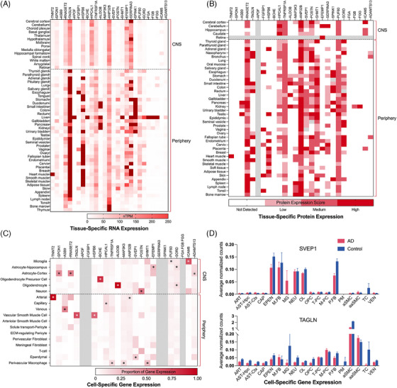
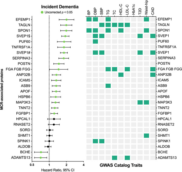
Results: Proteome-wide analysis (n = 4076) identified 25 MCR-associated proteins. Eight of these proteins remained associated with late-life MCR when measured during midlife. Two proteins (SVEP1 and TAGLN) were externally replicated. Compared to MCI, MCR had a distinct and much stronger proteomic signature enriched for cardiometabolic and immune pathways.

Discussion: Our findings highlight the divergent biology underlying two pre-dementia syndromes. Metabolic and immune dysfunction may be a primary driver of MCR.

Highlights: MCR is defined by concurrent cognitive and gait dysfunction. MCR protein biomarkers have key roles in cardiometabolic and vascular function. MCR biomarkers are also associated with cerebrovascular disease and dementia. MCR and MCI demonstrate overlapping but divergent proteomic signatures.

Keywords: dementia; mild cognitive impairment; motoric cognitive risk; pre‐dementia syndrome; proteomic.


Conflict of interest statement

J.C. (Josef Coresh) is a scientific advisor with personal fees to SomaLogic and Healthy.io. Proteomic assays in ARIC were conducted free of charge as part of a data exchange agreement with Soma Logic. The remaining authors declare no competing interests. Author disclosures are available in the Supporting information.

Figures

FIGURE 1
FIGURE 1
Study Design. ARIC, Atherosclerosis Risk in Communities study; BBB, blood–brain barrier; HPA, Human Protein Atlas; MCI, mild cognitive impairment; MCR, motoric cognitive risk; WMH, white matter hyperintensity.
FIGURE 2
FIGURE 2
Proteome‐wide associations with motoric cognitive risk and validation of candidate proteins. Proteomic analyses of MCR and MCI were performed using multivariable logistic regression models adjusted for age, sex, race‐center, education, eGFR‐creatinine, body mass index, diabetes, hypertension, smoking status, and APOEε4 status at the time of protein assessment. Volcano plots display the effect sizes (x‐axis) and p‐values (y axis) for the association of log2 protein level with (A) MCR and (B) MCI. Proteins in teal were significant at the FDR‐corrected < 0.05 threshold; proteins in light green were significant at a Bonferroni adjusted significance level of < 1.03E‐05. (C) Among the 25 MCR‐associated proteins, 8 were associated with MCI, 24 with SWS, and 4 with SCC. Additionally, several MCR‐associated proteins were also associated with neuroimaging markers of small vessel disease, including WMHs (14 proteins), lacunar infarcts (8 proteins), and CMHs (7 proteins) (< 0.05). (D) All proteins maintained a significant relationship with MCR in a sensitivity analysis additionally adjusting for stroke, and 7 proteins were associated with late‐life MCR when measured at midlife. (E) A fixed‐effect, inverse variance weighted meta‐analysis of the association between candidate MCR‐associated proteins and MCR in the ARIC study (discovery cohort) and LonGenity (validation cohort). ARIC, Atherosclerosis Risk in Communities study; CMH, cerebral microhemorrhage; eGFR, estimated glomerular filtration rate; FDR, false discovery rate; MCI, mild cognitive impairment; MCR, motoric cognitive risk; SCC, subjective cognitive complaints; SWS, slow walking speed; WMH, white matter hyperintensity.
FIGURE 3
FIGURE 3
MCR‐associated proteins are expressed across tissues and cell types. (A) RNA expression of genes coding for MCR‐associated proteins in brain, whole blood, and other tissues using data available from GTEx RNA‐Seq database. RNA expression is reported in transcripts per million. (B) Expression of MCR‐associated proteins in brain and other tissue using data available in the Tissue Atlas of the Human Protein Atlas. The Antibody‐based protein levels are qualitatively displayed as: not detected, low, medium, and high. https://www.proteinatlas.org/. (C) Expression of genes coding for MCR‐associated proteins in human brain vascular and perivascular cell types, derived from the Human BBB database, generated using the VINE‐seq method. Values are reported using the proportion of gene expression across cell types. (D) Expression of gene coding for SVEP1 and TAGLN derived from the Human BBB database. aaSMC, arteriolar smooth muscle cell; ART, arterial; aSMC, vascular smooth muscle cell; AST‐Ctx, astrocyte‐cortex; AST‐Hpc, astrocyte‐hippocampus; CAP, capillary; EPEN, ependymal; M.FB, meningeal fibroblast; MCR, motoric cognitive risk; MG, microglia; M‐PC, ECM‐regulating pericyte; NEU, neuron; OL, oligodendrocyte; OPC, oligodendrocyte precursor cell; P.FB, perivascular fibroblast; PM, perivascular macrophage; TC, T‐cell; T‐PC, solute transport‐pericyte; VEN, venous; VINE, vessel isolation and nuclei extraction for sequencing.
FIGURE 4
FIGURE 4
Protein modules are associated with motoric cognitive risk and mild cognitive impairment. (A) Hierarchical cluster tree of 4877 proteins measured at ARIC visit 5 (2011–13). As illustrated by the multicolor band, these proteins are divided into 15 modules using Netboost clustering. (B) A heatmap of the topological overlap matrix illustrating the protein networks. Lighter colors represent higher protein‐protein adjacency. (C) A heatmap depicting the association of module expression with MCR and MCI variables, in addition to small vessel disease neuroimaging characteristics (small vessel disease module associations have been published previously). Five MCR‐associated modules (ME10, ME8, ME7, ME15, ME11) and no MCI‐associated modules remained significant after correction for multiple comparisons (FDR < 0.05). p‐values: *< 0.05; **< 0.01; ***< 0.001. (D‐H) Gene set enrichment (using the g:Profiler toolkit) of each select MCR and MCI‐associated protein modules defined by Netboost., Functional enrichment for each protein set was assessed using GO, KEGG, Reactome, and WikiPathways databases. Left facing bars display pathway enrichment for proteins negatively associated with module expression. Right facing bars display pathway enrichment for proteins positively associated with module expression. The six proteins in each network with the highest correlation with overall network expression are designated as Hub Proteins. Elements of the data displayed in this figure have been previously published. (NCOMMS‐22‐39581A). * Gene shows significant differential expression in at least one brain region based on AMP‐AD consortium work. t Gene shows significant differential protein expression in at least one brain region based on the AMP‐AD consortium dataset (AMP‐AD Sage Bionetworks Agora platform); Gene has been nominated as an AD therapeutic target by the AMP‐AD consortium. ARIC, Atherosclerosis Risk in Communities study; CMH, cerebral microhemorrhage; FDG, false discovery rate; GO, gene ontology; KEGG, Kyoto Encyclopedia of Genes and Genomes; MCI, mild cognitive impairment; MCR, motoric cognitive risk; WMH, white matter hyperintensity.
FIGURE 5
FIGURE 5
Select candidate proteins predict 8‐year dementia risk and show genetic overlap with cardiometabolic traits. Hazard ratios were derived from Cox proportional hazard models adjusted for demographic variables (age, sex, race‐center, education), APOEε4 status, kidney function defined as eGFR‐creatinine, and cardiovascular risk factors (BMI, diabetes, hypertension, and smoking status). Green circles represent statistical significance at < 0.05. The GWAS Catalog Traits panel displays proteins significantly associated with MCR and coded for by genes associated in GWAS with cardiovascular and metabolic traits and disease. Gene lists were based on GWAS catalog summary statistics. APOEε4, apolipoprotein Eε4; BMI, body mass index; CI, confidence interval; eGFR, estimated glomerular filtration rate; GWAS, genome‐wide association study; MCR, motoric cognitive risk.
FIGURE 6
FIGURE 6
Summary of supportive evidence for Motoric Cognitive Risk‐associated proteins. MCI, mild cognitive impairment; MCR, motoric cognitive risk; SVD, small vessel disease.

References

    1. Montero‐Odasso M, Speechley M, Muir‐Hunter SW, et al. Motor and cognitive trajectories before dementia: results from gait and brain study. J Am Geriatr Soc. 2018;66(9):1676‐1683. - PubMed
    1. Chhetri JK, Chan P, Vellas B, Cesari M. Motoric cognitive risk syndrome: predictor of dementia and age‐related negative outcomes. Front Med. 2017;4:166. Accessed August 27, 2020. https://www.frontiersin.org/articles/10.3389/fmed.2017.00166/full - DOI - PMC - PubMed
    1. Verghese J, Ayers E, Barzilai N, et al. Motoric cognitive risk syndrome. Neurology. 2014;83(24):2278‐2284. - PMC - PubMed
    1. Verghese J, Wang C, Lipton RB, Holtzer R. Motoric cognitive risk syndrome and the risk of dementia. J Gerontol A Biol Sci Med Sci. 2013;68(4):412‐418. - PMC - PubMed
    1. Verghese J, Annweiler C, Ayers E, et al. Motoric cognitive risk syndrome. Neurology. 2014;83(8):718‐726. - PMC - PubMed