. 2025 Mar;12(11):e2410760.
doi: 10.1002/advs.202410760. Epub 2025 Jan 30.

Subcellular Cavitation Bubbles Induce Cellular Mechanolysis and Collective Wound Healing in Ultrasound-Inflicted Cell Ablation

Affiliations

Subcellular Cavitation Bubbles Induce Cellular Mechanolysis and Collective Wound Healing in Ultrasound-Inflicted Cell Ablation

Ziyue Bai et al. Adv Sci (Weinh). 2025 Mar.

Abstract

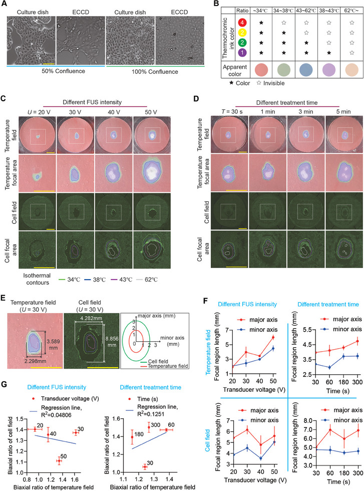
Focused ultrasound (FUS) has been widely adopted in medical and life science researches. Although various physical and biological effects of FUS have been well-documented, there is still a lack of understanding and direct evidence on the biological mechanism of therapeutic cell ablation caused by high-intensity ultrasound (HIFU) and the subsequent wound healing responses. This study develops an enclosed cell culture device that synergistically combines non-invasive FUS stimulation and real-time, on-the-fly live-cell imaging, providing an in vitro platform to explore short and long-term biological effects of ultrasound. The process, mechanism, and wound healing response of cell ablation induced by HIFU are elucidated, revealing a unique mechanism, termed ultrasound-inflicted cellular mechanolysis, that is mediated by growing subcellular cavitation air bubbles under confined contact with cells. This provides a previously unappreciated mechanism for understanding the biomechanical principles of ultrasound-based ablative therapy. A post-ablation phantom layer is also revealed that serves as a guiding cue for collective cell migration during wound healing, thereby providing a biomimetic model for studying wound healing after HIFU-inflicted damage. Together, this study provides theoretical and technological basis for advancing the understanding of the biological effects of ultrasound-based ablative therapy and inspiring clinically relevant applications in the future.

Keywords: cavitation bubbles; cell ablation; collective cell migration; focused ultrasound.


Conflict of interest statement

The authors declare no conflict of interest.

Figures

Figure 1
Figure 1
Design of the enclosed cell culture device (ECCD) compatible with focused ultrasound (FUS) stimulation and real‐time, on‐the‐fly imaging. A) Schematic of the ECCD design and the acoustic pathway of FUS stimulation. B) Structural components of the ECCD. C) Structural components of the FUS transducer coupler. D) Schematic showing the aligned assembly of the FUS transducer and ECCD with the acoustic pathway and expected focal area. E) Design of restraint structures for co‐axial transducer‐coupler‐ECCD alignment. F) Simulation results of the acoustic field elicited by FUS in the ECCD. Zoom‐in image displays the acoustic intensity field in the focal region. The acoustic intensity along the white dashed line (at the height of 0.05 mm above the bottom of ECCD) was extracted and plotted in (G). G) Line‐profile of acoustic intensity along the white dashed line shown in (F), showing the focused deployment of acoustic energy in the expected focal region.
Figure 2
Figure 2
An integrated platform for FUS stimulation and live‐cell imaging based on the ECCD. A) Flowchart showing the experimental setup and its operational procedure. B) Schematic showing the assembly of the integrated platform, including the first‐level (tri‐axial) and second‐level (uni‐directional) displacement control systems that are designed to control the 3D movement of the ECCD within the light path of the microscope and the alignment of the transducer to the ECCD, respectively. C) Photographs showing the ECCD and its carrier system. The ECCD holder is connected to the second‐level displacement system. Scale bars: 10 mm (left), 10 cm (right). D) Photographs of the coupler before (left) and after (right) assembled to the FUS transducer. Scale bar: 10 mm. E) Photograph showing the assembly of the first and second‐level displacement systems, carrier system, FUS transducer and coupler, cooling system for the transducer, and the ECCD. Scale bar: 10 cm. F) Photographs showing a fully assembled, integrated platform for FUS stimulation and real‐time microscopic imaging based on the ECCD.
Figure 3
Figure 3
FUS‐induced cell ablation does not show significant correlation with ultrasound‐caused thermal effect. A) Bright‐field images of MDCK cells cultured in regular 60 mm culture dishes and ECCD at confluence of 50% and 100%, respectively. Scale bar: 100 µm. B) Table showing the temperature‐dependent coloration of the four‐color thermochromic ink. The mixing ratio of mono‐chromic ink is red: yellow: green: purple = 4:2:2:1. C) Representative images showing the distribution of temperature and cell ablation, respectively, in the ECCD upon FUS treatment at different transducer voltage (U) while the frequency (1 MHz, continuous waves) and treatment duration (T = 3 min) remain constant. The gradient coloration of the thermosensitive tape was used to plot the isotherm contours of the temperature field. Live dye (green) was used to visualize living cells and the corresponding FITC fluorescence confocal micrographs (cell field) were used to analyze the cell ablation within a MDCK monolayer. Cell ablation area is reflected by the loss of fluorescence after FUS treatment. Square boxes indicated the region shown in zoom‐in images. Similar results were observed in n = 3 independent experiments. Scale bar: 5 mm. D) Representative images showing the distribution of temperature and cell ablation, respectively, in the ECCD upon FUS treatment for different duration (T) while the frequency (1 MHz, continuous waves) and transducer voltage (U = 40 V) remain constant. Similar results were observed in n = 3 independent experiments. Scale bar: 5 mm. E) Representative distributions of the temperature field (left) and cell field (right) at the focal domain of the ECCD upon FUS treatment at U = 30 V and T = 3 min. The contours of the temperature field and cell field were demarcated as indicated, respectively, with the dimensions of their major axis and minor axis labeled accordingly. F) Plots showing the change of the dimensions of the focal region of the temperature field (upper panels) and cell field (lower panels), respectively, under indicated conditions of transducer voltage (left) or treatment duration (right). Data were plotted as mean ± 95% confidence interval. n = 3 independent experiments. G) Plots showing the relationship between the aspect ratio of the focal region of the temperature field and that of the cell field, under indicated conditions of transducer voltage (left) or treatment duration (right). Linear fitting was applied using the least squares method. Data were plotted as mean ± s.e.m. n = 3 independent experiments.
Figure 4
Figure 4
Significant association between cell ablation zone and bubble generation zone under FUS. A) Confocal micrographs showing the distribution of living cells (tracked by FITC fluorescence) after FUS treatment (U = 40 V, T = 60 s, 1 MHz continuous waves). The black arrow indicates the direction of incident ultrasound waves. Live dye is used to label living cells, and non‐fluorescent regions reflect the cell ablation zone. Scale bar: 1 mm. The purple box marks the streak‐like ablation domains in the focal area. The area between the two orange ellipses is the ablation region where cavitation bubbles appear. The area enclosed by the red irregular lines is the area with a temperature greater than 62 °C during FUS treatment. B) Confocal fluorescence micrographs showing the distributions of cavitation bubbles, living cells, and cell nuclei, within the focal region after FUS treatment (U = 40 V, T = 3 min, 1 MHz continuous waves). Bubbles are opaque and spherical under bright field. Live dye (green) is used to detect living cell domain and non‐fluorescent cell ablation domain. H2B‐RFP MDCK cells are used to track the cell nuclei (red). Similar results were observed in n = 5 independent experiments. Scale bar: 100 µm. C) Time‐lapse micrographs showing the progressive cell ablation marked by Dead dye fluorescence within the focal domain of a monolayer of MDCK cells under FUS treatment (U = 40 V, continuous waves at 1 MHz). Yellow arrows indicate bands of bubbles. The time‐stamp represents the time lapsed from the first image. Scale bar: 100 µm. D) Time‐lapse micrographs showing the progressive rupture of the MDCK monolayer under continued FUS stimulation (U = 40 V, 1 MHz continuous waves). Yellow lines represent the boundary of the bubble‐enriched region. Red and green arrows indicate the expansion and retreat movement of bubble clusters. The time‐stamp represents the time lapsed from the first image. Scale bar: 100 µm. E) Representative images showing the post‐ablation phantom layer (PAPL) composed of FUS‐ablated dead cells and its neighboring living cell domain (U = 40 V, T = 3 min, 1 MHz continuous waves). Colored square boxes mark the living cell domain (blue), dead cell domain (green), and the boundary between living and dead cell domains (yellow dashed lines in red box) in zoom‐in images. Scale bar: 100 µm. F) Confocal fluorescence micrographs showing the post‐ablation MDCK monolayer (U = 40 V, T = 3 min, 1 MHz continuous waves). Living cells are marked by live dye (green). 3D reconstructed images of the PAPL and its adjacent living cell monolayer are also shown. Region 1 shows the PAPL that remained in its original place; region 2 shows PAPL ruptured and peeled off the substrate by bubbles, along with some live cell monolayer connected to it; region 3 shows the PAPL forming a “dome‐like” shape, with its central domain detached but peripheral domain connected to the live cell monolayer on the substrate. Color bar represents the z‐position within the 3D reconstructed image. Scale bar: 100 µm.
Figure 5
Figure 5
Subcellular cavitation bubble formation and FUS‐induced mechanolysis of the cell. A) Time‐lapse FITC fluorescence micrographs of MDCK cells under FUS treatment (U = 40 V, continuous waves at 1 MHz). Images shown in the three rows represent the formation of subcellular cavitation bubbles in a single cell (zoom‐in 1), three different cells (zoon‐in 2), and multiple cells (zoom‐out), respectively. Live dye is used on cells, and the disappearance of the fluorescent signal reflects cell ablation. Arrowheads indicate the subcellular cavitation bubbles. Similar results were observed in n = 10 independent experiments. Scale bar: 50 µm. B) Plots showing the change of live‐dye fluorescence intensity within the cell cytoplasm (excluding the bubble area) over time for the cell indicated by the arrowhead in the first row in (A). C) Plot of time duration from the moment of bubble formation to the moment when fluorescence intensity of the cell dropped to the background level (box: 25% – 75%, bar‐in‐box: median, and whiskers: min and max). n = 3 independent experiments; n cell = 350. D) Reconstructed 3D z‐stack FITC fluorescence images showing subcellular bubbles generated within a GFP‐expressing cell during FUS treatment. The xy, xz, and yz section images were all shown. The fluorescence‐depleted domain appearing in the GFP‐expressing cell represents the subcellular cavitation bubble, which is indicated by the white arrowhead. Scale bar: 10 µm. E) Analysis of relative fluorescence intensity at the location of the cavitation bubble. The fluorescence intensity at the center of the bubble is denoted as h 1 when it first appeared, h 0 before the bubble formed at the same location, and h 2 after the bubble caused cell ablation, escaped, and expanded to the space above neighboring cells. Data were plotted as mean ± s.d. n = 3 independent experiments; n cell = 59. F) Bright field and FITC fluorescence micrographs showing subcellular cavitation bubbles generated underneath the cell and eventually causing cell membrane rupture (indicated by arrowheads) during FUS treatment. Similar results were seen in n = 3 independent experiments. Membrane‐GFP MDCK cells were used. Scale bar: 10 µm. G) Time‐lapse FITC fluorescence micrographs showing bubbles appearing and disappearing underneath the cell during FUS treatment. Live dye is used to the cells. Scale bar: 10 µm. H) Schematics showing different types of interactions between FUS‐induced subcellular cavitation bubbles and the cell, including the unique type of cellular mechanolysis by subcellular cavitation bubbles stably growing under confined contact with the cell.
Figure 6
Figure 6
Observation of collective cell migration and wound healing after FUS‐induced ablation. The first three columns display time‐lapse bright‐field and RFP fluorescence micrographs showing the collective migration of living H2B‐RFP MDCK cells towards the PAPL area after FUS‐induced ablation. In the third column, the white area represents the PAPL (with remaining RFP‐labeled cell nuclei that showed no movement), with white arrows indicating the direction of collective cell migration. The yellow area represents secondary tissue gaps that appear during the primary wound healing process, largely due to the different directions and speeds of cell migration within the living cell monolayer, with yellow arrows indicating the cell migration direction. The fourth to sixth columns show the trajectories of representative cells during the collective wound healing. Colored lines represent the migration trajectory of each individual cells. The fifth to sixth columns show zoom‐in views of the boxed regions. Similar results were seen in n = 3 independent experiments. Scale bar: 100 µm.
Figure 7
Figure 7
Effect of the PAPL on the collective migration and wound healing after FUS‐induced ablation. A) Time‐lapse bright‐field and RFP fluorescence micrographs showing the cell migration after FUS‐induced cell ablation, featuring collective migration of living cells toward the PAPL (with remaining RFP‐labeled cell nuclei that showed no movement) or the free area (where the PAPL has detached and left a nuclei‐free region). The whitened area represents the PAPL or the free area, with white arrows indicating the direction of collective cell migration. The yellow area represents secondary gaps formed in the collectively migrating cell sheet nearby the wound. Yellow arrows indicate the cell migration direction near the edge of the free area. The trajectories of individual cell nuclei during the wound healing process are shown as colored lines. Blue and green boxes mark the region shown in zoom‐in images. Similar results were seen in n = 3 independent experiments. Scale bar: 100 µm. B) Confocal micrographs showing the distribution of individual cell nuclei at the beginning of collective migration. Scale bar: 100 µm. C) Trajectories of individual living cells during the collective wound healing after FUS‐induced ablation, which features a bifurcating migration path that is guided by the presence of PAPL flanking the clear area. The dashed line marks the neural line between the two cell groups that migrate towards the left (trajectories marked by green lines) and right (trajectories marked by purple lines) flanks of the PAPL, respectively. Similar results were seen in n = 3 independent experiments. θ 1 and θ 2 represent the orientation angle of the interface between the PAPL and the free area, which are defined and calculated as indicated in the graph. D) Plots showing the inclined angle between cell migration direction and the orientation angles of the interfaces between the PAPL and the free area shown in (C). The inclined angles were calculated for the cell groups migrating towards the left (trajectories marked by green lines in (C), shown in the left plot here) and the right (trajectories marked by purple lines in (C), shown in the right plot here) flanks of the PAPL, respectively, during time duration as indicated (T 1: 0.00‐0.67 h; T 2: 2.33‐3.00 h; T 3: 4.67‐5.33 h; T 4: 7.00‐7.67 h; T 5: 8.50‐9.17 h; T 6: 10.17‐10.83 h; T 7: 11.83‐12.50 h). Data were plotted as mean ± s.e.m. n cell = 34 for each indicated condition and time duration. Two‐tailed Student's t‐test was applied to analyze the statistical significance. * p < 0.05, ** p < 0.01, *** p < 0.001, **** p < 0.0001.

Similar articles

Cited by

References

    1. Krishna V., Sammartino F., Rezai A., JAMA Neurol 2018, 75, 246. - PubMed
    1. Izadifar Z., Izadifar Z., Chapman D., Babyn P., J. Clin. Med. 2020, 9, 460. - PMC - PubMed
    1. Meng Y., Hynynen K., Lipsman N., Nat. Rev. Neurol. 2020, 17, 7. - PubMed
    1. Recker F., Thudium M., Strunk H., Tonguc T., Dohmen S., Luechters G., Bette B., Welz S., Salam B., Wilhelm K., Egger E. K., Wüllner U., Attenberger U., Mustea A., Conrad R., Marinova M., Sci. Rep. 2021, 11, 22768. - PMC - PubMed
    1. Tang Y., Chen C., Jiang B., Wang L., Jiang F., Wang D., Wang Y., Yang H., Ou X., Du Y., Wang Q., Zou J., Int. J. Nanomed. 2021, 16, 4643. - PMC - PubMed

LinkOut - more resources