. 2025 Feb;13(2):e70140.
doi: 10.1002/iid3.70140.

Progranulin Plays a Protective Role in Pneumococcal Meningitis by Inhibiting Pyroptosis

Affiliations

Progranulin Plays a Protective Role in Pneumococcal Meningitis by Inhibiting Pyroptosis

Jingyao Wang et al. Immun Inflamm Dis. 2025 Feb.

Abstract

Objective: Pneumococcal meningitis is a serious infectious disease with a high mortality rate and a global presence, and survivors have different degrees of neurological sequelae as a consequence of the host response to the infection. Progranulin (PGRN) is a multifunctional autocrine growth factor that is also a major immunoregulator. We want to investigate the role for PGRN in Pneumococcal meningitis in vivo and in vitro.

Method: Mouse and cell models were established to explore the protective effect and mechanism of PGRN against pneumococcal meningitis.

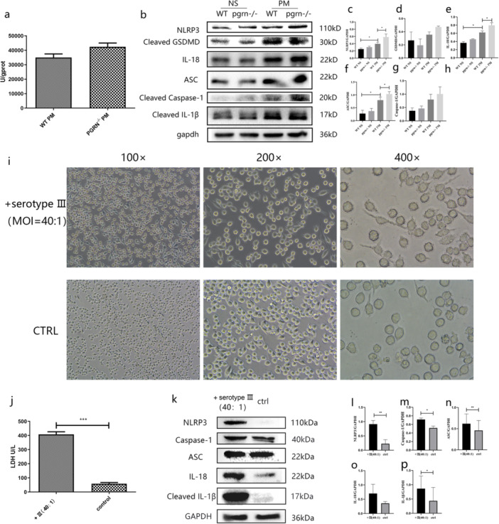
Results: Progranulin plays a protective role in pneumococcal meningitis by inhibiting pyroptosis. Pyroptosis resulted from exposure of BV-2 cells to the bacterium and this was confirmed in the in vivo model. Administration of the NLRP3 inflammasome inhibitor MCC950 to mice prior to infection inhibited pyroptosis and protected PGRN -/- mice and BV-2 cell model from meningitis.

Conclusion: This study implicates a protective role for PGRN in pneumococcal meningitis by inhibiting pyroptosis, indicating that PGRN may have therapeutic potential.

Keywords: pneumococcal meningitis; progranulin; pyroptosis.


Conflict of interest statement

The authors declare no conflicts of interest.

Figures

Figure 1
Figure 1
Histology and ELISA determinations of TNF‐α, IL‐1β, and IL‐6 in mice with experimental pneumococcal meningitis. (a) H&E staining of brain serial sections of the indicated mice (n = 4). (b) TNF‐α levels in brain homogenates at 24 h. (c) IL‐6 levels in plasma at 24 h. (d) IL‐1β expression in brain homogenates at 6 h. (e) Plasma levels of PGRN. (f) PGRN levels mice in brain serial sections (n = 4). Results of the above animal experiments are representative of three independent experiments.
Figure 2
Figure 2
A comparison of WT and PGRN ‐/‐ mice with experimental pneumococcal meningitis. (a) H&E staining of brain tissue sections (n = 4). (b) Loeffler's neurobehavioral scores for mice with meningitis at 24 h postinfection (n = 4). (c) Bacterial loads in brains at 24 h (n = 6). (d) Survival curves for infected and control mice (n = 13). Results of the above animal experiments are representative of three independent experiments. *p < 0.05; **p < 0.01.
Figure 3
Figure 3
LDH and NLRP3 inflammasome components in brains of mice with pneumococcal meningitis and BV‐2 cells exposed to Streptococcus pneumonia. (a) NLRP3 inflammasome components and GAPDH (internal control) Western blots of brain tissue homogenates from saline control (NS) and PM mice (n = 3). (b–g) Quantification of Western blots using relative densitometry. (h–i) Morphology of BV‐2 cells exposed to S. pneumoniae serotype III and LDH expression in tissue culture supernatants. (j) Western blot detection of NLRP3 inflammasome components and controls as indicated in BV‐2 cells exposed to S. pneumonia. (k–o) Quantification of Western blot bands by densitometry (n = 3). Results of the above animal experiments are representative of three independent experiments. *p < 0.05; **p < 0.01, ***p < 0.001.
Figure 4
Figure 4
MCC950 prevent on BV‐2 cell model and PGRN‐/‐ pneumococcal meningitis in mice. (a) Western blot detection of inflammasome components and controls as indicated in BV‐2 cells exposed to S. pneumoniae treated by PGRN or MCC950. (b) NLRP3 expression in PGRN‐/‐ mice with experimental pneumococcal meningitis (PM) (n = 3). (c) Quantification of Western blot bands by relative densitometry of NLRP3 and GAPDH internal control (n = 3). (d) Bacterial loads in brains of infected and control mice (n = 6). (e) IL‐6 levels in brain homogenates of mice as indicated (n = 3). *p < 0.05; **p < 0.01.
Figure 5
Figure 5
PGRN levels in CSF of patients with bacterial and viral meningitis are correlated with diagnostic markers of disease. (a) nucleated leukocytes (b) glucose and (c) albumin.

Similar articles

References

    1. Sigurdardóttir B., Björnsson Ó. M., and Jónsdóttir K. E., “Acute Bacterial Meningitis in Adults: A 20‐Year Overview,” Archives of Internal Medicine 157, no. 4 (1997): 425–430, 10.1001/archinte.1997.00440250077009. - DOI - PubMed
    1. Vestergaard H. H., Larsen L., Brandt C., et al., “Normocellular Community‐Acquired Bacterial Meningitis in Adults: A Nationwide Population‐Based Case Series,” Annals of Emergency Medicine 77 (2021): 11–18, 10.1016/j.annemergmed.2020.05.041. - DOI - PubMed
    1. D. van de Beek, , J. de Gans, , Spanjaard L., Weisfelt M., Reitsma J. B., and Vermeulen M., “Clinical Features and Prognostic Factors in Adults With Bacterial Meningitis,” New England Journal of Medicine 351, no. 18 (2004): 1849–1859, 10.1056/NEJMoa040845. - DOI - PubMed
    1. Grände P. O., Myhre E. B., Nordström C. H., and Schliamser S., “Treatment of Intracranial Hypertension and Aspects on Lumbar Dural Puncture in Severe Bacterial Meningitis,” Acta Anaesthesiologica Scandinavica 46, no. 3 (2002): 264–270, 10.1034/j.1399-6576.2002.460307.x. - DOI - PubMed
    1. Doran K. S., Fulde M., Gratz N., et al., “Host–Pathogen Interactions in Bacterial Meningitis,” Acta Neuropathologica 131, no. 2 (2016): 185–209, 10.1007/s00401-015-1531-z. - DOI - PMC - PubMed

Publication types

MeSH terms

Substances

LinkOut - more resources