. 2025 Jun;86(7):1091-1100.
doi: 10.1111/his.15414. Epub 2025 Jan 30.

Identification of intraductal-to-invasive spatial transitions in prostate cancer: proposal for a new unifying model on intraductal carcinogenesis

Affiliations

Identification of intraductal-to-invasive spatial transitions in prostate cancer: proposal for a new unifying model on intraductal carcinogenesis

Lucia L Rijstenberg et al. Histopathology. 2025 Jun.

Abstract

Aims: Intraductal carcinoma (IDC) is an independent pathological parameter for adverse prostate cancer (PCa) outcome. Although most IDC are believed to originate from retrograde spread of established PCa, rare IDC cases may represent precursor lesions. The actual transition areas between intraductal and invasive cancer, however, have not yet been identified. Our objective was to identify intraductal-invasive PCa transitions using 2- and 3-dimensional microscopy.

Methods and results: Seventy-five samples from 46 radical prostatectomies with PCa were immunohistochemically stained for basal cell keratins. In 35 samples, atypical glands that were indistinguishable from invasive adenocarcinoma (IAC) had focal 34BE12-positive basal cells. These IAC-like glands were present adjacent to IDC and prostatic intra-epithelial neoplasia (PIN) in 21 of 45 (46.7%) and 16 of 58 (27.6%) cases, respectively. Whole-mount confocal imaging of immunofluorescent Ker5/18 double-stained and cleared 1-mm-thick intact tissues revealed spatial continuity between IDC, IAC-like glands and IAC with a gradual loss of basal cells. In 24 of 35 (68.6%) samples more than one IAC-like focus (median 3.0) was present.

Conclusions: We identified areas of spatial transition between PIN, IDC and IAC, characterised by remnant basal cells in IAC-like glands. Based on the coexistence of IDC and PIN, the gradual loss of basal cells in IAC-like glands and IAC-like glands' multifocality, we propose a novel hypothesis on intraductal carcinogenesis, which we term 'repetitive invasion, precursor progression' (RIPP).

Keywords: IDC; PTEN; basal cell; intraductal; prostate cancer; three‐dimensional; transition.


Conflict of interest statement

None of the authors declare any conflicts of interest.

Figures

Figure 1
Figure 1
Prostate cancer with high‐grade prostatic intra‐epithelial neoplasia (PIN, green asterisk), atypical intraductal proliferation (AIP, red asterisk) and intraductal carcinoma (IDC, black asterisk) being present at close proximity. [Color figure can be viewed at wileyonlinelibrary.com]
Figure 2
Figure 2
A, High‐grade prostatic intra‐epithelial neoplasia (PIN, green asterisk), atypical intraductal proliferation (AIP, red asterisk) and intraductal carcinoma (IDC, black asterisk) surrounded by basal cells. BD, Putative transitions of IDC (black asterisk) and AIP (red asterisk) to invasive adenocarcinoma (IAC)‐like glands, which have an irregular invasive architecture with remnant basal cells (black arrowhead). The IAC‐like glands are located adjacent to IDC and AIP, and spatially situated in between IDC and AIP at one side and invasive adenocarcinoma without basal cells at the other side; 34BE12, original magnifications. [Color figure can be viewed at wileyonlinelibrary.com]
Figure 3
Figure 3
Phosphatase and tensin homologue deleted on chromosome 10 (PTEN) expression in high‐grade prostatic intra‐epithelial neoplasia (PIN, green asterisk), atypical intraductal proliferation (AIP, red asterisk), intraductal carcinoma (IDC, black asterisk), invasive adenocarcinoma (IAC)‐like glands and invasive adenocarcinoma. A, PTEN heterogeneity in invasive adenocarcinoma with retained (left) and lost (right) expression. B, PTEN heterogeneity in invasive adenocarcinoma with retained (left) and lost (right) expression; PTEN expression is retained in PIN and lost in AIP. C, PTEN loss in IAC‐glands (black arrowhead) and invasive adenocarcinoma. D, PTEN loss in IDC, IAC‐like glands and invasive adenocarcinoma; area of retained PTEN expression in right upper corner. Double‐staining PTEN (brown)/34BE12 (red), original magnifications. [Color figure can be viewed at wileyonlinelibrary.com]
Figure 4
Figure 4
Representations of atypical intraductal proliferation (AIP; AC) and intraductal carcinoma (IDC; DF) transitions to invasive prostate cancer (PCa). AC, Spatial transition (white box) from AIP to poorly formed Gleason pattern 4 PCa with gradual loss of basal cells (red). DF, Spatial transition (white box) of IDC to poorly formed Gleason pattern 4 PCa with abrupt complete loss of basal cells. A, B, D, E, Two‐dimensional microscopy of representative Z‐stack image. C, F, Three‐dimensional representation of complete Z‐stack. Keratin 5 red, keratin 8/18 green. [Color figure can be viewed at wileyonlinelibrary.com]
Figure 5
Figure 5
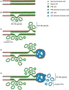
Schematic overview of the ‘repetitive invasion, precursor progression’ (RIPP) model. A, Within a pre‐existent prostate gland luminal epithelial cells (grey) acquire genomic alteration and develop into prostatic intra‐epithelial neoplasia (PIN) (green); the pre‐existent gland and PIN are surrounded by basal epithelial cells (red). B, Luminal PIN cells infiltrate adjacent stroma with gradual loss of basal cells, named invasive adenocarcinoma (IAC)‐like glands. C, Upon further proliferation, invasive tumour glands completely lose basal cells. As remnant luminal PIN cells had acquired infiltrative capacity, they invade adjacent stroma again at another location (‘repetitive invasion’). D, Intraglandular remnant PIN cells further accumulate genomic alterations finally resulting in progression towards intraductal carcinoma (IDC, blue) (‘precursor progression’). E, IDC cells subsequently infiltrate adjacent stroma with gradual loss of basal cells (‘repetitive invasion’). [Color figure can be viewed at wileyonlinelibrary.com]

Similar articles

References

    1. Moch H. WHO classification of tumours: urinary and male genital tumours. 5th ed. International Agency for Research on Cancer (IACR), 2022. Lyon, France.
    1. Guo CC, Epstein JI. Intraductal carcinoma of the prostate on needle biopsy: histologic features and clinical significance. Mod. Pathol. 2006; 19; 1528–1535. - PubMed
    1. Hollemans E, Verhoef EI, Bangma CH et al. Large cribriform growth pattern identifies isup grade 2 prostate cancer at high risk for recurrence and metastasis. Mod. Pathol. 2019; 32; 139–146. - PMC - PubMed
    1. Kweldam CF, Kümmerlin IP, Nieboer D et al. Disease‐specific survival of patients with invasive cribriform and intraductal prostate cancer at diagnostic biopsy. Mod. Pathol. 2016; 29; 630–636. - PubMed
    1. McKenney JK, Wei W, Hawley S et al. Histologic grading of prostatic adenocarcinoma can be further optimized: analysis of the relative prognostic strength of individual architectural patterns in 1275 patients from the canary retrospective cohort. Am. J. Surg. Pathol. 2016; 40; 1439–1456. - PubMed

Grants and funding