. 2025 Jan 31;11(5):eadp7066.
doi: 10.1126/sciadv.adp7066. Epub 2025 Jan 31.

Mechanisms of uropathogenic E. coli mucosal association in the gastrointestinal tract

Affiliations

Mechanisms of uropathogenic E. coli mucosal association in the gastrointestinal tract

Philippe N Azimzadeh et al. Sci Adv. .

Abstract

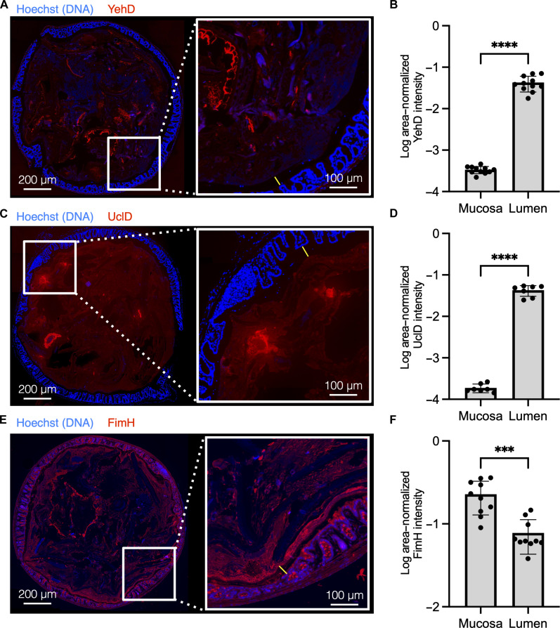
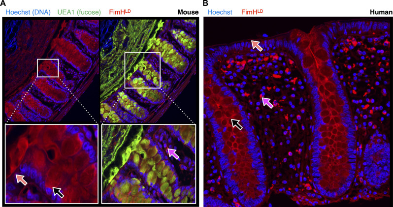
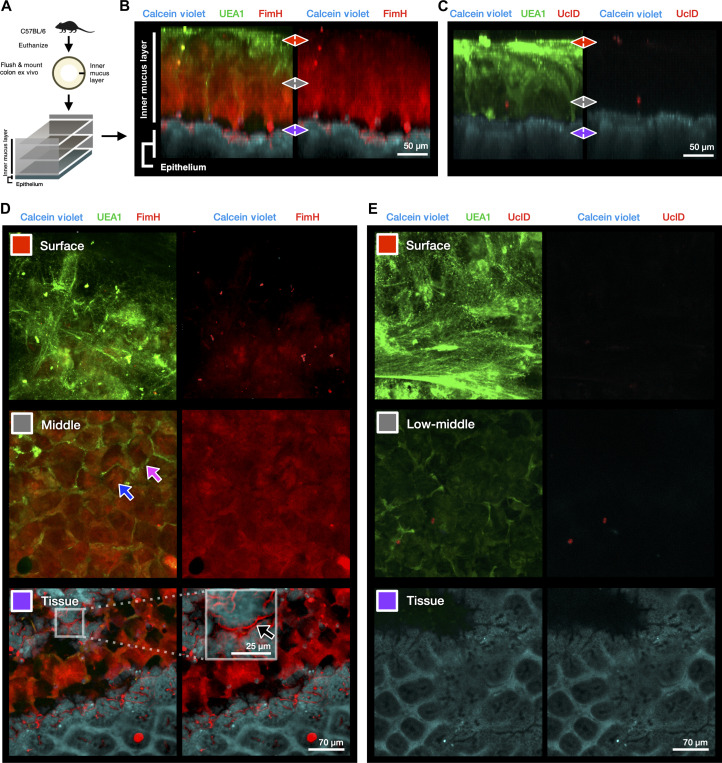
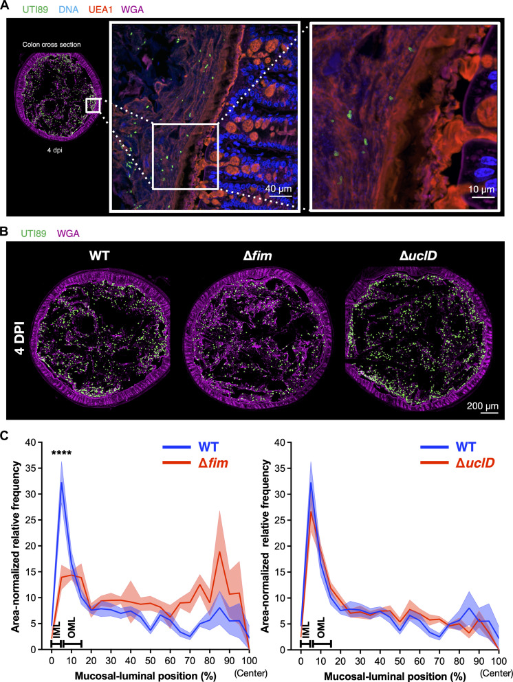
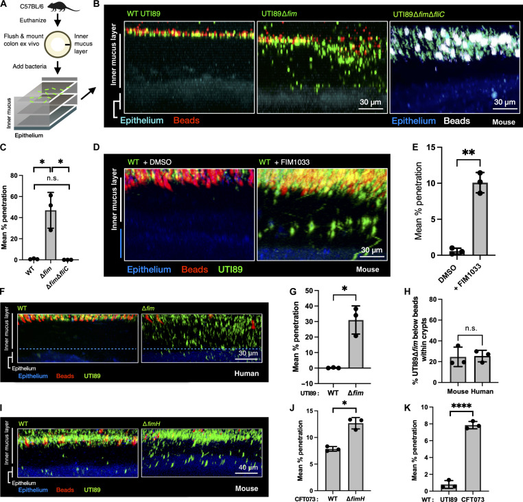
Urinary tract infections (UTIs) are highly recurrent and frequently caused by Uropathogenic Escherichia coli (UPEC) strains that can be found in patient intestines. Seeding of the urinary tract from this intestinal reservoir likely contributes to UTI recurrence (rUTI) rates. Thus, understanding the factors that promote UPEC intestinal colonization is of critical importance to designing therapeutics to reduce rUTI incidence. Although E. coli is found in high abundance in large intestine mucus, little is known about how it is able to maintain residence in this continuously secreted hydrogel. We discovered that the FimH adhesin of type 1 pili (T1P) bound throughout the secreted mucus layers of the colon and to epithelial cells in mouse and human samples. Disruption of T1P led to reduced association with colon mucus. Notably, this mutant up-regulated flagellar production and infiltrated the protective inner mucus layer of the colon. This could explain how UPEC resists being washed off by the continuously secreted mucus layers of the colon.


Figures

Fig. 1.
Fig. 1.. Differential binding of UTI89 CUP adhesins to colon mucosa and lumen.
(A, C, and E) Purified lectin domains of Yeh, (YehD), F17-like (UclD), and type 1 (FimH) pili were bound to methacarn-fixed C57BL/6NJ (The Jackson Laboratory) female mouse colon sections. Lectin domain is labeled in red, and DNA is labeled in blue (Hoechst stain). On the left of (A), (C), and (E) is a representative tile scan of the entire colon section, and on the right is a magnified view of the colon mucosa. The IML is denoted by a yellow line. Left scale bars, 200 μm; right scale bars, 100 um. (B, D, and F) Segmentation analysis was performed on whole-colon sections to quantify the intensity of FimH binding signal in the colon mucosa and the lumen (fig. S2). [(A) and (B)] YehD binds in the colon lumen but does not bind within mucosa, n = 4 mice, two sections per mouse. [(C) and (D)] UclD binds within the colon lumen but does not bind within mucosa, n = 4 mice, two sections per mouse. (C) FimH binds both the mucosa and lumen of the colon, n = 5 mice, two sections per mouse.
Fig. 2.
Fig. 2.. FimH binds broadly within the mouse and human colon mucosa.
Fixed colon tissue was stained with FimH for high-resolution imaging of the mucosa. (A) C57BL/6NJ (Taconic Biosciences) female mouse distal colon was stained with FimH (red), UEA1 (green), and Hoechst (blue). IML denotes the inner mucus layer, and OML denotes the outer mucus layer, n = 3 mice, two sections each. (B) Fixed colon tissue sections from healthy human donors were stained with FimH (red) and Hoechst (blue), n = 3 individuals, two sections each. [(A) and (B)] Black arrows mark FimH signal on goblet cells. Orange arrows mark FimH signal on the epithelial surface. Magenta arrows mark FimH signal within the lamina propria.
Fig. 3.
Fig. 3.. FimH binds the IML and the colon epithelial surface ex vivo.
(A) Experimental design of 3D ex vivo mucus imaging procedure with female C57BL6/NJ (The Jackson Laboratory). (B) Representative side view (x/z projection) of FimH-stained reconstruction of the IML, n = 2 mice, and two pieces of colon per mouse. (C) Representative side view (x/z projection) of UclD-stained reconstruction of the IML, n = 4 mice, and two pieces of colon per mouse. (D) Selected slices (x/y projections) of three areas of interest (IML surface (red), IML middle (gray), and tissue (purple) representative of FimH-stained colon explants. Magenta arrow highlights UEA1-stained intercrypt mucus. Blue arrow highlights FimH-bound main mucus plume. Black arrow and white box inset denote FimH binding within the glycocalyx at the epithelial surface beneath the IML (fig. S5, A and B). (E) Selected slices (x/y projections) of IML surface (red), IML lower middle area (gray), and tissue (purple) representative of UclD-stained colon explants.
Fig. 4.
Fig. 4.. UTI89∆fim::kanR is defective for mucus association during colonization.
Female C3H/HeN mice (Envigo) were colonized with WT, ∆fim, or ∆uclD mutant UTI89 and euthanized 4 days postinfection (dpi). Fixed colons were stained for UTI89 by using an antibody specific for the O18 lipopolysaccharide (LPS) antigen (green) and co-stained with fucose-binding UEA1 (red) and GlcNAc-binding WGA (pink) lectins to visualize mucus layers (fig. S7, A and B). DNA from epithelial cells was visualized by Hoechst stain (blue). (A) WT UTI89 (green) is mainly found in the lumen and outer mucus layer of the colon at 4 dpi (fig. S7C). (B) Representative images from tiled mouse colon sections were stained for UTI89 (green) and WGA (pink). (C) Quantification of radial distribution of UTI89∆fim and UTI89∆uclD (red line) compared to WT UTI89 (blue line). n = 5 mice per group, two sections per mouse. ****P < 0.0001.
Fig. 5.
Fig. 5.. Flagella-mediated motility permits UPEC penetration of the IML ex vivo.
(A) Experimental design of ex vivo mucus imaging procedure, with GFP-expressing UTI89 mutants added on top of freshly prepared mouse distal colon explants (female C57BL/6NJ mice, The Jackson Laboratory). Fluorescent beads (white; 1-μm size) were also added on top of each sample to determine the top of the IML. (B) WT UTI89 is unable to penetrate the IML, whereas UTI89∆fim (green) is detected below the fluorescent beads (white). Deletion of FliC allele from UTI89∆fim restores non-penetration phenotype. (C) Quantification of the proportion of each mutant penetrating below the bead-containing portion of the IML, n = 3 mice per group, two pieces of colon per mouse. (D) WT UTI89 cultures in the presence of compound FIM1033 recapitulate penetration phenotype of UTI89∆fim, whereas vehicle control [dimethyl sulfoxide (DMSO)] does not. (E) Quantification of the proportion of each group from (D), which penetrate below the bead-containing portion of the IML, n = 3 mice per group, two samples per colon. (F) UTI89∆fim penetrates the IML of healthy human colon explants. (G) Quantification of the proportion of WT UTI89 and UTI89∆fim, which penetrated past the bead-containing portion of the human IML, represented in (I); WT and ∆fim: n = 3 piece of colon from two individual donors. (H) The proportion of UTI89∆fim found below the bead-containing portion of the IML that is found in association with epithelial tissue is equivalent between mouse and human colons, n = 3 mice, two pieces per colon. (I) CFT073∆fimH penetrates the IML to a greater degree than WT CFT073. (J) Quantification of the proportion of WT CFT073 and CFT073∆fimH below the beads; WT: n = 3 mice, two pieces per colon; ∆fimH: n = 3 mice, two pieces per colon. (K) Quantitative comparison of penetration of WT UTI89 and CFT073. [(D), (G), and (I) to (K)] *P < 0.05, **P < 0.005, and ****P < 0.00005. n.s., not significant.

References

    1. Mazzulli T., Resistance trends in urinary tract pathogens and impact on management. J. Urol. 168, 1720–1722 (2002). - PubMed
    1. Franco A. V., Recurrent urinary tract infections. Best Pract. Res. Clin. Obstet. Gynaecol. 19, 861–873 (2005). - PubMed
    1. Foxman B., Urinary tract infection syndromes: occurrence, recurrence, bacteriology, risk factors, and disease burden. Infect. Dis. Clin. North Am. 28, 1–13 (2014). - PubMed
    1. Maddali N., Cantin A., Koshy S., Eiting E., Fedorenko M., Antibiotic prescribing patterns for adult urinary tract infections within emergency department and urgent care settings. Am. J. Emerg. Med. 45, 464–471 (2021). - PubMed
    1. Seymour C. W., Gesten F., Prescott H. C., Friedrich M. E., Iwashyna T. J., Phillips G. S., Lemeshow S., Osborn T., Terry K. M., Levy M. M., Time to treatment and mortality during mandated emergency care for sepsis. N. Engl. J. Med. 376, 2235–2244 (2017). - PMC - PubMed

MeSH terms