. 2025 Mar:458:109203.
doi: 10.1016/j.heares.2025.109203. Epub 2025 Jan 28.

Rational design of a Lfng-enhancer AAV construct drives specific and efficient gene expression in inner ear supporting cells

Affiliations

Rational design of a Lfng-enhancer AAV construct drives specific and efficient gene expression in inner ear supporting cells

Richard Seist et al. Hear Res. 2025 Mar.

Abstract

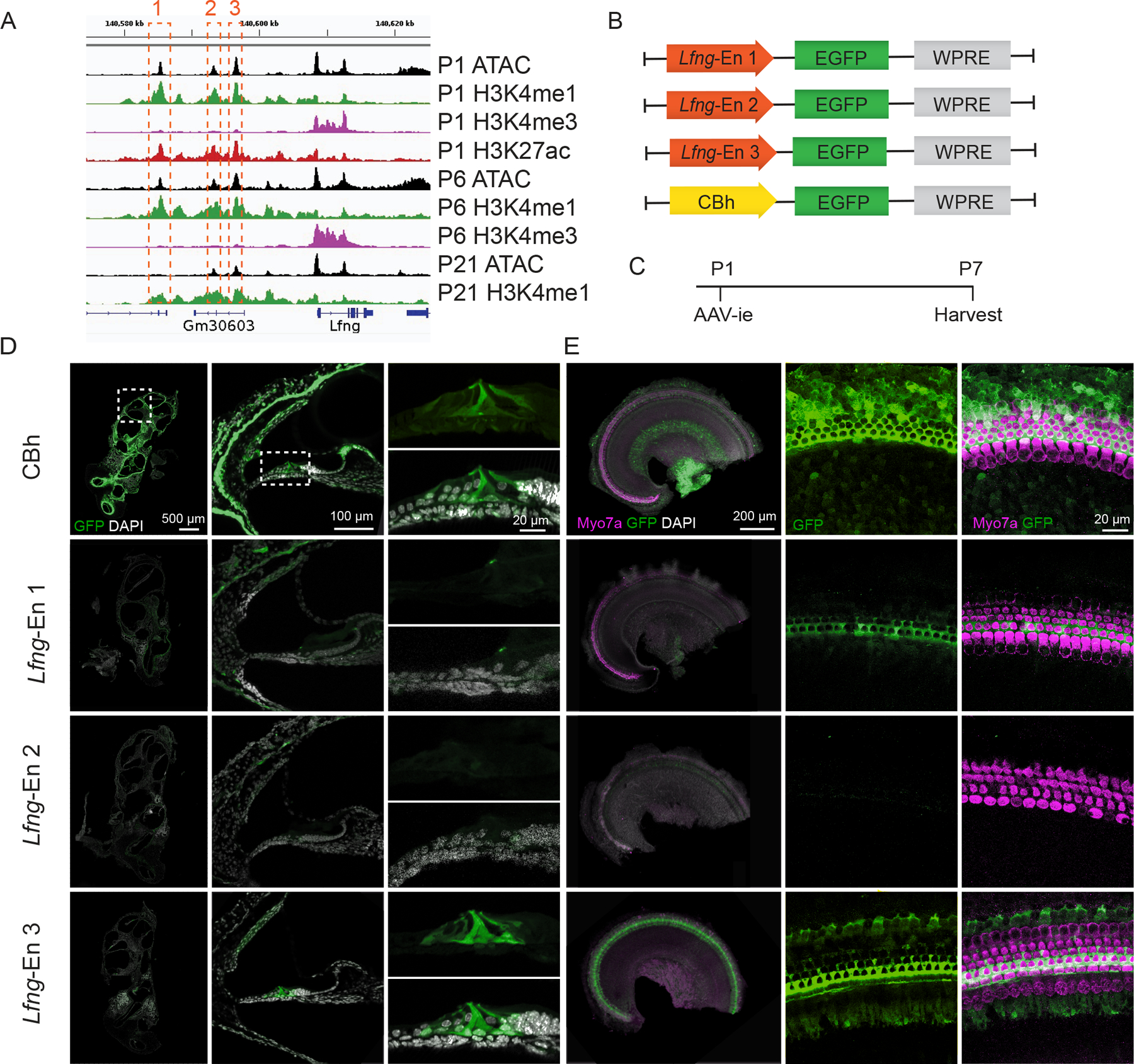
Achieving cell-specific gene expression is crucial in the design of safe and efficacious gene therapies for the treatment of sensorineural hearing loss. Although a variety of adeno-associated virus (AAV) serotypes have been used to deliver genes to inner ear hair cells, few serotypes currently allow specific targeting of supporting cells. We sought to specifically target supporting cells by combining an AAV serotype with high tropism for the inner ear with enhancer sequences from the supporting cell-specific gene Lunatic Fringe (Lfng). We identified three candidate Lfng enhancer sequences using bioinformatic analysis to identify accessible chromatin and histone marks associated with active transcription of the Lfng locus in supporting cells. Candidate Lfng enhancers or the ubiquitous CBh promoter driving an EGFP reporter gene were packaged into the AAV-ie capsid, and the virus was introduced into the inner ear of neonatal mice. AAV-CBh-EGFP transduced multiple sensory and non-sensory inner ear cell types, as well as cells in the brain. One of the three Lfng enhancers gave robust EGFP expression in border cells, inner phalangeal cells, pillar cells, and all three rows of Deiters' cells along the entire cochlear duct, as well as in vestibular organ supporting cells. Significantly, no fluorescently labeled cells were detected in the brains of mice injected with this virus. We further designed an AAV-Lfng-CreERT2 vector that drove strong recombination in Cre reporter mice supporting cells after tamoxifen treatment. Our results provide a tool to specifically target supporting cells of the juvenile and adult inner ear.

Keywords: Aav; Cre Recombinase; Enhancer; Gene therapy; Inner ear; Lunatic Fringe; Supporting cells.


Conflict of interest statement

Declaration of competing interest The authors disclose no competing interests.

Figures

Figure 1.
Figure 1.
(A) Chromatin accessibility (ATAC-seq), and histone modifications at the Lfng locus in cochlear supporting cells at P1, P6, and P21. Enhancer sequences 1–3 are marked by orange boxes upstream of Lfng. (B) Plasmid maps of AAV constructs. (C) Experimental timeline. Neonatal day P1 mice were injected with AAV, and their cochleae harvested on day P7. Gene expression was evaluated by immunofluorescence microscopy in inner ears prepared as cross sections (D) as well as whole mount preparations (E). White dashed boxes indicate magnified areas: mid-apical turn, and organ of Corti (D). Representative images of n = 3 ears. Green: EGFP, Magenta: Myo7a, White: DAPI.
Figure 2.
Figure 2.
(A) Experimental timeline. Cochlear whole mount preparations and sections of (C) AAV-Lfng-En3-EGFP or (D) AAV-CBh-EGFP injected animals on day P1 and harvested on day P21. AAV-Lfng-En3-EGFP selectively and robustly induces gene expression in SCs from apex to base. AAV-CBh-EGFP induces gene expression in most sensory and non-sensory inner ear cells. (B) Quantification of EGFP gene expression in Sox2+ SCs. Data are presented as mean ± SD. Representative images of n = 3 ears. Green: EGFP, Red: Myo7a, Magenta: Sox2.
Figure 3.
Figure 3.
(A) Experimental timeline. Transduction and gene expression in vestibular organs of P21 mice infected with (C) AAV-Lfng-En3-EGFP or (D) AAV-CBh-EGFP at P1. Asterisk marks a Myo7a+/Sox2+ type 2 hair cell exhibiting weak GFP signal. (B) Quantification of EGFP gene expression in Sox2+ SCs in the extrastriolar region. Data are presented as mean ± SD. Representative images of n = 3 ears. Green: EGFP, Magenta: Sox2, Red: Myo7a.
Figure 4.
Figure 4.
(A) Rosa26 locus of Cre reporter mouse line and AAV-Lfng-En3-CreER plasmid map. (B) Experimental timeline. Neonatal day P1 mice were injected with AAV-Lfng-En3-CreER. Animals were treated with tamoxifen (TM) on days P18 and P19 and collected on day P21. Inner ears were processed as whole mount preparations (C) and crosssections (D). Most IBC, IPhC, IPC, OPC, and DCs were fluorescently labeled along the cochlear duct indicating strong recombination after tamoxifen treatment. Representative images from n = 3 ears. Green: EGFP, Magenta: Myo7a (C) or Sox2 (D). Red: Myo7a (D), White: DAPI.
Figure 5.
Figure 5.
(A) Chromatin accessibility at the Lfng locus in cortical astrocytes and hippocampus neural stem cells (NSCs), central nervous system (CNS) cells known to express Lfng, as well as cochlear supporting cells. The three enhancer regions are marked with orange dashed rectangles. CNS transduction and gene expression of P21 mice infected with (B) AAV-CBh-EGFP, (C) AAV-Lfng-En3-EGFP, or (D) AAV-Lfng-En3-CreER at P1/2. AAV-CBh-EGFP infects and drives gene expression in cells of the cortex, hippocampus and cerebellum (B). We did not detect any fluorescently labeled cells in the CNS of AAV-Lfng-En3-EGFP (C) or AAV-Lfng-En3-CreER inoculated mice that were treated with tamoxifen (D). Representative images of n = 3 ears. Green: EGFP, Magenta: DAPI.

References

    1. Abascal Federico, Acosta Reyes, Addleman Nicholas J., Adrian Jessika, Afzal Veena, Aken Bronwen, Akiyama Jennifer A., et al. 2020. “Expanded Encyclopaedias of DNA Elements in the Human and Mouse Genomes.” Nature 583 (7818): 699–710. 10.1038/s41586-020-2493-4. - DOI - PMC - PubMed
    1. Akdemir Ekin Su, Woo Junsung, Bosquez Huerta Navish A., Lozzi Brittney, Groves Andrew K., Harmanci Akdes Serin, and Deneen Benjamin. 2022. “Lunatic Fringe-GFP Marks Lamina-Specific Astrocytes That Regulate Sensory Processing.” Journal of Neuroscience 42 (4): 567–80. 10.1523/JNEUROSCI.1392-21.2021. - DOI - PMC - PubMed
    1. Basch Martin L., Brown Rogers M., Jen Hsin I., Semerci Fatih, Depreux Frederic, Edlund Renée K., Zhang Hongyuan, et al. 2016. “Fine-Tuning of Notch Signaling Sets the Boundary of the Organ of Corti and Establishes Sensory Cell Fates.” eLife 5 (DECEMBER2016): 1–23. 10.7554/eLife.19921. - DOI - PMC - PubMed
    1. Bulcha Jote T., Wang Yi, Ma Hong, Tai Phillip W.L., and Gao Guangping. 2021. “Viral Vector Platforms within the Gene Therapy Landscape.” Signal Transduction and Targeted Therapy 6 (1). 10.1038/s41392-021-00487-6. - DOI - PMC - PubMed
    1. Cox Brandon C, Chai Renjie, Lenoir Anne, Liu Zhiyong, Zhang Lingli, Nguyen D-H, Chalasani Kavita, et al. 2014. “Spontaneous Hair Cell Regeneration in the Neonatal Mouse Cochlea in Vivo.” Development 141 (7): 1599–1599. 10.1242/dev.109421. - DOI - PMC - PubMed

MeSH terms

Substances

LinkOut - more resources