. 2025 Feb 14;12(2):ENEURO.0114-24.2024.
doi: 10.1523/ENEURO.0114-24.2024. Print 2025 Feb.

Temporal Lobectomy Evidence for the Role of the Amygdala in Early Emotional Face and Body Processing

Affiliations

Temporal Lobectomy Evidence for the Role of the Amygdala in Early Emotional Face and Body Processing

Eleanor Moses et al. eNeuro. .

Abstract

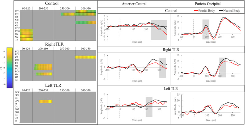
The amygdala is believed to make invaluable contributions to visual emotion processing. Yet how this subcortical body contributes to emotion perception across time is contended. Here, we measured differences in the perceptual processing of emotional stimuli after unilateral temporal lobe and amygdala resection (TLR) in humans, using EEG. Through mass univariate analysis of brain activity, we compared responses to fearful and neutral faces (left TLR N = 8, right TLR N = 8, control N = 8), and fearful and neutral bodies (left TLR N = 9, right TLR N = 9, control N = 9). We found that TLR impaired the early-stage perceptual processing of emotional stimuli seen in the control group. Indeed, in controls a heightened responses to fearful faces was found in the 140-170 ms time window, over temporoparietal electrodes. This effect was also present in the left TLR group but disappeared in the right TLR group. For emotional bodies, brain activity was differentially sensitive to fearful stimuli at 90-120 ms in the control group, but this effect was eliminated in both TLR groups. Collectively, these results reveal the amygdala contributes to the early stages of perceptual processing that discriminate emotional stimuli from neutral stimuli. Further, they emphasize the unique role of the right medial temporal structures such as the amygdala in emotional face perception.

Keywords: EEG; amygdala; bodily emotion; emotion; facial emotion; temporal lobe resection.


Conflict of interest statement

The authors declare no competing financial interests.

Figures

Figure 1.
Figure 1.
Results for the MUA facial emotion comparison. Left, Condensed raster plots for epochs and channels following the MUA cluster permutation analysis. Note all 64 electrodes were included in the analysis but only channels that were significantly different in any of the three groups have been shown in this representation. Orange areas indicate epochs and channels where a significant emotion based difference was found. Right, ERPs represent the pooling of clusters of electrodes that were found to be significantly different in any of the three groups for the facial expression comparison. These have been separated into temporoparietal (TP7, P5, P7, P9, PO7) and occipital (Oz, Iz) groupings. Shaded areas indicate where at least one of the pooled electrodes were found to be significantly different between the emotion condition for that group.
Figure 2.
Figure 2.
Results for the MUA bodily emotion comparison. Left, Condensed raster plots for epochs and channels following the MUA cluster permutation analysis. Note all 64 electrodes were included in the analysis but only channels that were significantly different in any of the three groups have been shown in this representation. Orange areas indicate epochs and channels where a significant emotion-based difference was found. Right, ERPs represent the pooling of clusters of electrodes that were found to be significantly different in any of the three groups for the bodily expression comparison. These have been separated into anterior-central (CP1, C5, FC1, FC3, F8) and parieto-occipital (PO8, PO7, O1, O2, Oz, Iz) groupings. Shaded areas indicate where at least one of the pooled electrodes were found to be significantly different between the emotion condition for that group.

References

    1. Amaral DG, Behniea H, Kelly JL (2003) Topographic organization of projections from the amygdala to the visual cortex in the macaque monkey. Neuroscience 118:1099–1120. 10.1016/S0306-4522(02)01001-1 - DOI - PubMed
    1. Anderson AK, Phelps EA (2001) Lesions of the human amygdala impair enhanced perception of emotionally salient events. Nature 411:305–309. 10.1038/35077083 - DOI - PubMed
    1. Armony JL, Quirk GJ, LeDoux JE (1998) Differential effects of amygdala lesions on early and late plastic components of auditory cortex spike trains during fear conditioning. J Neurosci 18:2592–2601. 10.1523/jneurosci.18-07-02592.1998 - DOI - PMC - PubMed
    1. Batty M, Taylor MJ (2003) Early processing of the six basic facial emotional expressions. Brain Res Cogn Brain Res 17:613–620. 10.1016/S0926-6410(03)00174-5 - DOI - PubMed
    1. Bertini C, Cecere R, Ladavas E (2019) Unseen fearful faces facilitate visual discrimination in the intact field. Neuropsychologia 128:58–64. 10.1016/j.neuropsychologia.2017.07.029 - DOI - PubMed

LinkOut - more resources