. 2025 Jan 31;16(1):1217.
doi: 10.1038/s41467-025-55969-w.

Lymphatic-derived oxysterols promote anti-tumor immunity and response to immunotherapy in melanoma

Affiliations

Lymphatic-derived oxysterols promote anti-tumor immunity and response to immunotherapy in melanoma

Mengzhu Sun et al. Nat Commun. .

Abstract

In melanoma, lymphangiogenesis correlates with metastasis and poor prognosis and promotes immunosuppression. However, it also potentiates immunotherapy by supporting immune cell trafficking. We show in a lymphangiogenic murine melanoma that lymphatic endothelial cells (LECs) upregulate the enzyme Ch25h, which catalyzes the formation of 25-hydroxycholesterol (25-HC) from cholesterol and plays important roles in lipid metabolism, gene regulation, and immune activation. We identify a role for LECs as a source of extracellular 25-HC in tumors inhibiting PPAR-γ in intra-tumoral macrophages and monocytes, preventing their immunosuppressive function and instead promoting their conversion into proinflammatory myeloid cells that support effector T cell functions. In human melanoma, LECs also upregulate Ch25h, and its expression correlates with the lymphatic vessel signature, infiltration of pro-inflammatory macrophages, better patient survival, and better response to immunotherapy. We identify here in mechanistic detail an important LEC function that supports anti-tumor immunity, which can be therapeutically exploited in combination with immunotherapy.


Conflict of interest statement

Competing interests: The authors declare no competing interests.

Figures
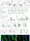
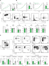
Fig. 1
Fig. 1. LECs express Ch25h and control extracellular 25-hydroxycholesterol (25-HC) levels in lymphangiogenic B16F10-OVA tumors.
A B16F10-OVA VEGF-C tumors were inoculated in C57BL/6 mice. LECs were sorted by flow cytometry (CD45negCD31+GP38+) from tumors, tumor-draining LNs (TdLN) and non-draining LNs (NdLN) after 11 days. Ch25h mRNA levels (RPKM) provided by RNA sequencing. n = 5 mice/group. Data were presented as mean values ± SD. B Correlation between Ch25h expression and lymphatic vessel (LV) signature (LV signature 1: pdpn, vegfc, lyve1, and LV signature 2: prox1, flt4, lyve1, pdpn, vegfc) in SKCM (Skin cutaneous melanoma) patients (TPM transcript per million). SKCM patient overall survival (OS) of high and low Ch25h expression. Data extracted from TCGA. C Predictive activity (OS) of Ch25h high and Ch25h low-expressors SKCM patients treated with anti-PD-1 and anti-CTLA-4, from an integrated dataset of multiple clinical trial studies. Significance was determined by log-rank analysis. DF B16F10-OVA VEGF-C tumor cells were injected into Ch25h-GFP and WT mice. D, E Ch25h expression by tumor infiltrated cells on day 14. (DC dendritic cells, CAF cancer-associated fibroblasts, BEC blood endothelial cells). Results are representative of two independent experiments, with n = 4 mice/ group. F Ch25h expression by LECs in indicated organs at different time points. Results are pooled from two independent experiments, with n = 2–3 mice/group each. Two-way ANOVA, ****P < 0.0001. G, H B16F10-OVA VEGF-C tumor cells were injected in LECΔCh25h and LECWT mice. G Ch25h expression by LECs and BECs in tumors on day 11. Results are representative of two independent experiments, with n = 5 mice/group. DG Ch25h expression is represented as MFI Ch25h-GFP – MFI-WT mice. H 25-HC levels in tumor interstitial fluid measured by liquid chromatography–mass spectrometry. Results are pooled from two experiments, with n = 4–13 mice /group. G, H Two-tailed unpaired t-test. *P < 0.05; ****P < 0.0001. I VEGF-C expression in human melanoma cell lines by Q-PCR. Histograms depict technical triplicates from one experiment. EI Data were presented as mean values ± SD. J, K Human melanoma VEGF-Chigh T362C and VEGF-Clow T618A cells were injected in NSG (WT), NSG-LECΔCh25h, and NSG-LECWT mice. Ch25h expression (MFI Ch25h-GFP-MFI-WT mice) by LECs from tumors and skin was assessed by flow cytometry. Data were presented as mean values ± SD. Results are representative of two independent experiments. L Ch25h mRNA and PDPN staining on human melanoma sections (n = 3 patients). Representative images are shown for patients 1 (Pt1) and 2 (Pt2). Scale bar, 20 and 5 µm (zoomed).
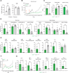
Fig. 2
Fig. 2. Loss of Ch25h expression by tumor LECs enhances tumor growth by dampening anti-tumor immunity.
AD B16F10-OVA VEGF-C cells were injected in LECΔCh25h and LECWT mice (A, C, D), in NSG-LECΔCh25h and NSG-LECWT mice, or in Rag2KO-LECΔCh25h and Rag2KO-LECWT mice (B). A, B Tumor growth was followed and normalized to the size of tumors in LECWT, NSG-LECWT, and Rag2KO-LECWT mice, respectively, at day 6 for tumor growth, two-way ANOVA, *P < 0.05. Data were presented as mean values ± SEM. Tumor cell proliferation (Ki67+) was evaluated by flow cytometry on day 11. Data were presented as mean values ± SD. Results are pooled from two independent experiments (n = 4–8 mice per group each) and normalized to WT (A), and Ch25h expression (MFI Ch25h-GFP – MFI-WT mice) was measured by flow cytometry at day 12 (B). C, D Tumors were harvested on day 11. C CD4+ and CD8+ T cells were analyzed for their expression of indicated markers. Representative flow cytometry dot plots and histograms provide the frequency of positive cells among T cell subsets. Data are presented as mean values ± SD. Results are pooled from two independent experiments (n = 4–8 mice/ group each) and normalized to WT. D Myeloid cells (CD11b+CD68+) were separated into monocytes (Ly6C+F4/80-) and macrophages (Ly6C-F4/80+) and were analyzed for their expression of indicated markers. Representative flow cytometry dot plots provide the frequency of positive cells among indicated cells. Data were presented as mean values ± SD. Results are representative of two independent experiments, n = 5–7 mice/group each. AD For FACS analysis, two-tailed unpaired t-test. *P < 0.05; **P < 0.01; ***P < 0.0001.
Fig. 3
Fig. 3. Loss of Ch25h in LECs dampens immunotherapy efficacy in lymphangiogenic melanoma.
AE LECΔCh25h and LECWT mice were inoculated with B16F10-OVA VEGF-C (B16-OVA-VC) cells and vaccinated with OVA protein and CpG-B at day 5 (A) or at day 4 and day 8 (BE). A Ch25h expression (GFP MFI) was measured by flow cytometry on day 11. n = 5 mice/ group. B Tumor growth was followed and compared to unvaccinated B16F10-OVA VEGF-C tumor-bearing WT mice. Representative of four independent experiments, n = 5–6 mice/group each. CE Tumor were harvested on day 24 and analyzed by flow cytometry. C CD45+/tumor cell ratio and tumor cell proliferation (Ki67+) and CD45+ cell frequency in living cells. D Myeloid cells (CD11b+CD68+) were separated into monocytes (Ly6C+F4/80-) and macrophages (Ly6C-F4/80+) and were analyzed for their expression of indicated markers. E CD4+ and CD8+ T cells were analyzed for their expression of indicated markers. CE Representative of two independent experiments, n = 5–8 mice/group each. Data were presented as mean values ± SD (F, G) LECΔCh25h and LECWT mice were inoculated with B16F10-OVA VEGF-C cells and adoptively transferred 8 days later with OT-1 effector T cells. Tumor growth was followed and normalized to the size of WT at day 6 (F), and OT-1 cells were analyzed in tumors 2 days after transfer for indicated markers (G). Data were pooled from two independent experiments, n = 2–5 mice/group each, and are presented as mean values ± SEM (F) or as mean values ± SD (G). B, F Two-way ANOVA, *P < 0. 05. (A, C, D, E, G) Two-tailed unpaired t-test. *P < 0.05; **P < 0.01; ***P < 0.0001.
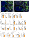
Fig. 4
Fig. 4. 25-HC treatment reverses tumor-associated macrophage immunosuppressive phenotype and functions in vitro.
A LECΔCh25h and LECWT mice were inoculated with B16F10-OVA VEGF-C cells and tumors were harvested at day 11. Montages of maximum projected 3D confocal images of representative sections (n = 3 mice/group) immunostained for lymphatic vessels (Lyve-1, green), T cells (CD8, red), myeloid cells (CD68, white), and nuclei (blue). Images were obtained using a 10x objective, including a 3x relative magnification. Selected regions of interest are indicated by dashed squares and denoted magnified areas are shown in images beside. Scale bars, 1 mm, 100 μm (zoomed). B BMDMs were incubated with B16F10-OVA VEGF-C tumor-conditioned medium (TCM) and treated or not with 25-HC for 48 h. BMDM phenotype was analyzed by flow cytometry. Data were representative of three independent experiments with n = 3–6 replicates each. Data were presented as mean values ± SD. C BMDM were treated or not with IFN-γ or IL-4 and further treated or not with 25-HC for 48 h. BMDM phenotype was analyzed by flow cytometry. Data were representative of two independent experiments with n = 4–8 replicates each. Data were presented as mean values ± SD. D TCM-exposed BMDM were treated or not with 25-HC for 48 h, washed, and incubated with OT-1 effector cells (5:1 ratio) for 48 h. T-cell phenotype was analyzed by flow cytometry. Data were representative of two independent experiments with n = 3–5 replicates each. Data were presented as mean values ± SD. B Two-tailed unpaired t-test. C Two-way ANOVA, D One-way ANOVA. *P < 0.05; **P < 0.01; ***P < 0.001; ****P < 0.0001.
Fig. 5
Fig. 5. LEC-derived 25-HC modulates tumor growth by impacting tumor-associated macrophages and monocytes.
A LECΔCh25h and LECWT mice were inoculated with B16F10-OVA VEGF-C cells and vaccinated with OVA protein and CpG-B at day 4 and day 8. Mice were additionally injected with anti-CSF1R (1 mg / mouse at day 10, followed by 500 µg/mouse every 3 days or PBS), as represented in the scheme. Tumor growth was followed. Two-way ANOVA, **P < 0.01. Data were representative of two independent experiments with n = 5–7 mice/group each. B NSG-LECΔCh25h and NSG-LECWT mice were inoculated with B16F10-OVA VEGF-C cells mixed or not with BMDM. Tumor-bearing mice were further adoptively transferred with OT-1 effectors on day 8, as represented in the scheme. Tumor growth was followed and normalized to the size individually from day 6. Two-way ANOVA, **P < 0.01. Data were pooled from two independent experiments with n = 5–8 mice/group each. A, B Data were presented as mean values ± SEM.
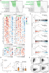
Fig. 6
Fig. 6. Altered monocyte and macrophage transcriptomes in the absence of Ch25h expression by LECs in lymphangiogenic melanoma.
AC LECΔCh25h and LECWT mice were inoculated with B16F10-OVA VEGF-C cells and vaccinated with OVA protein and CpG-B on day 4 and day 8. Tumors were harvested on day 24, and monocytes and macrophages were sorted by flow cytometry (see Fig. 2D for gating strategy). RNA sequencing was performed (Illumina). A Top 10 Hallmark pathways expressed in monocytes and macrophages in tumors from LECΔCh25h and LECWT mice. B Heatmap showing expression levels of genes implicated in “anti-tumor” or “pro-tumor” immune signatures in monocytes and macrophages and in tumors from LECΔCh25h and LECWT mice. C GSEA enrichment of M1 and M2 signatures in monocytes and macrophages from tumor in LECΔCh25h and LECWT mice using gene set from ref. . D B16F10-OVA VEGF-C tumor-bearing C57BL/6 mice were treated with 25-HC every 2 days, starting at day 4. Tumor growth was followed. Two-way ANOVA, *P < 0.05. Data were a pool of three independent experiments with n = 4–6 mice/group each. On day 11, tumors were harvested, and the CD68+CD11b+ myeloid cell (macrophage, F4/80+Ly6C- and monocytes, F4/80-Ly6C+) phenotype was assessed by flow cytometry. Ratio iNOS+/CD206+ cells are provided. Two-tailed unpaired t-test, ***p < 0.001. E Correlation between Ch25h expression and M1/M2 signatures in normal skin and in SKCM patients. Data were extracted from the TCGA database.
Fig. 7
Fig. 7. LEC-derived 25-HC dampens the immunosuppressive functions of tumor-associated macrophages by inhibiting PPAR-γ expression.
A BMDM were incubated with B16F10-OVA VEGF-C tumor-conditioned medium (TCM), treated or not with 25-HC for 48 h, and analyzed by Q-PCR for PPAR-γ mRNA levels. Data were representative of two experiments with n = 2–3 replicates each. Data were presented as mean values ± SD. Two-tailed unpaired t-test. *P < 0.05. B BMDMs were incubated with tumor-conditioned medium (TCM), treated or not with 25-HC, Troglitazone, or the combination of 25-HC and Troglitazone for 48 h. BMDM phenotype was analyzed by flow cytometry. Data were representative of two independent experiments with n = 3 replicates each. Data were presented as mean values ± SD. One-way ANOVA. ***P < 0.001; ****P < 0.0001. C TCM-exposed BMDM were treated or not with 25-HC, Troglitazone, or the combination of 25-HC and Troglitazone for 48 h, washed, and incubated with OT-1 effector cells for 48 h. T-cell phenotype was analyzed by flow cytometry. Data were representative of two independent experiments with n = 6 replicates each. Data were presented as mean values ± SD. One-way ANOVA. *P < 0.05; ***P < 0.001; ***P < 0.0001. D LECΔCh25h and LECWT mice were inoculated with B16F10-OVA VEGF-C cells mixed with either WT or PPAR-γ deficient (PPAR-γKO) BMDM and vaccinated with OVA protein and CpG-B on day 4 and day 8. Tumor growth was followed. Data are presented as mean values ± SEM. Two-way ANOVA, **P < 0.01. Data were representative of two independent experiments with n = 4–5 mice/group each.

Similar articles

Cited by

References

    1. Jones, D., Pereira, E. R. & Padera, T. P. Growth and immune evasion of lymph node metastasis. Front. Oncol.8, 36 (2018). - PMC - PubMed
    1. Stacker, S. A. et al. Lymphangiogenesis and lymphatic vessel remodelling in cancer. Nat. Rev. Cancer14, 159–172 (2014). - PubMed
    1. Hirakawa, S. et al. VEGF-A induces tumor and sentinel lymph node lymphangiogenesis and promotes lymphatic metastasis. J. Exp. Med.201, 1089–1099 (2005). - PMC - PubMed
    1. Qian, C. N. et al. Preparing the “soil”: the primary tumor induces vasculature reorganization in the sentinel lymph node before the arrival of metastatic cancer cells. Cancer Res.66, 10365–10376 (2006). - PubMed
    1. Garnier, L., Gkountidi, A. O. & Hugues, S. Tumor-associated lymphatic vessel features and immunomodulatory functions. Front. Immunol.10, 720 (2019). - PMC - PubMed

MeSH terms

LinkOut - more resources