. 2025 Jan 31;16(1):1235.
doi: 10.1038/s41467-025-56626-y.

Glucomannan engineering highlights roles of galactosyl modification in fine-tuning cellulose-glucomannan interaction in Arabidopsis cell walls

Affiliations

Glucomannan engineering highlights roles of galactosyl modification in fine-tuning cellulose-glucomannan interaction in Arabidopsis cell walls

Yoshihisa Yoshimi et al. Nat Commun. .

Abstract

Widely found in most plant lineages, β-mannans are structurally diverse polysaccharides that can bind to cellulose fibrils to form the complex polysaccharide architecture of the cell wall. How changes in polysaccharide structure influence its cell wall solubility or promote appropriate interaction with cellulose fibrils is poorly understood. Glucomannan backbones acquire variable patterns of galactosyl substitutions, depending on plant developmental stage and species. Here, we show that fine-tuning of galactosyl modification on glucomannans is achieved by the differing acceptor recognition of mannan α-galactosyltransferases (MAGTs). Biochemical analysis and 13C solid-state nuclear magnetic resonance spectroscopy of Arabidopsis with cell wall glucomannan engineered by MAGTs reveal that the degree of galactosylation strongly affects the interaction with cellulose. The findings indicate that plants tailor galactosyl modification on glucomannans for constructing an appropriate cell wall architecture, paving the way to convert properties of lignocellulosic biomass for better use.


Conflict of interest statement

Competing interests: The authors declare no competing interests.

Figures

Fig. 1
Fig. 1. MAGTs from different plant species display diverse acceptor recognition.
a Model structure of β-mannans modified with Gal side chains, which are found in different plant species. A single-letter nomenclature for the residues in the backbone and possible side chains are indicated. b An MAGT clade of GT34 phylogenetic tree. Amino acid sequences of MAGTs in different plant species were determined from the GT34 phylogenetic tree in Supplementary Fig. 1. AtXXT1 was selected as an outgroup. Highlighted MAGTs were characterised in the present study. Plant species were as followed: Pab, Picea abies; Pme, Pseudotsuga menziesii; Pta, Pinus taeda; Amt, Amborella trichopoda; At, Arabidopsis thaliana; Br, Brassica rapa; Ct, Cyamopsis tetragonoloba; Gm, Glycine max; Md, Malus domestica; Ptr, Populus trichocarpa; Solyc, Solanum lycopersicum; Bd, Brachypodium distachyon; Hv, Hordeum vulgare; Os, Oryza sativa; Sb, Sorghum bicolor; Spipo, Spirodela polyrhiza; Zosma, Zostera marina. Scale bar represents 0.5 substitutions per site. In vitro activity of MAGTs towards homomannan (c), glucomannan (d), and patterned glucomannan (e). Two independent attempts yielded comparable results. The presence of Gal on the products was confirmed by α-galactosidase (α-Galase) treatment. S, standard of Man and mannooligosaccharides with D.P. 2-6. Structures of oligosaccharides in (d) were determined individually by further analysis shown in Supplementary Fig. 3. Source data are provided as a Source Data file.
Fig. 2
Fig. 2. In vivo activity of MAGTs towards patterned glucomannan.
a Complementation of seed mucilage capsule of magt1 mutant by four MAGTs. Bar = 100 µm. b Area of mucilage capsules. Open circles indicate individual measurements; the white lines represent the median of the group. One-way ANOVA (two-tailed) indicated a significant effect of genotype on mucilage area (p = 1.87 × 10-94; F13,335 = 80.24). Results of post hoc multiple comparisons (Dunnet’s method compared with wild-type; wild-type, n = 26; magt1, n = 25; AtMAGT1#1, n = 24; AtMAGT1#2, n = 25; AtMAGT1#3, n = 29; AtMAGT2#2, n = 25; AtMAGT2#7, n = 25; AtMAGT2#16, n = 25; PtMAGT#1, n = 27; PtMAGT#3, n = 27; PtMAGT#5, n = 24; CtMAGT#5, n = 20; CtMAG#6, n = 26; CtMAGT#8, n = 21) are indicated by asterisks (*p < 0.05; **p < 0.01) with p values. c CjMan26A-digestion profile of glucomannan in seed mucilage of the complementation lines. Note that GMGM and GM were produced from PtMAGT seed mucilage, indicating that no galactosylation occurred. No product was detected in AtMAGT2 despite the recovery of the mucilage capsule. S, standards of Man and mannooligosaccharides with D.P. 2-6. d AnGH5-digestion profile of seed mucilage of complementation lines. AtMAGT2 produced almost only GA, indicating galactosylation occurred at almost all mannosyl residues. Two independent attempts yielded comparable results. Data from two independent biological replicates were provided in Supplementary Fig. 6. e Proportion of GA and GM determined from band intensity. Source data are provided as a Source Data file.
Fig. 3
Fig. 3. Engineering of AcGGM in Arabidopsis secondary cell walls by MAGTs.
a Gal/Man ratio of AcGGM in pIRX3::MAGT lines determined from the ratio of AnGH5-digested products. Data are mean values and standard deviation of three biological replicates. One-way ANOVA (two-tailed) indicated a significant effect of genotype on mucilage area (p = 1.21 × 10⁻⁵; F8,18 = 11.41). Results of post hoc multiple comparisons (Dunnet’s method compared with wild-type) are indicated by asterisks (*p < 0.05; **p < 0.01) with p values. Digestion profile and the measured proportion of products were shown in Supplementary Fig. 7. b Galactosylated oligosaccharide released from AcGGM in pIRX3::MAGTs expressing Arabidopsis. Unique bands found in pIRX3::MAGTs lines (I-IV) were further analysed (Supplementary Fig. 8). c Summary of galactosylated oligosaccharides found in the regions I-IV. Dotted squares indicate oligosaccharides with clustered galactosylation. Source data are provided as a Source Data file.
Fig. 4
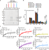
Fig. 4. Galactosylation changes the interaction of AcGGM with cellulose.
a AnGH5-digestion profile of hot water extract from bottom stem AIR of pIRX3::MAGT lines. The presence of products indicates the high extractability of AcGGM. S, standards of Man and mannooligosaccharides with D.P. 2-6. b Monosaccharides composition of the hot water extract. Data are mean values and standard deviation of three biological replicates. The significant difference was determined by one-way ANOVA on each monosaccharide, followed by Dunnets multiple comparisons where pRIX3::MAGT lines were compared with wild-type (*p < 0.05; **p < 0.01). F values and P values were provided in Supplementary Data 6. c In vitro cellulose-binding isotherms. KOH extracts of pRIX3::MAGT lines of a range of concentrations were incubated with and without Avicel. The amount of Man in the supernatant was determined by HPAEC-PAD, from which the bound Man was calculated. Data from three biological replicates were plotted in the graphs. Each point indicates a mean value with SEM of three technical replicates. Non-linear regression was used to fit the curves. The area with the shaded colour indicates the 95% confidence interval. Bmax and Kd were calculated from the fitted curves and shown in the graphs with SEM. Source data are provided as a Source Data file.
Fig. 5
Fig. 5. Altered molecular behaviour of highly galactosylated AcGGM in secondary cell walls analysed by solid-state NMR.
a Overlay of 13C CP- and DP-refocused INADEQUATE MAS ssNMR spectra of pIRX3::AtMAGT2. AcGGM peaks are labelled: Man (M), substituted Man (sM), α-Gal (Gal). Cellulose (domain 1, 1C; domain 2, 2C), xylose of xyloglucan (XgX), arabinose (A), and pectic galactan (G) are also labelled. Most AcGGM peaks were found in the DP spectrum, indicating its high mobility in the cell walls. b Comparison of C1, C2, and C5/6 regions of the DP spectra between wild-type and pIRX3::AtMAGT2. The spectra are normalised by C1 peaks at 105 ppm. Much less intensity of AcGGM peaks in the DP spectra of wild-type compared to pIRX3::AtMAGT2. Chemical shifts of the annotated peaks are listed in Supplementary Data 7. Spectra were acquired at a 13C Larmor frequency of 213.8 MHz and a MAS frequency of 12.5 kHz. The spin-echo duration used was 2.24 ms.
Fig. 6
Fig. 6. Summary of substrate specificity of MAGT and effect of galactosylation on AcGGM-cellulose interaction.
a MAGTs characterised here showed different acceptor recognition. Circles indicate the substrates that MAGTs can recognise, while Crosses are substrates that MAGTs cannot act on. 1) AtMAGT1/2 and PtMAGT are specific to glucomannan while CtMAGT has a promiscuous activity towards any (gluco)mannan. 2) PtMAGT prefers Glc residues on the acceptor at subsite 3, and shows no activity on the patterned glucomannan. 3) AtMAGT2 has the additional pocket for Gal adducts, which allows it to transfer Gal near Gal branches on the acceptor. b Low galactosylation does not affect the interaction of AcGGM with cellulose in muro whereas high galactosylation ends up incomplete binding to cellulose presumably due to the loss of proper conformation to bind to cellulose and a steric hindrance of the high number of branches.

References

    1. Bar-On, Y. M., Phillips, R. & Milo, R. The biomass distribution on Earth. Proc. Natl Acad. Sci.115, 6506–6511 (2018). - PMC - PubMed
    1. Scheller, H. V. & Ulvskov, P. Hemicelluloses. Annu Rev. Plant Biol.61, 263–289 (2010). - PubMed
    1. Cosgrove, D. J. Building an extensible cell wall. Plant Physiol.189, 1246–1277 (2022). - PMC - PubMed
    1. Voiniciuc, C. Modern mannan: a hemicellulose’s journey. New Phytol.234, 1175–1184 (2022). - PubMed
    1. Berglund, J. et al. Wood hemicelluloses exert distinct biomechanical contributions to cellulose fibrillar networks. Nat Commun11, 4692 (2020). - PMC - PubMed

MeSH terms

LinkOut - more resources