. 2025 Jan 31;8(1):150.
doi: 10.1038/s42003-025-07590-2.

Selective activation of SIGMAR1 in anterior cingulate cortex glutamatergic neurons facilitates comorbid pain in depression in male mice

Affiliations

Selective activation of SIGMAR1 in anterior cingulate cortex glutamatergic neurons facilitates comorbid pain in depression in male mice

Xianlei Wang et al. Commun Biol. .

Abstract

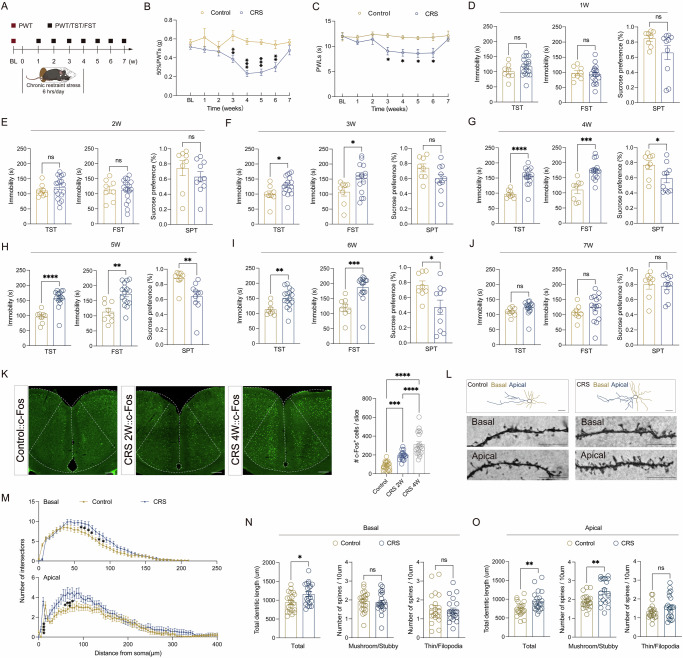
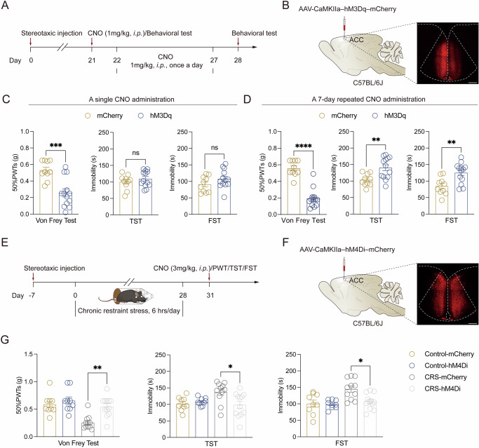
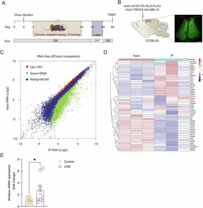
Depression and comorbid pain are frequently encountered clinically, and the comorbidity complicates the overall medical management. However, the mechanism whereby depression triggers development of pain needs to be further elucidated. Here, by using the chronic restraint stress (CRS) mouse model of depression and comorbid pain, we showed that CRS hyperactivated the glutamatergic neurons in the anterior cingulate cortex (ACC), as well as increasing the dendrite complexity and number. Chemogenetic activation of these neurons can induce depression and pain, while chemogenetic blockade can reverse such depression-induced pain. Moreover, we utilized translating ribosome affinity purification (TRAP) in combination with c-Fos-tTA strategy and pharmacological approaches and identified SIGMAR1 as a potential therapeutic molecular target. These results revealed a previously unknown neural mechanism for depression and pain comorbidity and provided new mechanistic insights into the antidepressive and analgesic effects of the disease.


Conflict of interest statement

Competing interests: The authors declare no competing interests.

Figures

Fig. 1
Fig. 1. ACC neurons are activated in CRS mice with depression-pain comorbidity.
A Experimental procedure for mice with CRS treatment and behavioral tests. B 50% PWTs at different time points after CRS (Control, n = 8 mice; CRS, n = 16–20 mice). C PWLs at different time points after CRS (Control, n = 8 mice; CRS, n = 8 mice). D–J Behavioral tests of TST, FST and SPT in Control and CRS mice at 1 W (D), 2 W (E), 3 W (F), 4 W (G), 5 W (H), 6 W (I), and 7 W (J) (Control, n = 8 mice; CRS, n = 10–18 mice). K Representative confocal images (left) of c-Fos protein expression in ACC of Control, CRS 2 W and CRS 4 W mice and the quantification (right) of c-Fos positive neurons in ACC (n = 20 slices, 4–5 mice/group). Scale bar, 200 μm. L Representative Golgi-staining images of dendritic spine morphology of the ACC in Control and CRS 4 W mice (Scale bar, top: 50 μm; middle: 10 μm; bottom:10 μm). M Sholl analysis of dendritic branching complexity in the basal and apical dendrites of Control and CRS 4 W mice (n = 18–22, 4 mice/group). N, O Summary of the total dendritic length (left), the number of mushroom/stubby type dendritic spines (middle) and the number of thin/filopodia type dendritic spines (right) on basal (N) and apical (O) dendrites of ACC pyramidal neurons in Control and CRS 4 W mice (n = 18–22, 4 mice/group). Data were analyzed by one-way (K) or two-way ANOVA with post hoc Bonferroni’ s multiple comparisons test (B, C, M) or two-tailed unpaired Student’s t-test (DJ, NO). All data are presented as the mean ± s.e.m. *P < 0.05, **P < 0.01, ***P < 0.001, ****P < 0.0001, ns not significant. CRS chronic restraint stress, ACC anterior cingulate cortex, BL baseline, W week, PWL paw withdrawal latency, PWT paw withdrawal threshold, TST tail suspension test, FST forced swimming test, SPT sucrose preference test.
Fig. 2
Fig. 2. Chemogenetic modulation of ACC glutamatergic neurons bidirectionally regulate depression-like and pain behaviors in mice.
A Experimental procedure for chemogenetic activation of naive C57BL/6 J mice. B Schematic illustration for virus injection and representative confocal images of AAV-CaMKIIa–hM3Dq–mCherry expression in the ACC. Scale bar, 200 µm. C 50% PWTs (left), TST immobility time (middle) and FST immobility time (right) 30 min after a single CNO administration (Control, n = 10 mice; hM3Dq, n = 13 mice). D 50% PWTs (left), TST immobility time (middle) and FST immobility time (right) 30 min after a 7-day repeated CNO treatment (Control, n = 10 mice; hM3Dq, n = 13 mice). E Experimental procedure for chemogenetic inhibition of CRS mice. F Schematic illustration for virus injection and representative confocal images of AAV-CaMKIIa–hM4Di–mCherry expression in the ACC. Scale bar, 200 µm. G 50% PWTs (left), TST immobility time (middle) and FST immobility time (right) upon chemogenetic inhibition of ACC glutamatergic neurons in CRS mice with depression-pain comorbidity. Behavioral tests were performed 30 min after CNO administration (Control-mCherry, n = 9 mice; Control-hM4Di, n = 9 mice; CRS-mCherry, n = 11 mice; CRS-hM4Di, n = 12 mice). Data were analyzed by two-tailed unpaired Student’s t-test (C, D) or two-way ANOVA with post hoc Bonferroni’ s multiple comparisons test (G). All data are presented as the mean ± s.e.m. *P < 0.05, **P < 0.01, ***P < 0.001, ****P < 0.0001, ns not significant. CRS chronic restraint stress, ACC anterior cingulate cortex, PWT paw withdrawal threshold, TST tail suspension test, FST forced swimming test. CNO clozapine n-oxide.
Fig. 3
Fig. 3. Identification of Sigmar1 in depression-pain comorbidity-related ACC engram neurons as a potential receptor target for regulating the comorbidity.
A Experimental procedure. B Schematic illustration for virus injection and a representative confocal image for virus expression. Scale bar, 200 µm. C RNA-seq scatterplots for depression-pain comorbidity-related neurons in the ACC with normalized expression. Significantly different genes (q < 0.05) are displayed in red (enriched) and green (reduced), and all other genes are displayed in grey. Black lines represent unity and 1.5-fold change in enrichment. D Relative expression levels are shown for upregulated genes in depression and pain comorbidity-related neurons as compared between the IP and Input (Input, n = 12 mice/repeat, 3 repeats in total; IP, n = 12 mice/repeat, 3 repeats in total). E Fold change of mRNA expression of Sigmar1 in the ACC (Control, n = 9 mice; CRS, n = 9 mice). Data were analyzed by two-tailed unpaired Student’s t-test. All data are presented as the mean ± s.e.m. *P < 0.05. CRS chronic restraint stress, ACC anterior cingulate cortex, TRAP translating ribosome affinity purification, IP immunoprecipitation.
Fig. 4
Fig. 4. Effects of intra-ACC infusion of Sigmar receptor 1 agonist (SA4503) on depression-like and pain behaviors.
A Experimental procedure. B Schematic illustration for cannula implantation and a representative confocal image showing cannula implantation site in naive mice. Scale bar, 1 mm. C 50% PWTs (left), TST immobility time (middle) and FST immobility time (right) of naive mice following a single dose of bilateral intra-ACC infusion of Sigma receptor 1 agonist SA4503 (Vehicle, n = 9 mice; SA4503, n = 9 mice). D 50% PWTs (left), TST immobility time (middle) and FST immobility time (right) of naive mice following a 7-day repeated of bilateral intra-ACC infusion of Sigma receptor 1 agonist SA4503 (Vehicle, n = 9 mice; SA4503, n = 8–9 mice). E Representative Golgi-staining images of dendritic spine morphology in naive mice subjected to local infusion of vehicle/SA4503 in the ACC (Scale bar, top: 50 μm; middle: 10 μm; bottom:10 μm). F Sholl analysis of dendritic branching complexity in the basal and apical dendrites of naive mice subjected to local infusion of vehicle/SA4503 (n = 18–22, 4 mice/group). G, H Summary of the total dendritic length (left), the number of mushroom/stubby type dendritic spines (middle), and the number of thin/filopodia type dendritic spines (right) on basal (G) and apical (H) dendrites of ACC pyramidal neurons in naive mice subjected to local infusion of vehicle/SA4503 (n = 18–22, 4 mice/group). Data were analyzed by two-tailed unpaired Student’s t-test (C, D, G, H) or two-way ANOVA with post hoc Bonferroni’ s multiple comparisons test (F). All data are presented as the mean ± s.e.m. *P < 0.05, **P < 0.01, ***P < 0.001, ns not significant, ACC anterior cingulate cortex, PWT paw withdrawal threshold, TST tail suspension test, FST forced swimming test.
Fig. 5
Fig. 5. Effects of intra-ACC infusion of Sigmar receptor 1 antagonist (NE-100) on depression-like and pain behaviors.
A Experimental procedure. B Schematic illustration for cannula implantation and a representative confocal image showing cannula implantation site. Scale bar, 1 mm. C 50% PWTs (left), TST immobility time (middle) and FST immobility time (right) of mice with depression-pain comorbidity following a single dose of bilateral intra-ACC infusion of Sigma receptor 1 antagonist NE-100 (Control-Vehicle, n = 7 mice; Control-NE-100, n = 7 mice; CRS-Vehicle, n = 7 mice; CRS-NE-100, n = 7 mice). D 50% PWTs (left), TST immobility time (middle) and FST immobility time (right) of mice with depression-pain comorbidity following a 7-day repeated of bilateral intra-ACC infusion of Sigma receptor 1 antagonist NE-100 (Control-Vehicle, n = 7 mice; Control-NE-100, n = 7 mice; CRS-Vehicle, n = 7 mice; CRS-NE-100, n = 7 mice). E Representative Golgi-staining images of dendritic spine morphology in Control- and CRS-treated mice subjected to local infusion of vehicle/NE-100 in the ACC (Scale bar, top: 50 μm; middle: 10 μm; bottom:10 μm). F Sholl analysis of dendritic branching complexity in the basal and apical dendrites of Control- and CRS-treated mice subjected to local infusion of vehicle/NE-100 (n = 18–22, 4 mice/group). G, H Summary of the total dendritic length (left), the number of mushroom/stubby type dendritic spines (middle), and the number of thin/filopodia type dendritic spines (right) on basal (G) and apical (H) dendrites of ACC pyramidal neurons in Control- and CRS-treated mice subjected to local infusion of vehicle/NE-100 (n = 18–22, 4 mice/group). Data were analyzed by two-way ANOVA with post hoc Bonferroni’ s multiple comparisons test. All data are presented as the mean ± s.e.m. *P < 0.05, **P < 0.01, ns not significant, CRS chronic restraint stress, ACC anterior cingulate cortex, PWT paw withdrawal threshold, TST tail suspension test, FST forced swimming test.

References

    1. Armbrecht, E. et al. Economic and humanistic burden associated with noncommunicable diseases among adults with depression and anxiety in the United States. J. Med. Econ.23, 1032–1042 (2020). - PubMed
    1. Jaracz, J. et al. Unexplained Painful Physical Symptoms in Patients with Major Depressive Disorder: Prevalence, Pathophysiology and Management. CNS Drugs30, 293–304 (2016). - PMC - PubMed
    1. Von Knorring, L. et al. Pain as a symptom in depressive disorders. II. Relationship to personality traits as assessed by means of KSP. PAIN17, 377–384 (1983). - PubMed
    1. Gureje, O., Simon, G. E. & Von Korff, M. A cross-national study of the course of persistent pain in primary care. PAIN92, 195–200 (2001). - PubMed
    1. Carroll, L. J., Cassidy, D. J. & Côté, P. Depression as a risk factor for onset of an episode of troublesome neck and low back pain. PAIN107, 134–139 (2004). - PubMed

LinkOut - more resources