. 2025 Jan 31;15(1):3913.
doi: 10.1038/s41598-025-87122-4.

Discovery of a sushi domain-containing protein 2-positive phenotype in circulating tumor cells of metastatic breast cancer patients

Affiliations

Discovery of a sushi domain-containing protein 2-positive phenotype in circulating tumor cells of metastatic breast cancer patients

Kai Bartkowiak et al. Sci Rep. .

Abstract

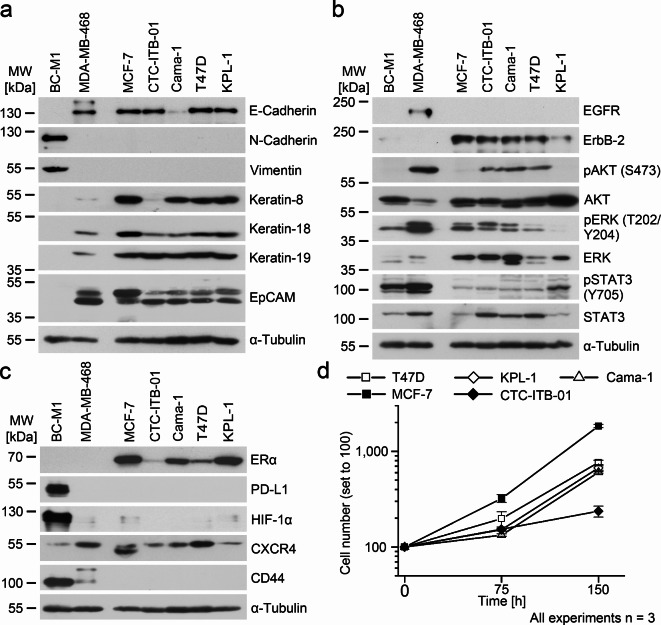
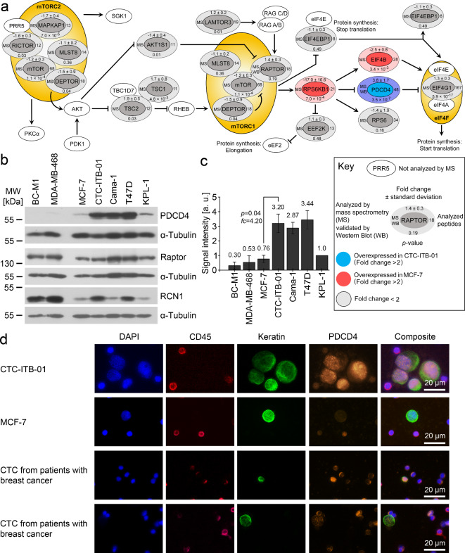
Cell lines derived from circulating tumor cells (CTCs) in the blood provide important biological information on cancer metastasis. CTC-ITB-01 is a CTC cell line derived from a patient with metastatic estrogen receptor-alpha (ER-alpha) positive breast cancer two months before the death of the patient. After a LC-MC/MS based proteomics analysis of CTC-ITB-01, we found extraordinary high levels of the poorly characterized protein SUSD2 (sushi domain-containing protein 2) in CTC-ITB-01. Expression of SUSD2 on subsets of CTCs was validated on clinical blood samples of patients with metastatic breast cancer. SUSD2-positive CTCs could be captured specifically by a MACS-based approach. We overexpressed SUSD2 in the poorly-metastatic cell line MCF-7. This resulted in upregulation of ER-alpha, the tumor progression protein GRP78 (78-kDa glucose-regulated protein) and downregulation of the tumor suppressor protein PDCD4 (programmed cell death protein 4). We observed downregulation of SUSD2 and PDCD4 after hypoxia and simulation of re-oxygenation in the blood in MCF-7 and MDA-MB-468, while in CTC-ITB-01 SUSD2 levels remained unchanged, and only PDCD4 was downregulated under hypoxia. In conclusion, we show, for the first time, that SUSD2 is expressed in CTCs and appears to affect key proteins in tumor progression and survival.

Keywords: Breast cancer; Circulating tumor cells; Estrogen receptor positive; Metastasis; PDCD4; SUSD2.


Conflict of interest statement

Competing interests: The authors declare no competing interests.

Figures

Fig. 1
Fig. 1
Characterization of the CTC cell line CTC-ITB-01, analyses by Western Blot using three biological replicates.A: Determination of the epithelial phenotype.B: Analysis of receptor tyrosine kinase signaling.C: Other breast cancer relevant proteins.D: Growth curves of the denoted cell lines. At the time point 0 h, 40,000 cells of each cell lines were counted and were set to 100. Vertical error bars show the standard deviation of three independent measurements. Uncropped X-ray film images are shown in supplementary information.
Fig. 2
Fig. 2
Detection and validation of differentially expressed proteins from the SILAC approach. A: Detection of SUSD2 by SILAC LC-MS/MS. Left image: MS survey scan containing the peptides around m/z 1001. Right: enlarged section of the SUSD2 peptides. CTC-ITB-01 was cultured in presence of light amino acids (light);13C6-labelled peptides from MCF-7 were not detected in this analysis. B: Positive ion mode HCD (higher-energy collisional dissociation) fragment spectrum of m/z 1001.52 [M + 2 H]2+ from the SILAC approach. Relevant fragments of the y-ion series are assigned with their masses. C: Validation of the denoted proteins by Western Blot. Alpha-tubulin served as loading controls. D: SDS-PAGE of protein extract from CTC-ITB-01 and marking of the excised gel area analyzed by LC-MS/MS (left). Right: Assignment of the identified peptides by LC-MS/MS in the amino acid sequence of SUSD2 (black background). Underlined and bold: GDPH cleavage sentence. Bold: predicted N-glycosylation sites. Italic: signal peptide. E: schematic representation of the posttranslational processing of SUSD2. (1) Full-length 130-kDa SUSD2 is anchored with the C-terminus in the cell membrane and contains a GDPH cleavage site at amino acid 452. (2) Disulfide-bridges are formed so that the N-terminal end is bent back towards the C-terminus of SUSD2. (3) Cleavage at the GDPD sequence results in a separate polypeptide chain that is linked to the N-terminal fragment by disulfide bridges. (4) Application of a reducing agent like DTT cleaves the disulfide bridges. Both the N-terminal (light grey) and the C-terminal fragment (dark gray) migrate at about 55 kDa by SDS-PAGE. The processing of SUSD2 was resolved by Patrick and Egland. Uncropped X-ray film images are shown in supplementary file S1.
Fig. 3
Fig. 3
Detection and validation of differentially expressed proteins between CTC-ITB-01 vs. MCF-7.A: Analysis of the mTOR pathway based on SILAC-based LC-MS/MS proteomic data. Synonyms: MAPKAP1: SIN1, MLST8: GβL, AKT1S1: PRAS40, RPS6KB1: P70S6K1. For detailed values of the protein identification and quantification, see Supplementary Table S3.B: Western Blot analysis of the denoted proteins (n = 3). BC-M1 was selected as an example of a disseminated tumor cell line from the bone marrow of a breast cancer patient. MDA-MB-468 was selected as an example of a triple negative breast cell line.C: Quantitative Western Blot analysis of PDCD4 (n = 3). The average value is shown as numbers and the standard deviation as vertical error bars. The p-value was calculated using Student’s two-sided t-test with p < 0.05 considered significant.D: Detection of PDCD4 in tumor cells of breast cancer patients (n = 3). The composite images show overlays of the DAPI, CD45, keratin and PDCD4 signals. Uncropped X-ray film images are shown in supplementary file F1.
Fig. 4
Fig. 4
Analysis of the heterologous SUSD2 overexpression in MCF-7.A: Principal component analysis of LC-MS/MS data from the three samples without SUSD2-insert (Ctrl) and carrying the SUSD2 gene in the vector (OE, overexpression). Clone OE2 was analyzed.B: Visualization of a Pearson-correlation based supervised hierarchical clustering. The color legend displays the log2 foldchanges. Analyzed peptides: 24 (PDCD4), 5 (RCN1), 144 (GRP78, HSPA5). For all proteins, the differential expression was > 2 and significant (p < 0.05, Student’s t-test).C: Western Blot analysis of the control cell line and two SUSD2 overexpressing clones for the denoted proteins.D: Quantitative Western Blot analyses of the X-ray films shown in (C). The signals were normalized to α-tubulin, in case of p70 S6 kinase (Thr389) the signals were normalized to total p70 S6 kinase. Detailed information is shown in Supplementary Table S4.E: Response of the cells to treatment with 17β-Estradiol. Cells were cultured in hormone depleted medium (HD) or in presence of 100 nmol/ml 17β-Estradiol (E2). The diagrams show the quantitative analysis of the BCL2 levels (left: control, right: clone OE2). PR A/B: progesterone receptor A/B). D,E: Average values are shown as numbers, and the standard deviation as vertical error bars. The p-values were calculated using Student’s two-sided t-test where p < 0.05 was considered significant. Three biological replicates were analyzed (A-E). Uncropped X-ray film images are shown in supplementary file F1.
Fig. 5
Fig. 5
Modification of SUSD2 levels and detection of SUSD2 in CTC of breast cancer patients.A: Downregulation of the SUSD2 protein in CTC-ITB-01 by siRNA, analyses by Western Blot.B: Quantitative Western Blot analysis of the PDCD4 and GRP78 levels shown in (A).C: Response of SUSD2 and PDCD4 to 14 days of hypoxia (1% O2; 14 d) and reoxygenation (14 days of hypoxia, followed by 10% O2 for 4 h; + 4 h). ERα (MCF-7) and EGFR (MDA-MB-468) were analyzed as an established reaction to hypoxia. S: standard cell culture condition.D: Quantitative Western Blot analysis of the SUSD2 and PDCD4 levels shown in (C).E: Detection of SUSD2 in CTC of breast cancer patients using the CellSearch system.F: Isolation of SUSD2 positive CTCs from the blood of breast cancer patients by MACS. Slides were stained with anti-mouse IgG to detect the MicroBeads bound tumor cells (IgG). The top row shows a SUSD2 positive CTC cluster isolated by MACS. The middle row shows an overview of an elution fraction on a cytospin. The SUSD2 positive CTCs are labelled with the white box and are enlarged in the bottom row.A-D,F: All experiments were performed in biological triplicates. B, D: Average values are shown as numbers, and the standard deviation as vertical error bars. The p-values were calculated using Student’s two-sided t-test where p < 0.05 was considered significant.E: The CellSearch Results are specified in Supplementary Table S5 (E). Uncropped X-ray film images are shown in supplementary file F1.
Fig. 6
Fig. 6
Correlation of PDCD4 levels with clinicopathological data. The data were taken from the breast invasive carcinoma TCGA PanCancer Atlas data set.A: Correlation with breast cancer subtypes (basal: n = 171, HER2 n = 78, LumA, luminal A: n = 499, LumB, lumimal B:n = 197, normal: n = 36). B: Correlation with staging (stage I: n = 180, stage II: n = 615, stage III: n = 213, stage IV n = 19).C: Correlation with dissemination status (M0: n = 895, CM0, M0 with CTC in the blood (cM0(1+)): n = 6, M1: n = 21).D: Survival analysis of all breast cancer patients. PDCD4 high: n = 271, PDCD4 low: n = 271. E: Survival analysis of the luminal subtypes. PDCD4 high: n = 174, PDCD4 low: n = 174.

References

    1. Burstein, H. J. et al. Adjuvant endocrine therapy for women with hormone receptor-positive breast cancer: American society of clinical oncology clinical practice guideline focused update. J. Clin. Oncol.32, 2255–2269. 10.1200/JCO.2013.54.2258 (2014). - PMC - PubMed
    1. Pantel, K. & Hayes, D. F. Disseminated breast tumour cells: biological and clinical meaning. Nat. Rev. Clin. Oncol.15, 129–131. 10.1038/nrclinonc.2017.174 (2018). - PubMed
    1. Waks, A. G. & Winer, E. P. Breast Cancer Treatment: A review. JAMA321, 288–300. 10.1001/jama.2018.19323 (2019). - PubMed
    1. Ma, C. X., Reinert, T., Chmielewska, I. & Ellis, M. J. Mechanisms of aromatase inhibitor resistance. Nat. Rev. Cancer. 15, 261–275. 10.1038/nrc3920 (2015). - PubMed
    1. Meng, S. et al. Circulating tumor cells in patients with breast cancer dormancy. Clin. Cancer Res.10, 8152–8162 (2004). - PubMed

Publication types

MeSH terms