. 2025 Jan 31;22(1):13.
doi: 10.1186/s12987-025-00621-4.

Endothelial and neuronal engagement by AAV-BR1 gene therapy alleviates neurological symptoms and lipid deposition in a mouse model of Niemann-Pick type C2

Affiliations

Endothelial and neuronal engagement by AAV-BR1 gene therapy alleviates neurological symptoms and lipid deposition in a mouse model of Niemann-Pick type C2

Charlotte Laurfelt Munch Rasmussen et al. Fluids Barriers CNS. .

Abstract

Background: Patients with the genetic disorder Niemann-Pick type C2 disease (NP-C2) suffer from lysosomal accumulation of cholesterol causing both systemic and severe neurological symptoms. In a murine NP-C2 model, otherwise successful intravenous Niemann-Pick C2 protein (NPC2) replacement therapy fails to alleviate progressive neurodegeneration as infused NPC2 cannot cross the blood-brain barrier (BBB). Genetic modification of brain endothelial cells (BECs) is thought to enable secretion of recombinant proteins thereby overcoming the restrictions of the BBB. We hypothesized that an adeno-associated virus (AAV-BR1) encoding the Npc2 gene could cure neurological symptoms in Npc2-/- mice through transduction of BECs, and possibly neurons via viral passage across the BBB.

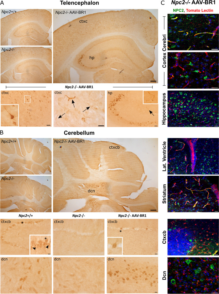
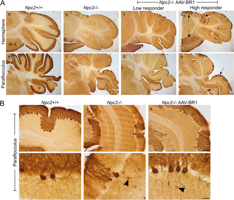
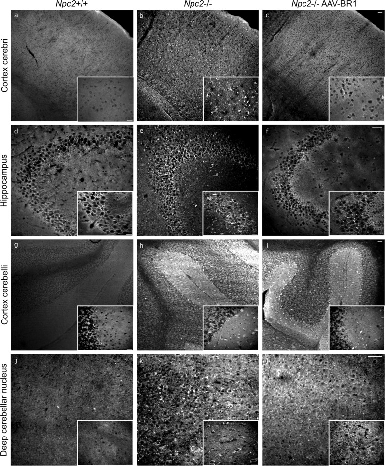
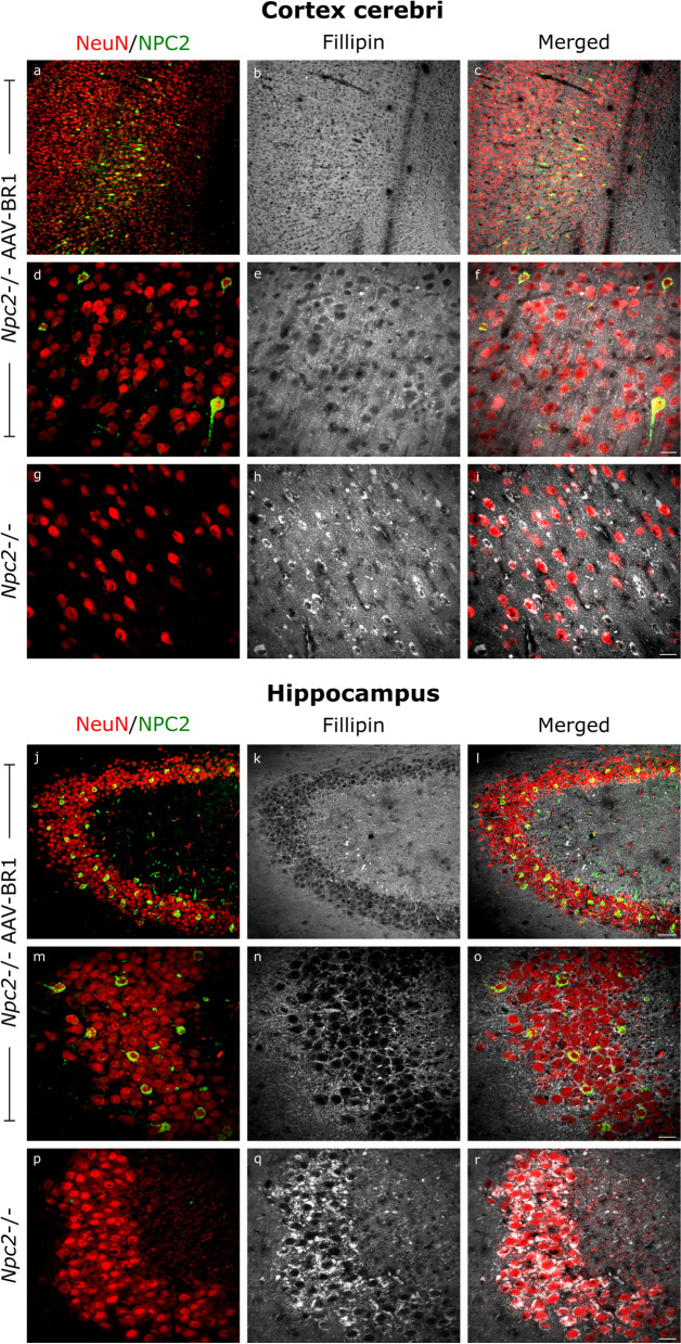
Methods: Six weeks old Npc2-/- mice were intravenously injected with the AAV-BR1-NPC2 vector. Composite phenotype scores and behavioral tests were assessed for the following 6 weeks and visually documented. Post-mortem analyses included gene expression analyses, verification of neurodegeneration in Purkinje cells, determination of NPC2 transduction in the CNS, assessment of gliosis, quantification of gangliosides, and co-detection of cholesterol with NPC2 in degenerating neurons.

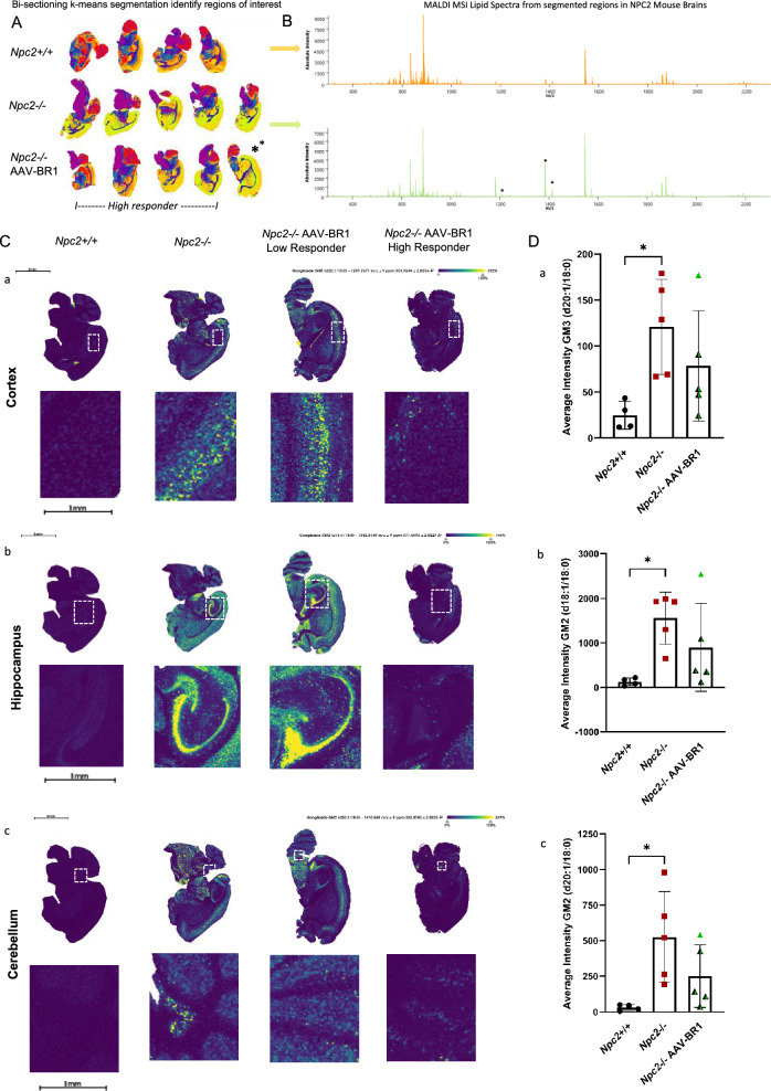
Results: Treatment with the AAV-BR1-NPC2 vector improved motor functions, reduced neocortical inflammation, and preserved Purkinje cells in most of the mice, referred to as high responders. The vector exerted tropism for BECs and neurons resulting in a widespread NPC2 distribution in the brain with a concomitant reduction of cholesterol in adjacent neurons, presumably not transduced by the vector. Mass spectrometry imaging revealed distinct lipid alterations in the brains of Npc2-/- mice, with increased GM2 and GM3 ganglioside accumulation in the cerebellum and hippocampus. AAV-BR1-NPC2 treatment partially normalized these ganglioside distributions in high responders, including restoration of lipid profiles towards those of Npc2+/+ controls.

Conclusion: The data suggests cross-correcting gene therapy to the brain via delivery of NPC2 from BECs and neurons.

Keywords: AAV-BR1; Blood–brain barrier; Cholesterol; Cross-correction; Gangliosides; NPC2; Niemann-Pick type C2 disease; Viral gene therapy.


Conflict of interest statement

Declarations. Ethics approval and consent to participate: The animal studies were performed according to the Danish Animal Experimentation Act (BEK no. 2028 of 14/12/2020) and the European directive (2010/63/EU) and carried out by licensed staff. The Danish Animal Experiments Inspectorate under the Ministry of Food, Agriculture and Fisheries has approved all animal experiments and breeding of NPC2-deficient mice (license no. 2018-15-0201-01467 and 2019-15-0202-00056). Consent for publication: Not applicable. Competing interests: JK is listed as an inventor on a patent on AAV-BR1, held by Boehringer Ingelheim International. All other authors have no competing interests to declare.

Figures

Fig. 1
Fig. 1
Study design. After weaning, at 3 weeks of age, all mice were socialized (S). At 4 weeks of age, the mice were trained in the rotarod protocol. The first rotarod test was conducted at 5 weeks of age, and afterwards once a week until the end of the study (12 weeks). The NPC2-deficient mice (Npc2−/−) were randomly allocated to the treatment group, and at 6 weeks of age, the Npc2−/− mice were either injected in the tail vein with the brain-specific adeno-associated viral vector encoding the Npc2 gene (AAV-BR1-NPC2) or with phosphate-buffered saline (PBS). Untreated wild-type (Npc2+/+) mice were included as controls. At 6, 9, and 12 weeks of age, gait and phenotype score was evaluated. At 12 weeks of age, the mice were euthanized, a blood sample was drawn, and organs of interest (brain, lung, spleen, and liver) were weighed and collected for further analyses. Created with BioRender.com
Fig. 2
Fig. 2
Effects of AAV-BR1-NPC2 on the Niemann Pick type C2 disease phenotype. A The body weight was assessed weekly in wild-type (Npc2+/+), Npc2−/−, and AAV-BR1-NPC2-treated Npc2−/− mice. There was no significant difference in body weight between the three groups at any time point during the study period analyzed with a REML mixed-effects model with Greenhouse–Geisser correction (F[2,26] = 2.073, p = 0.1461). B From 4 to 6 weeks of age, all mice increased their body weight, and there was no difference between the three groups (one-way ANOVA (F[2,26] = 0.0891, p = 0.9151). When examined from the time-point of treatment (6 weeks of age) until the time of euthanasia (12 weeks of age), the Npc2−/− and AAV-BR1-NPC2-treated Npc2−/− mice had a significantly lower increase in body weight compared to Npc2+/+ mice (one-way ANOVA (F[2,25] = 12.88, ***p ≤ 0.005). C Composite phenotype scores were assessed at 6, 9, and 12 weeks of age. The data were analyzed using the Kruskal–Wallis test (6 weeks: H(2) = 1.022, p = 0.5998, 9 weeks: H(2) = 17.95, p = 0.0001, 12 weeks: H(2) = 20.08, p < 0.0001). Dunn’s multiple comparisons test with Bonferroni correction (due to multiple tests being carried out) was used for the data sets from 9 and 12 weeks. Significant differences between Npc2+/+ and untreated Npc2−/−, and Npc2+/+ and AAV-BR1-NPC2-treated Npc2−/− mice are reported with # and § respectively, §p = 0.0483, ###p ≤ 0.0003. Data are presented with a median with interquartile ranges, n = 8 mice/group. D Video Stills (see supplemental video material) of Npc2+/+, Npc2−/−, and AAV-BR1-NPC2-treated Npc2−/− mice performing on the ledge test at 12 weeks of age, included as a part of the composite phenotype score. Wild-type mice receive a score of 0, as these mice can walk along the ledge without losing their balance. However, Npc2−/− mice struggle to keep their balance, move forward, and are nearly falling off the ledge, and therefore receive a score of 2. 4/8 AAV-BR1-NPC2-treated Npc2−/− mice receive a score of 1, due to some trouble keeping their balance, and not using their hindlimbs effectively. However, the mice can still walk along the ledge. E Gait analysis was performed at 6, 9, and 12 weeks of age. The front paws were painted with red dye, and the hind paws in blue dye. Representative images are shown for Npc2+/+ n = 8/8 mice, Npc2−/− n = 8/8, and AAV-BR1-NPC2-treated Npc2−/− n = 6/8 mice. F Rotarod’s performance was assessed weekly from 5 weeks of age until the end of the study at 12 weeks of age. From 8 weeks of age, the time the Npc2−/− and AAV-BR1-NPC2-treated Npc2−/− mice spent on the rotarod (sec) was significantly lower compared to the age-matched Npc2+/+ mice (REML mixed-effects model with Greenhouse–Geisser correction (F[2,26] = 13.89, p < 0.0001)). Significant differences between Npc2+/+ and untreated Npc2−/−, and Npc2+/+ and AAV-BR1-NPC2-treated Npc2−/− mice are reported with # and § respectively, #, §p < 0.05, ##p < 0.005. The rotarod performance at 12 weeks of age is visualized with individuals and exact p-values in G (***p = 0.0007, **p = 0.002). The results in AB and FG, are presented as mean ± SD, and the number in each group are as follows: Npc2+/+ = 10 mice, Npc2−/− = 9 mice, from 11 weeks of age n = 8 mice, AAV-BR1-NPC2-treated Npc2−/− mice = 10 mice. 6/10 AAV-BR1 treated mice (high responders) can perform on the rotarod at 12 weeks of age (individual high responder mice are indicated with green/black triangles)
Fig. 3
Fig. 3
AAV-BR1 vector distribution and its effect on organ weight and cholesterol accumulation in peripheral tissue. A Biodistribution of AAV-BR1-NPC2 [viral genomes (vg)] in brain, lung, liver, and spleen, analyzed by quantitative qPCR at 12 weeks of age. B Relative Npc2 gene expression in the cerebrum and lung tissue was analyzed by RT-qPCR. The Npc2 gene expression was significantly lower in the cerebrum of untreated Npc2−/− compared to Npc2+/+, whereas no significant difference was seen between AAV-BR1-NPC2-treated Npc2−/− mice and Npc2+/+. The Npc2 gene expression in the lungs of treated and untreated Npc2−/− mice was significantly lower compared to wild-type littermates. Data are analyzed with a one-way ANOVA (FCerebrum[2,6] = 9.64, p = 0.013, FLung[2,6] = 74.52, p < 0.0001) with Tukey’s multiple comparisons test (*p = 0.011, ***p ≤ 0.0001). A, B Data are presented as mean ± SD (n = 3 mice/group). High responders (n = 2) are indicated with green/black triangles, while low responders (n = 1) are indicated with green triangles. C Injection with the AAV-BR1-NPC2 vector had no significant effect on organ size, and both Npc2−/− (n = 9 mice) and AAV-BR1 treated Npc2−/− mice (n = 10 mice) had significantly lower brain weight and a significantly larger lung and spleen compared to wild-type (WT) mice (Npc2+/+) (n = 10 mice) analyzed with a one-way ANOVA (FBrain[2,26] = 20.00, p < 0.0001, FLung[2,26] = 19.49, p < 0.0001), FSpleen[2,26] = 24.34, p < 0.0001) with Tukey’s multiple comparisons tests (***p < 0.001, ****p < 0.0001). There was no significant difference in the size of the liver between the three groups (FLiver[2,26] = 20.1866, p = 0.8309). Data for the brain, liver, and spleen are presented as mean ± SD, whereas data for the lungs are presented as geometric mean with a 95% confidence interval. D, E Cholesterol concentrations were evaluated in the liver, lung, spleen, and serum in the three groups (n = 7 mice/group). The cholesterol levels in the investigated organs were significantly higher in Npc2−/− and AAV-BR1-NPC2-treated Npc2−/− mice compared to Npc2+/+ age-matched controls (****p < 0.0001). However, the cholesterol concentration in the spleen of treated mice was significantly lower compared to untreated Npc2−/− mice (**p = 0.0089). No significant differences were observed in serum. All data were analyzed with one-way ANOVA (FLiver[2,18] = 36.37, p < 0.0001), FLung[2,18] = 23.93, p < 0.0001, FSpleen[2,18] = 83.17, p < 0.0001, FSerum[2,18] = 0.31, p = 0.7351) with Tukey’s multiple comparisons test. Data are presented as geometric mean with a 95% confidence interval for D and as mean ± SD for E. High responders (n = 4) are indicated with green/black triangles, while low responders (n = 3) are indicated with green triangles. F Visceral pathology was analyzed with hematoxylin and eosin staining. Images are representative of n = 5–7 mice/group. Arrows point to lipid-laden macrophages in the liver (top panel), lung (mid panel), and spleen (bottom panel). Asterisks indicate the accumulation of eosinophilic granular material in the alveolar lumen, shown in higher magnification in the black boxes. Scale bars are 100 µm and 20 µm (black box)
Fig. 4
Fig. 4
NPC2 distribution in the brain after brain-endothelial-directed gene therapy. Immunohistochemical stainings reveal NPC2-positive cells in the brain of Npc2−/− mice after AAV-BR1-NPC2 gene therapy. A NPC2 is particularly prominent in neurons of the cortex cerebri (ctxc) and the hippocampus (hp) (CA3 region) in the AAV-BR1-NPC2-treated Npc2−/− mice. Although not intensely stained, NPC2 was also seen in microvessels (arrows). B A high expression of NPC2 is also found in the deep cerebellar nuclei (dcn) of AAV-BR1-NPC2-treated Npc2−/− mice. Only sparse NPC2-positive cells are seen in the cortex cerebelli (ctxcb) of the treated Npc2−/− group. No NPC2-positive cells are seen in untreated Npc2−/− mice. Granular labeling is seen in Bergmann glia (arrowheads) in Npc2+/+ mice. Asterisks indicate Purkinje cells. C AAV-BR1-NPC2-treated Npc2−/− mice were further stained with NPC2, and counterstained with tomato lectin to label the vasculature and analyzed with confocal microscopy. Transduced microvessels are seen throughout the brain, however more pronounced in certain areas like ctxc and striatum. Throughout the brain areas with a high occurrence of NPC2 positive neurons and NPC2 negative microvessels can likewise be found, especially the cortex, and cerebellum. All images are representative of n = 4 mice in Npc2+/+ and n = 3/5 in Npc2−/− AAV-BR1-NPC2-treated group (high responders) and n = 5 in the non-treated Npc2−/− group. Scale bar = 50 µm (cortex cerebri and hippocampus), 25 µm (microvessels in the cortex cerebri), 20 µm (cerebellum, and all images in C), 10 µm [Purkinje cells (magnification)]
Fig. 5
Fig. 5
Effect of the AAV-BR1-NPC2 gene therapy on Purkinje cell pathology. Cerebellar sections evaluated using immunohistochemical staining for the Purkinje cell marker calbindin. A The wild-type (Npc2+/+) mice exhibit normal Purkinje cell patterns (a, e). In contrast, severe Purkinje cell degeneration is seen in untreated Npc2−/− mice (b, f). In AAV-BR1-NPC2-treated Npc2−/− mice, the Purkinje cell degeneration is variable (c, d, g, h), but in 4/8 mice assessed, preservation of Purkinje cells is clearly visualized (arrows), especially in the flocculus (F) and paraflocculus (PF) (h) (high responders). Purkinje cell preservation is seen in lobules IX and X of Npc2−/− mice independent of treatment (b, c, d). B The Purkinje cell degeneration in both Npc2−/− and AAV-BR1-NPC2-treated Npc2−/− mice is accompanied by axonal swellings (arrowheads) caused by storage accumulation. Images are representative of n = 8 Npc2+/+ mice, n = 5 Npc2−/− mice, and n = 8 AAV-BR1-NPC2-treated Npc2−/− mice, Scale bar A: 300 µm (ah), B: 50 µm (ik), 25 µm (ln)
Fig. 6
Fig. 6
Effect of the AAV-BR1-NPC2 gene therapy on gliosis in the cortex cerebri and hippocampus. A Brain sections evaluated using immunohistochemical staining of the astrocytic marker GFAP (al) and microglial marker IBA1 (mx). In wild-type mice (Npc2+/+) only sparse GFAP-positive astrocytes (a, e, i) and resting ramified microglia (m, q, u) are seen in cortex cerebri (ctxc), and hippocampus (hp). Diffuse astrogliosis is seen in untreated Npc2−/− mice (b, f, j), whereas a reduction in GFAP-positive astrocytes is visualized in some of the AAV-BR1-NPC2-treated mice (high responders) (d, h, l), especially in neocortical regions just above corpus callosum (cc). The same pattern is evident for the distribution of reactive microglia in the cortex cerebri and hippocampus (p, t, x). Images are representative of n = 4 Npc2+/+ mice, n = 4 Npc2−/− mice, and n = 7 AAV-BR1-NPC2-treated Npc2−/− mice, where 4 out of 7 mice presented with lower gliosis (high responders) compared with untreated Npc2−/− mice. Scale bar = 100 µm (ad and mp), 50 µm (el and qx). The density of astrocytes (B) and microglia (C) cells per 10,000 µm2 in ctxc and hp. Npc2+/+ mice (black circles) (n = 4), Npc2−/− mice (n = 4) (red squares), and AAV-BR1-NPC2-treated Npc2−/− mice categorized as high responders (n = 5) (black/green triangles) and low responders (n = 2–3) (green triangles). The data were analyzed with a one-way ANOVA (FGFAP Ctxc[2,12] = 7.694, p = 0.007, FIBA1 Ctxc[2,13] = 3.220, p = 0.073, FIBA1 Hp[2,13] = 2.380, p = 0.1316) with Tukey’s multiple comparisons test (**p = 0.005) except for GFAP in hp were the Kruskal–Wallis test was used (H(2) = 5,645, p = 0,0510). Data for astrocytes in ctxc, microglia in ctxc, and hp are presented as mean ± SD, whereas data for GFAP in hp, are presented as median with IQR
Fig. 7
Fig. 7
Cholesterol storage in the brain after AAV-BR1-directed gene therapy. The cholesterol accumulation (visualized using Filipin staining) is reduced in the cortex cerebri (ac) and the CA3 region of the hippocampus (d- f) of AAV-BR1-NPC2-treated Npc2−/− mice (c, f, i, l) compared to untreated Npc2−/− mice (b, e, h, k). There is no obvious difference when comparing cholesterol storage in the cortex cerebelli (gi) and deep cerebellar nucleus (jl) of the treated vs. untreated Npc2−/− groups. No cholesterol storage is seen in Npc2+/+ control littermates (a, d, g, j). Images are representative of n = 4 mice/group (Npc2+/+ and Npc2−/−) and 3 out of 4 AAV-BR1-NPC2-treated Npc2−/− mice (high responders). Scalebar = 50 µm and 20 µm (white boxes)
Fig. 8
Fig. 8
Reduced cholesterol in the cortex cerebri and hippocampus corresponds to areas with many NPC2-positive cells. The high occurrence of NPC2-positive neurons [visualized with neuronal nuclei antigen (NeuN) immunolabeling (red) and NPC2 immunolabeling (green)] observed in the cortex cerebri (ai) and the CA3 region of the hippocampus (jk) correspond with lower levels of cholesterol accumulation [visualized using Filipin staining (white)] in the high responder group of AAV-BR1-NPC2-treated Npc2−/− mice in both brain regions assessed. In comparison untreated Npc2−/− mice show extensive neuronal accumulation of cholesterol (only shown in high magnifications). Reduced cholesterol accumulation in the high responder AAV-BR1-NPC2-treated Npc2−/− mice is not limited to the NPC2 expressing cells, but also obvious in neighboring cells, suggesting uptake of secreted NPC2 in these cells, equivalent to cross-correction. Representative images from cortex cerebri and CA3 region of the hippocampus of the high responder group (n = 3). Scale bar = 25 µm
Fig. 9
Fig. 9
Segmentation and differential ganglioside expression in the brain using MALDI-MS. A Unsupervised statistical analysis employing bi-sectioning k-means in SCiLS Lab reveals distinct regions in mouse brains, with segmentation analysis identifying green and orange regions of interest (ROIs) characterized by unique lipid profiles between wildtype Npc2+/+ and untreated Npc2−/−. High responder AAV-BR1-NPC2-treated Npc2−/− mice show lipid profiles comparable to Npc2+/+, while low responders are comparable to the untreated Npc2−/− mice. B Average spectra from the green and orange segmented regions, normalized for RMS intensity, highlight distinct lipid profiles characteristic of each segment. C Spatial distribution maps of gangliosides GM3(d20:1/18:0) at 1207.76 m/z (a), GM2(d18:1/18:0) at 1381.81 m/z (b), and GM2(d20:1/18:0) at 1410.84 m/z (c) illustrate their presence in the cortex, hippocampus, and cerebellum across different genotypes and treatment groups. Representative images are shown for Npc2+/+ (n = 4), Npc2−/− (n = 5), and AAV-BR1-NPC2-treated Npc2−/− high responders (n = 3/4), low responders (n = 1) mice. D Quantitative analysis of average ganglioside intensities: a GM3 (1207.76 m/z) in the cortex, b GM2 (1381.81 m/z) in the hippocampus, c GM2 (1410.84 m/z) in the cerebellum. Significant differences (*p < 0.05) are observed between Npc2+/+ and Npc2−/− mice. No significant differences are observed between Npc2+/+ and AAV-BR1-NPC2-treated Npc2−/− mice, indicating a therapeutic impact of AAV-BR1 treatment and that the variation in ganglioside accumulation is influenced by genotype and treatment efficiency. All data were analyzed with one-way ANOVA (FGM3 (20) (1207.76 m/z)[2,11] = 4.394, p < 0.0396), FGM2 (18) (1381.81 m/z)[2,11] = 4.735, p < 0.0328, F GM2(18) (1410.84 m/z)[2,11] = 5.067, p < 0.0276, with Tukey's multiple comparisons test. Data are presented as mean ± SD (n = 4–5). High responders (n = 4) are indicated with green/black triangles, while low responders (n = 1) are indicated with green triangles

References

    1. Platt FM. Emptying the stores: lysosomal diseases and therapeutic strategies. Nat Rev Drug Discov. 2018;17:133–50. - PubMed
    1. Verot L, Chikh K, Freydière E, Honoré R, Vanier MT, Millat G. Niemann-Pick C disease: functional characterization of three NPC2 mutations and clinical and molecular update on patients with NPC2. Clin Genet. 2007;71:320–30. - PubMed
    1. Geberhiwot T, Moro A, Dardis A, Ramaswami U, Sirrs S, Marfa MP, et al. Consensus clinical management guidelines for Niemann-Pick disease type C. Orphanet J Rare Dis. 2018;13:50. - PMC - PubMed
    1. Vanier MT. Niemann-Pick disease type C. Orphanet J Rare Dis. 2010;5:16. - PMC - PubMed
    1. Berzina Z, Solanko LM, Mehadi AS, Jensen MLV, Lund FW, Modzel M, et al. Niemann-Pick C2 protein regulates sterol transport between plasma membrane and late endosomes in human fibroblasts. Chem Phys Lipids. 2018;213:48–61. - PubMed

Substances

Supplementary concepts

LinkOut - more resources