. 2025 Jan 31;23(1):65.
doi: 10.1186/s12951-025-03158-8.

Real-world of Limosilactobacillus reuteri in mitigation of acute experimental colitis

Affiliations

Real-world of Limosilactobacillus reuteri in mitigation of acute experimental colitis

Ningning Yue et al. J Nanobiotechnology. .

Abstract

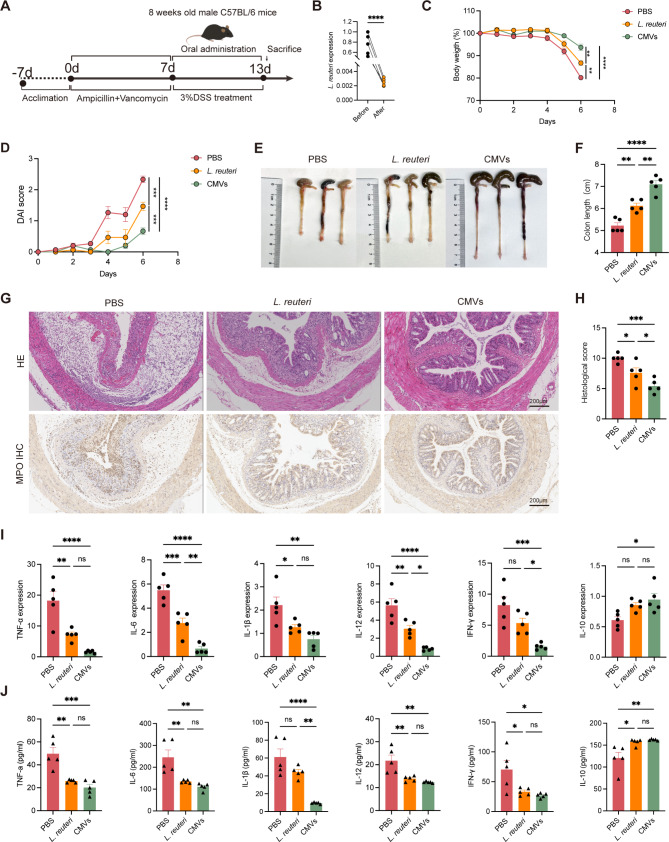
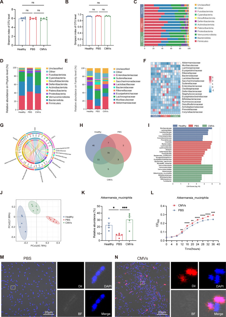
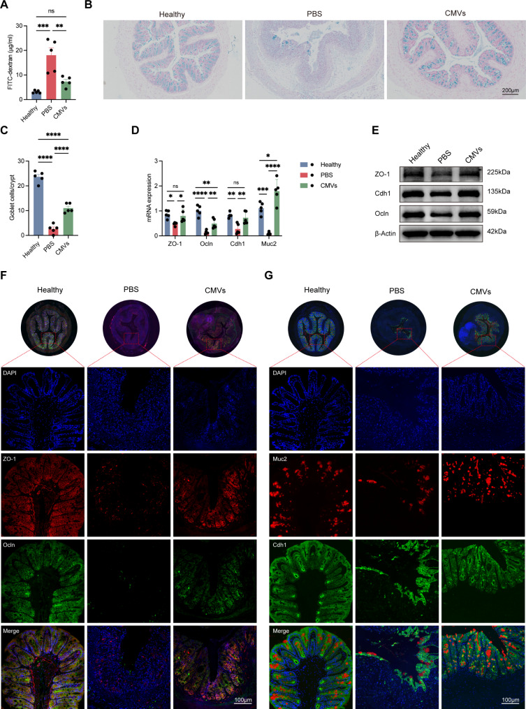
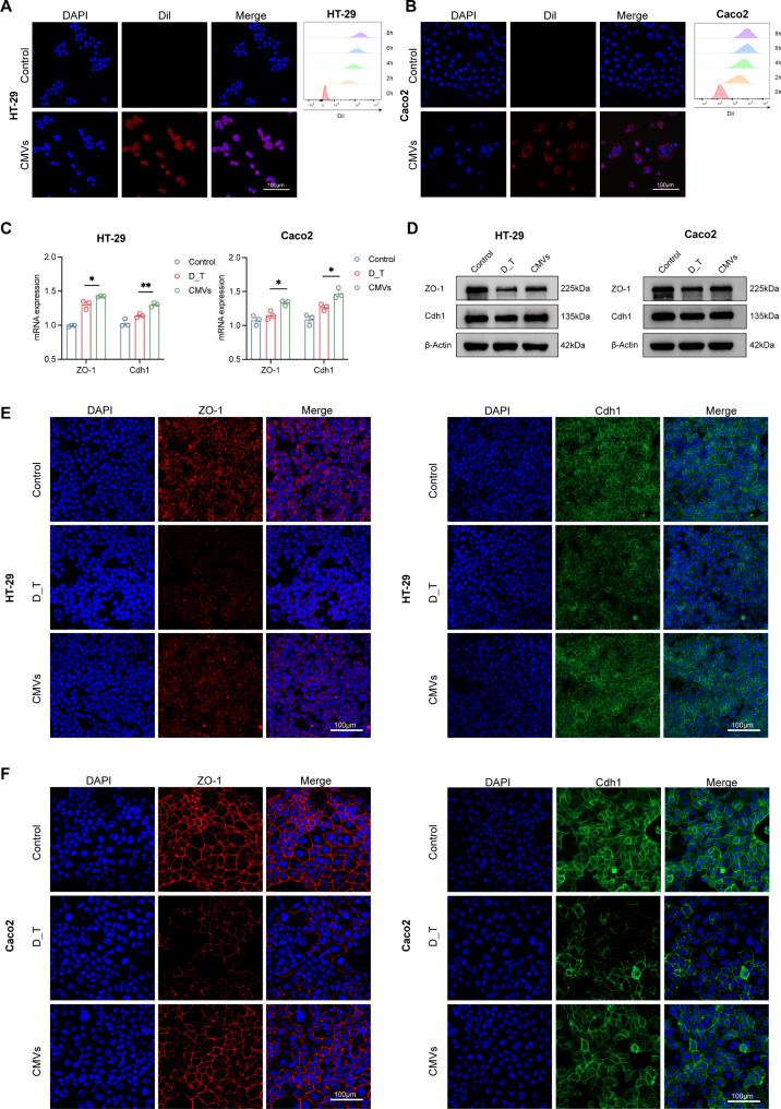
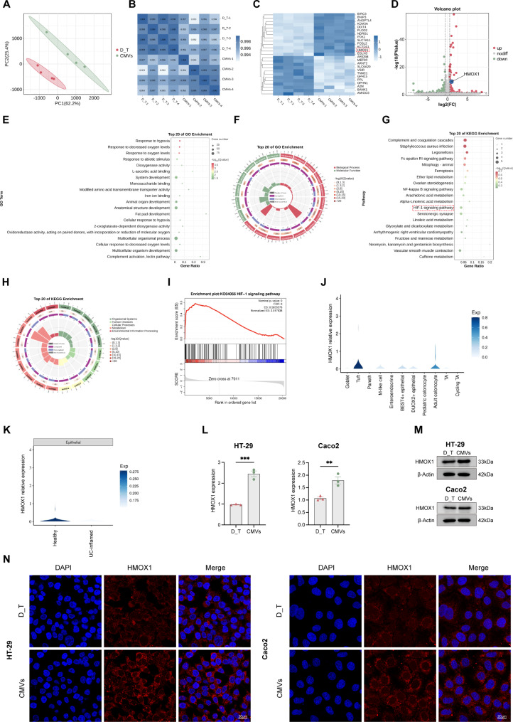
Probiotics have been proposed as a potential strategy for managing ulcerative colitis (UC). However, the underlying mechanisms mediating microbiota-host crosstalk remain largely elusive. Here, we report that Limosilactobacillus reuteri (L. reuteri), as a probiotic, secretes cytoplasmic membrane vesicles (CMVs) that communicate with host cells, alter host physiology, and alleviate dextran sulfate sodium (DSS)-induced colitis. First, L. reuteri-CMVs selectively promoted the proliferation of the beneficial bacterium Akkermansia muciniphila (AKK) by upregulating the expression of glycosidases (beta-N-acetylhexosaminidase and alpha-N-acetylglucosaminidase) involved in glycan degradation and metabolic pathways and restored the disrupted gut microbiota balance. Second, L. reuteri-CMVs were taken up by intestinal epithelial cells (IECs), elevated the expression of ZO-1, E-cadherin (Cdh1), and Occludin (Ocln), decreased intestinal permeability, and exerted protective effects on epithelial tight junction functionality. RNA sequencing analysis demonstrated that L. reuteri-CMVs repaired intestinal barrier by activating the HIF-1 signaling pathway and upregulating HMOX1 expression. Third, L. reuteri-CMVs increased the population of double positive (DP) CD4+CD8+ T cells in the intestinal epithelial layer, suppressing gut inflammation and maintaining gut mucosal homeostasis. Finally, L. reuteri-CMVs exhibited satisfactory stability and safety in the gastrointestinal tract and specifically targeted the desired sites in colitis mice. Collectively, these findings shed light on how L. reuteri interact with the host in colitis, and provide new insights into potential strategies for alleviating colitis.

Keywords: Limosilactobacillus reuteri; Colitis; Cytoplasmic membrane vesicles; Double positive CD4+CD8+ T cells; Gut microbiota; Intestinal barrier.


Conflict of interest statement

Declarations. Ethics approval and consent to participate: All the animal experiments were approved by the Animal Care Committee of the Shenzhen People’s Hospital, Shenzhen, China (No. 2024-118). Consent for publication: Not applicable. Competing interests: The authors declare no competing interests.

Figures

Fig. 1
Fig. 1
L. reuteri-CMVs alleviate dextran sulfate sodium (DSS)-induced colitis. (A) Schematic diagram for the treatment of DSS-induced C57BL/6 colitis mice. (B) Daily body weight over 7 days. (C) Disease activity index (DAI) scores. (D) Photographs of colons. (E) Colon length. (F) H&E and myeloperoxidase (MPO) IHC staining for colons. (G) Histological scores. (H) The mRNA expression levels of pro-inflammatory cytokines (TNF-α, IL-6, IL-1β, IL-12, and IFN-γ) and anti-inflammatory cytokine (IL-10) in colons. (I) The concentrations of pro-inflammatory cytokines (TNF-α, IL-6, IL-1β, IL-12, and IFN-γ) and anti-inflammatory cytokine (IL-10) in the serum quantified by the ELISA method. Data are mean ± SEM (n = 5). *p < 0.05; **p < 0.01; ***p < 0.001; ****p < 0.0001; ns, non-significance
Fig. 2
Fig. 2
L. reuteri-CMVs mitigate DSS-induced colitis in L. reuteri-depleted mice. (A) Schematic illustration for the administration regimen of DSS-induced colitis in L. reuteri-depleted C57BL/6 mice. (B) The mRNA expression levels of L. reuteri in mice treated with vancomycin plus ampicillin for 7 days. (C) Daily body weight over 6 days. (D) DAI scores. (E) Photographs of colons. (F) Colon length. (G) H&E and MPO IHC staining for colons. (H) Histological scores. (I) The mRNA expression levels of pro-inflammatory cytokines (TNF-α, IL-6, IL-1β, IL-12, and IFN-γ) and anti-inflammatory cytokine (IL-10) in colon tissues. (J) The concentrations of pro-inflammatory cytokines (TNF-α, IL-6, IL-1β, IL-12, and IFN-γ) and anti-inflammatory cytokine (IL-10) in the serum quantified by the ELISA method. Data are mean ± SEM (n = 5). *p < 0.05; **p < 0.01; ***p < 0.001; ****p < 0.0001; ns, non-significance
Fig. 3
Fig. 3
L. reuteri-CMVs restore gut microbiota dysbiosis in DSS-induced colitis. (A, B) 𝛼-diversity analysis by Shannon index and Simpson index. (C) Microbial composition analysis of different species on Phylum level. (D, E) Histograms of microbial community composition on Phylum and Family levels. (F) Community heatmap analysis on Family level. (G) Circos plot of the gut microbiota in different groups on Family level. (H) Venn diagram of the gut microbiota in different groups. (I) Linear discriminant analysis (LDA) for demonstrating dominant bacterial communities in different groups. (J) Principal Co-ordinates Analysis (PCoA) of the gut microbiota in different groups. (K) The relative abundance of Akkermansia_muciniphila (AKK) in different groups. (n = 5). (L) The growth curves of AKK in the PBS and CMVs groups measured by recording the OD value at 600 nm. (M, N) 3D CLSM images of AKK after incubation with PBS and Dil-labeled L. reuteri-CMVs. (n = 3). Data are mean ± SEM. *p < 0.05; **p < 0.01; ***p < 0.001; ****p < 0.0001; ns, non-significance
Fig. 4
Fig. 4
The regulation mechanism of L. reuteri-CMVs promoting AKK proliferation. (A) Principal component analysis (PCA) of the gene expression level (FPKM) in different samples between the PBS group and the CMVs group. (B, C) Heatmap and volcano plot analysis of the total differentially expressed genes (DEGs) in the PBS-treated and CMVs-treated AKK. (D, E) Gene Ontology (GO) and Kyoto Encyclopedia of Genes and Genomes (KEGG) enrichment analysis based on DEGs. (F) Gene Set Enrichment Analysis (GSEA) for genes associated with the other glycan degradation signaling pathway. (G) Heatmap of DEGs related to metabolic pathways and glycan degradation between in PBS and CMVs groups. (H) The growth curves of AKK between in different media measured by recording the OD value at 600 nm. (I) The mRNA expression levels of beta-N-acetylhexosaminidase and alpha-N-acetylglucosaminidase in AKK between in PBS and CMVs groups. Data are mean ± SEM (n = 3). *p < 0.05; **p < 0.01; ***p < 0.001; ****p < 0.0001
Fig. 5
Fig. 5
L. reuteri-CMVs repair intestinal barrier dysfunction in DSS-induced colitis. (A) FITC-dextran levels in serum. (B) AB staining of goblet cells in colons. (C) The goblet cell count. (D) The mRNA expression levels of ZO-1, Occludin (Ocln), E-cadherin (Cdh1), and Mucin2 (Muc2). (E) Western blot analysis of ZO-1, Ocln, and Cdh1. (F, G) Immunofluorescence (IF) images of ZO-1, Ocln, Cdh1, and Muc2. Data are mean ± SEM (n = 5). *p < 0.05; **p < 0.01; ***p < 0.001; ****p < 0.0001; ns, non-significance
Fig. 6
Fig. 6
L. reuteri-CMVs promote tight junction proteins in vitro. (A, B) Dil-labeled L. reuteri-CMVs are internalized by HT-29 and Caco2 cells, and the Dil fluorescence intensity increased with prolonged co-incubation time. (C) The mRNA expression levels of ZO-1 and Cdh1 in HT-29 and Caco2 cells. (D-F) The protein levels of ZO-1 and Cdh1 in HT-29 and Caco2 cells using Western blot and IF analysis. Data are mean ± SEM (n = 3). *p < 0.05, **p < 0.01
Fig. 7
Fig. 7
RNA-seq analysis of L. reuteri-CMVs treating DSS_hrTNF-treated (D_T) epithelial cell line HT-29. (A) PCA showing distinct transcriptional variations in the D_T group and the CMVs group. (n = 4). (B) Pearson correlation coefficient among each sample. (C, D) Heatmap and volcano plot analysis displaying the DEGs (p < 0.05 and fold change > 1.5). (E, F) GO enrichment analysis based on DEGs. (G, H) KEGG enrichment analysis based on DEGs. (I) GSEA involved in the HIF-1 signaling pathway. (J) Expression of HMOX1 in the intestinal epithelial cells (IECs) subset based on single-cell datasets. (K) The relative expression levels of HMOX1 in colon tissues of UC patients and the healthy control. (L) HMOX1 mRNA expression levels of HT-29 and Caco2 cells in D_T and CMVs groups. (M, N) HMOX1 protein levels of HT-29 and Caco2 cells in D_T and CMVs groups by Western blot and IF analysis. Data are mean ± SEM (n = 3). **p < 0.01, ***p < 0.001
Fig. 8
Fig. 8
L. reuteri-CMVs promote the differentiation of double positive (DP) CD4+CD8+ T cells. (A) IF images showing the population of DP CD4+CD8+ T cells increased in colon samples of L. reuteri-CMVs treating DSS-induced C57BL/6 colitis mice. (n = 3). (B) L. reuteri-CMVs treatment promoting the differentiation of DP CD4+CD8+ T cells for DSS-induced colitis in L. reuteri-depleted C57BL/6 mice. (n = 3)

References

    1. de Vos WM, Tilg H, Van Hul M, et al. Gut microbiome and health: mechanistic insights. Gut. 2022;71:1020–32. - PMC - PubMed
    1. Mars RAT, Yang Y, Ward T, et al. Longitudinal multi-omics reveals subset-specific mechanisms underlying irritable bowel syndrome. Cell. 2020;183:1137–40. - PubMed
    1. Tilg H, Adolph TE, Gerner RR, et al. The intestinal microbiota in Colorectal Cancer. Cancer Cell. 2018;33:954–64. - PubMed
    1. Lloyd-Price J, Arze C, Ananthakrishnan AN, et al. Multi-omics of the gut microbial ecosystem in inflammatory bowel diseases. Nature. 2019;569:655–62. - PMC - PubMed
    1. van Nood E, Vrieze A, Nieuwdorp M, et al. Duodenal infusion of donor feces for recurrent Clostridium difficile. N Engl J Med. 2013;368:407–15. - PubMed

MeSH terms

Supplementary concepts

LinkOut - more resources