. 2025 Feb 1;50(2):93.
doi: 10.1007/s11064-025-04341-x.

MTOR Promotes Astrocyte Activation and Participates in Neuropathic Pain through an Upregulation of RIP3

Affiliations

MTOR Promotes Astrocyte Activation and Participates in Neuropathic Pain through an Upregulation of RIP3

Bingru Dong et al. Neurochem Res. .

Abstract

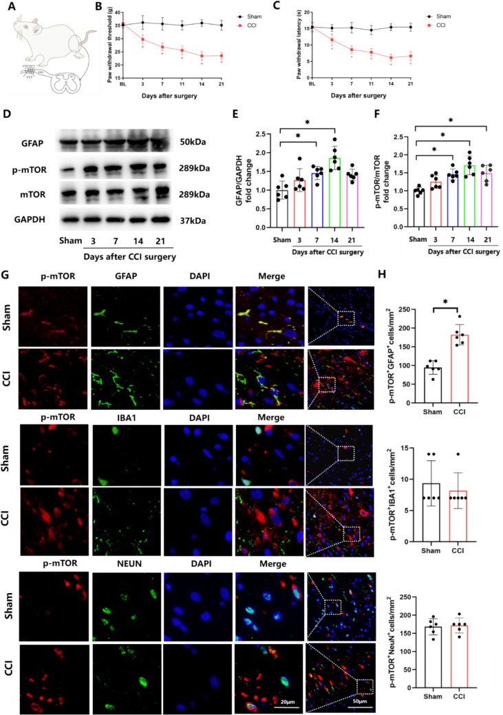
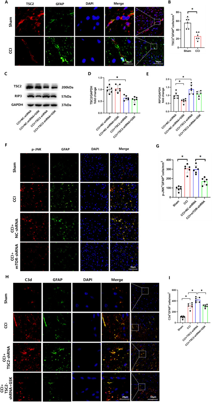
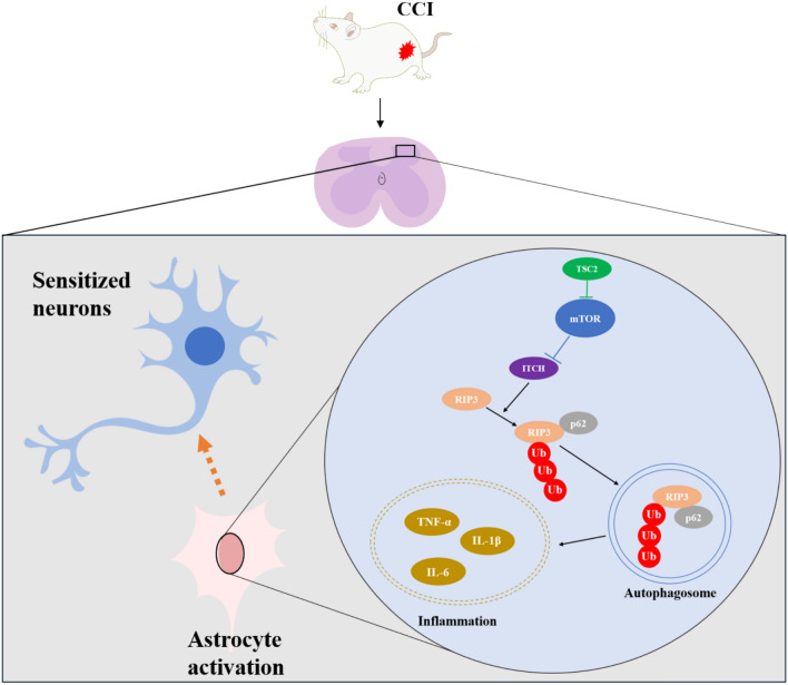
Neuropathic pain (NP), a chronic pain condition, is the result of abnormalities in both central and peripheral pain conduction pathways. Here, we investigated the underlying mechanisms associated with this effect. We found that following chronic constriction injury (CCI) surgery, there was an increase of mTOR in astrocytes and an activation of astrocytes within the spinal cord. Pharmacological inhibition of mTOR reversed CCI-induced hyperalgesia and neuroinflammation. Moreover, knockdown of astrocytic mTOR rescued the downregulation of spinal glutamate metabolism-related protein expression, underscoring the pivotal role of mTOR in modulating this pathway. Intriguingly, we observed that overexpression of mTOR, achieved via intrathecal administration of TSC2-shRNA, led to an upregulation of RIP3. Notably, pharmacological inhibition of RIP3, while ineffective in modulating mTOR activation, effectively eliminated the mTOR-induced astrocyte activation. Mechanistically, we found that mTOR controlled the expression of RIP3 in astrocytes through ITCH-mediated ubiquitination and an autophagy-dependent degradation. Taken together, our results reveal an unanticipated link between mTOR and RIP3 in promoting astrocyte activation, providing new avenues of investigation directed toward the management and treatment of NP.

Keywords: Astrocyte; MTOR; Neuroinflammation; Neuropathic pain; RIP3.


Conflict of interest statement

Declarations. Competing Interests: The authors declare no competing interests. Ethics Approval: All study protocols were reviewed and approved by Laboratory Animal Ethical and Welfare Committee of Shandong University Cheeloo College of Medicine (Approval No. 23056). Consent to Participate: Not applicable. Consent for Publication: Consent to publish was obtained from the participants.

Figures

Fig. 1
Fig. 1
p-mTOR is upregulated and astrocytes are activated in a CCI-induced rat model of NP. A Protocol for generating the CCI-induced rat model of NP. B, C Mechanical allodynia and thermal hyperalgesia in the ipsilateral hind paw were determined by calculating the paw withdrawal threshold (PWT) and latency (PWL) on days 0, 3, 7, 11, 14 and 21 post-CCI. DF Western blotting and quantification for the ratios of p-mTOR to mTOR and GFAP to GAPDH in sham and CCI rats. F(E) = 14.67, F(F) = 15.26. G, H Double-immunofluorescence of p- mTOR (red staining) and GFAP, IBA1 and NEUN (green staining) in the spinal cord of sham and CCI rats. Histogram illustrated that p- mTOR co-localization with GFAP was increased in CCI rats. (*p < 0.05, n = 6 in each group)
Fig. 2
Fig. 2
Inhibition of MTOR prevents CCI-induced allodynia and neuroinflammation. A An illustration of the experimental scheme. B, C Mechanical allodynia and thermal hyperalgesia in the ipsilateral hind paw was determined using the PWT and PWL on days 0, 3, 7 and 14 post-CCI in rats treated with rapamycin. D, E Western blotting and quantification for the ratios of p-mTOR to mTOR in sham and CCI rats treated with the mTOR inhibitor, rapamycin. F = 15.95. F, G mRNA levels of IL-6 (F) and IL-1β (G) in CCI rats treated with rapamycin. F(F) = 30.98, F(G) = 24.72. H, I TNF-α was assessed using immunohistochemistry in sham and CCI rats treated with rapamycin. F = 19.22. (*p < 0.05, n = 6 in each group)
Fig. 3
Fig. 3
Astrocytic reduction of mTOR increases glutamate clearance and attenuates spinal dorsal horn neuron activity induced by CCI. A An illustration of the experimental scheme. B, C Effect of a reduction in the expression of astrocytic mTOR on mechanical allodynia and thermal hyperalgesia as evaluated using PWT and PWL. D, E Western blotting and quantification of the mTOR to GAPDH ratio in the spinal dorsal horn of rats with a reduction in the expression of astrocytic mTOR. F = 11.57. F mRNA levels of the mTOR in the spinal dorsal horn of rats with a reduction in the expression of astrocytic mTOR. F = 6.698. G, H The double-immunofluorescence of mTOR (green staining) with GFAP (red staining) in the spinal dorsal horn. Histogram illustrated that mTOR co-localization with GFAP was decreased in rats treated with mTOR-shRNA. I The colocalization immunofluorescence of GFP (green staining) with GFAP (red staining), IBA1 (red staining), and NEUN (red staining). J, K Spinal glutamine synthetase (GS) expressions were determined and quantified using Western blotting. F = 11.68. L, M Double-immunofluorescence of c-fos (red staining) and NEUN (green staining) in each group. c-fos–positive neurons were quantified and normalized to that of the vehicle group. F = 121.3. (*p < 0.05, n = 6 in each group)
Fig. 4
Fig. 4
Pharmacological inhibition of RIP3 blocks the mTOR overexpression-induced astrocyte activation. A mRNA levels of TSC2 in astrocytes before and after transfection with TSC2-shRNA. F = 32.76. B, C Protein expression of TSC2 in astrocytes before and after transfection with TSC2-shRNA. F = 21.21. D GFP fluorescence in astrocytes transfected with TSC2-shRNA. EG Western blotting and quantification of ratios of TSC2 and RIP3 to GAPDH in astrocytes treated with TSC2-shRNA or add GSK872 (5 μM) for 24 h. F(F) = 13.89, F(G) = 15.51. HJ Immunofluorescent staining of p-mTOR (J) and RIP3 (I) in astrocytes treated with TSC2-shRNA or add GSK872 (5 μM) for 24 h. F(I) = 11.68, F(J) = 12.97. K, L mRNA levels of S100A10 (K) and C3d (L) in astrocytes treated with TSC2-shRNA or add GSK872 (5 μM) for 24 h. F(K) = 17.61, F(L) = 12.15. (*p < 0.05, n = 3 in each group)
Fig. 5
Fig. 5
RIP3 is required for mTOR-induced neuroinflammation and astrocyte activation. A, B Double-immunofluorescence of TSC2 (red staining) and GFAP (green staining) in the spinal dorsal horn of sham and CCI rats. CE Western blotting and quantification of the ratios of TSC2 and RIP3 to GAPDH in CCI rats treated with TSC2-shRNA or add GSK872. F(D) = 20.34, F(E) = 12.47. F, G Double-immunofluorescence showed the expression of p-JNK (red staining) and GFAP (green staining) in sham and CCI rats treated with NC-shRNA or mTOR-shRNA. F = 43.03. H, I Double-immunofluorescence images showing colocalization of GFAP-labeled astrocytes (green staining) and C3d-labeled A1 astrocytes (red staining) in CCI rats treated with TSC2-shRNA alone or with GSK872. F = 71.40. (*p < 0.05, n = 6 in each group)
Fig. 6
Fig. 6
MTOR modulates the ubiquitinated degradation of RIP3 via ITCH. A mRNA levels of RIP3 in astrocytes before and after transfection with TSC2-shRNA. F = 1.501. B Cell lysates from astrocytes transfected with NC-shRNA or TSC2-shRNA were immunoprecipitated with normal IgG or anti-RIP3 antibodies, then immunoblotted with their respective antibodies. C The top 20 E3 ligases of RIP3. D Prepare cell lysates and immunoprecipitate with normal IgG or anti-RIP3 antibodies. Co-IP showed the interaction of ITCH with RIP3. E Endogenous RIP3 (red staining) colocalized with ITCH (green staining) in WT astrocytes. F Lysates of cells treated with NC-siRNA or ITCH-siRNA were subjected to immunoprecipitation with normal IgG or anti-RIP3 antibodies. GI Western blotting demonstrated the ITCH dependency of mTOR mediated RIP3 ubiquitination. (*p < 0.05, n = 3 in each group)
Fig. 7
Fig. 7
The mTOR/ITCH axis modulates the ubiquitination and degradation of RIP3 via the autophagy pathway. AC RIP3 and p62 protein levels were determined using western blotting of cell lysates from NC-shRNA and TSC2-shRNA astrocytes treated with amino acid–free Earle’s balanced salt solution (EBSS) medium at the various times indicated. D Prepare cell lysates and immunoprecipitate with normal IgG or anti-RIP3 antibodies. E Endogenous p62 (red staining) colocalized with ITCH (green staining) in WT astrocytes. F HEK 293 T cells overexpressing FLAG-RIP3 alone or with HA-ITCH were either treated or not with 10 μM chloroquine (CQ, 18 h) or 10 μM (MG132, 6 h), respectively, with cell lysates then probed with anti-RIP3. (*p < 0.05, n = 3 in each group)
Fig. 8
Fig. 8
A functional model demonstrating how neuropathic pain is induced by astrocytic mTOR. Note: mTOR is overactived following the loss of TSC2’s negative regulation of it after chronic constriction injury, which results in the inhibition of ITCH activity and autophagy biogenesis. Due to the lack of degradation, RIP3 can be highly accumulated in the astrocytes, leading to the release of inflammatory factors. Finally, neuroinflammation induced the increased neuronal activity and enhancement of neuropathic pain

Similar articles

References

    1. Finnerup NB, Kuner R, Jensen TS (2021) Neuropathic pain: from mechanisms to treatment. Physiol Rev 101(1):259–301. 10.1152/physrev.00045.2019 - PubMed
    1. Bannister K et al (2020) Neuropathic pain: mechanism-based therapeutics. Annu Rev Pharmacol Toxicol 60:257–274. 10.1146/annurev-pharmtox-010818-021524 - PubMed
    1. Baron R, Binder A, Wasner G (2010) Neuropathic pain: diagnosis, pathophysiological mechanisms, and treatment. Lancet Neurol 9(8):807–819. 10.1016/S1474-4422(10)70143-5 - PubMed
    1. Woolf CJ (2011) Central sensitization: implications for the diagnosis and treatment of pain. Pain 152(3 Suppl):S2–S15. 10.1016/j.pain.2010.09.030 - PMC - PubMed
    1. Wen Q, Wang Y, Pan Q, Tian R, Zhang D, Qin G, Zhou J, Chen L (2021) MicroRNA-155–5p promotes neuroinflammation and central sensitization via inhibiting SIRT1 in a nitroglycerin-induced chronic migraine mouse model. J Neuroinflamm 18(1):287. 10.1186/s12974-021-02342-5 - PMC - PubMed

MeSH terms

Substances

LinkOut - more resources