. 2025 Apr;43(4):756-769.
doi: 10.1002/jor.26049. Epub 2025 Feb 2.

Carpal Kinematics in the Normal, Scapholunate Ligament Deficient, and Surgically Reconstructed Wrist

Affiliations

Carpal Kinematics in the Normal, Scapholunate Ligament Deficient, and Surgically Reconstructed Wrist

Xin Zhang et al. J Orthop Res. 2025 Apr.

Abstract

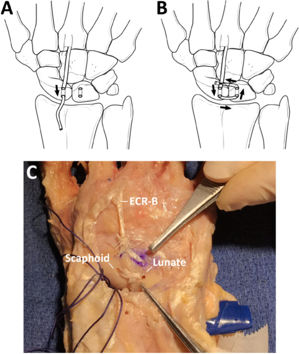
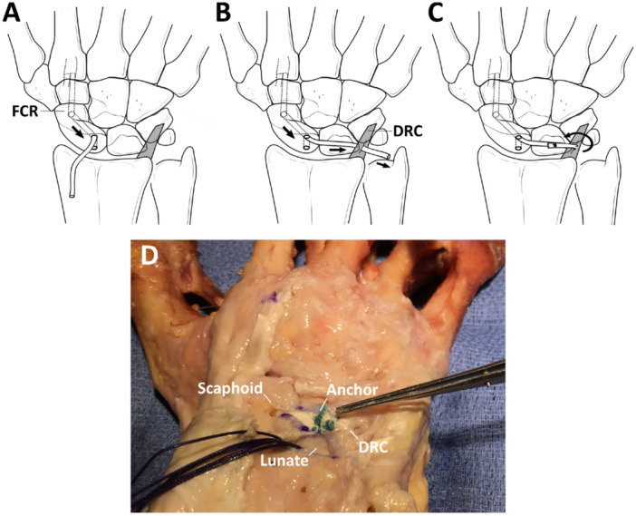
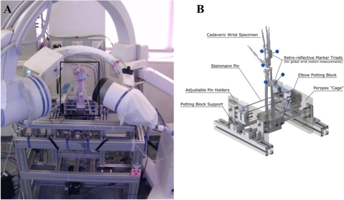
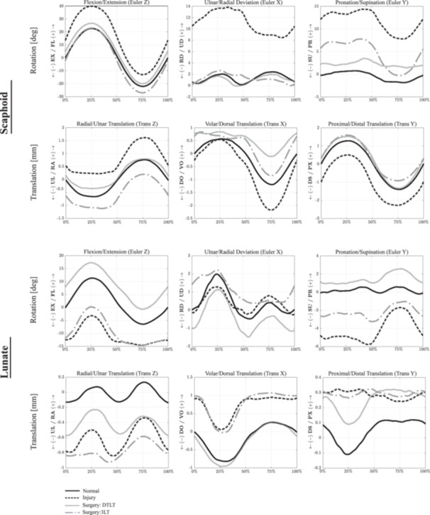
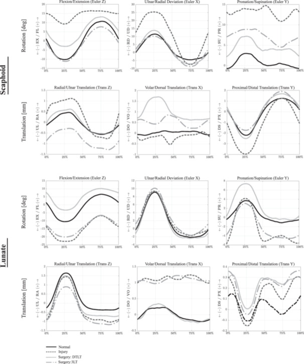
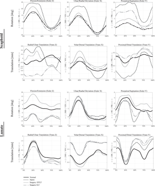
The objective of this study was to evaluate scaphoid, lunate and capitate kinematics after disruption to the primary and secondary scapholunate ligamentous stabilizers, and to assess the effectiveness of scapholunate ligament reconstruction in restoring carpal kinematics post-operatively. Seven upper extremities were harvested, and the scapholunate interosseous ligament (SLIL) was divided. Specimens were mounted onto a computer-controlled dynamic wrist simulator, and simulations of flexion-extension, radial-ulnar deviation, and dart-thrower's motion were undertaken by simulated force application to the wrist tendons. Three-dimensional kinematics of the scaphoid, lunate and capitate were measured using bi-plane X-ray fluoroscopy in the native and ligament deficient state. The SLIL was then reconstructed by either dorsal transarticular loop tenodesis (DTLT), or by the three-ligament tenodesis (3LT) technique, and re-evaluated. SLIL deficiency resulted in significant differences in carpal kinematics compared to that in the healthy wrist across all wrist motions (p < 0.05). The DTLT procedure corrected increased scaphoid ulnar deviation and pronation in the SLIL deficient wrist, but did not significantly improve scaphoid flexion or volar translation of the scaphoid. The 3LT reconstructive technique restored scaphoid flexion and ulnar deviation but did not correct pronation, the increased lunate extension, nor the volar and ulnar translation observed in the ligament deficient wrist. Three-dimensional scaphoid, lunate and capitate motion depends on SLIL integrity, with tears to this ligament resulting in pathological kinematics, which may be partially mitigated with DTLT and 3LT surgical reconstruction. These findings suggest that this surgical reconstruction of the SLIL may not mitigate long-term degenerative joint conditions at the wrist.

Keywords: carpal; kinematic model; scapholunate instability; scapholunate interosseous ligament; stereophotogrammetric analysis.


Conflict of interest statement

The authors declare no conflicts of interest.

Figures

Figure 1
Figure 1
Diagram illustrating the DTLT procedure for reconstruction of the scapholunate ligament deficient wrist, performed on the neutrally positioned wrist. Reconstruction was achieved by realigning and reducing the scapholunate joint, then stabilizing it with two Kirschner wires (K‐wire) placed across the scapholunate interval and one across the scaphocapitate (SC) joint. Two parallel connected burr holes, each measuring 2 mm in width (A), were created on the dorsum and adjacent surfaces of both the scaphoid and lunate in the region of the d‐SLIL insertion. The previously prepared ECR‐B graft was then passed dorsally, initially through the scaphoid and then the lunate in a box‐shaped fashion. The tendon graft was finally sutured back on itself, completing a transarticular passage and resulting in two parallel tendon grafts across the dorsal scapholunate interval, after which the K‐wires were removed (B). A photograph of the technique performed on a representative cadaveric specimen is also given (C).
Figure 2
Figure 2
Diagram illustrating the 3LT procedure for reconstruction of the scapholunate deficient wrist. A 3 mm wide, obliquely directed tunnel through the scaphoid was made under fluoroscopic guidance (A). This hole entered the scaphoid proximally and dorsally from one of the drill holes created for the DTLT technique and exited volarly at the scaphoid tubercle. The previously harvested FCR tendon strip was then passed through the tunnel. Sutures were used to assist the passage of the tendon strip. The tendon graft was then guided through a slit made on the distal and ulnar portion of the remaining DRC ligament, allowing it to be folded back onto the lunate (B). By using the DRC as a pivot, the tendon graft was then tightened until satisfactory reduction of the scapholunate joint was achieved, after which two scapholunate and one scaphocapitate wires K‐wires were inserted to maintain the reduction. The FCR graft was secured to the dorsal lunate using a radiolucent suture anchor (JuggerKnot Mini 1.0 mm; Zimmer Biomet, Indiana, USA), and the K‐wires were then removed (C). K‐Y jelly was applied to promote gliding between the ligament graft and the dorsal rim of the distal radius, mimicking the moist, low‐friction ligament gliding that occurs in vivo. A photograph of the technique performed on a representative cadaveric specimen is also given (D).
Figure 3
Figure 3
Dynamic cadaveric wrist testing apparatus (A), and specimen mounting platform (B), used to simulate dynamic motion of the wrist. Cadaveric wrist specimens had retro‐reflective marker triads inserted into the radius and third metacarpal so that global wrist motion could be measured using a video motion analysis system during testing. Wrist muscle‐tendon forces were then controlled in real‐time to reproduce the desired dynamic wrist motion trajectories. Specimens were rigidly fixed to an elbow potting block, which was mounted on a support and surrounded by a rigid Perspex cage for additional support to the mounting fixtures. Steinman pins passing through the radius and ulna to adjustable pin holders were employed to provide additional support to each specimen. Reprinted with permission from Elsevier.
Figure 4
Figure 4
Mean scaphoid and lunate kinematics during wrist flexion and extension in the normal, ligament deficient and surgically reconstructed wrist following the DTLT and 3LT procedures. Given are the three Euler angle rotations (degrees), including flexion–extension (Z), ulnar‐radial deviation (X), and pronation‐supination (Y), as well as bone translations (mm). Data are given as a percentage of the sinusoidal wrist flexion–extension motion cycle. Symbol definitions are as follows: EX, extension; FL, flexion; PR, pronation; RD, radial deviation; SU, supination; UD, ulnar deviation.
Figure 5
Figure 5
Mean scaphoid and lunate kinematics during wrist radial‐ulnar deviation in the normal, ligament deficient and surgically reconstructed wrist following the DTLT and 3LT procedures. Given are the three Euler angle rotations (degrees), including flexion–extension (Z), ulnar‐radial deviation (X) and pronation‐supination (Y), as well as bone translations (mm). Data are given as a percentage of the sinusoidal wrist ulnar‐radial deviation motion cycle. See Figure 4 Caption for more information.
Figure 6
Figure 6
Mean scaphoid and lunate kinematics during wrist dart‐thrower's motion in the normal, ligament deficient and surgically reconstructed wrist following the DTLT and 3LT procedures. Given are the three Euler angle rotations (degrees), including flexion–extension (Z), ulnar‐radial deviation (X) and pronation‐supination (Y), as well as bone translations (mm). Data are given as a percentage of the sinusoidal wrist dart‐thrower's motion cycle. See Figure 4 Caption for more information.

Similar articles

Cited by

References

    1. Kitay A. and Wolfe S. W., “Scapholunate Instability: Current Concepts in Diagnosis and Management,” Journal of Hand Surgery 37, no. 10 (2012): 2175–2196. - PubMed
    1. Jones W. A., “Beware the Sprained Wrist. The Incidence and Diagnosis of Scapholunate Instability,” Journal of Bone and Joint Surgery British Volume 7–B, no. 2 (1988): 293–297. - PubMed
    1. Pappou I. P., Basel J., and Deal D. N., “Scapholunate Ligament Injuries: A Review of Current Concepts,” Hand 8, no. 2 (2013): 146–156. - PMC - PubMed
    1. Watson H. K. and Ballet F. L., “The SLAC Wrist: Scapholunate Advanced Collapse Pattern of Degenerative Arthritis,” Journal of Hand Surgery 9, no. 3 (1984): 358–365. - PubMed
    1. Kleinman W. B., Steichen J. B., and Strickland J. W., “Management of Chronic Rotary Subluxation of the Scaphoid by Scapho‐Trapezio‐Trapezoid Arthrodesis,” Journal of Hand Surgery 7, no. 2 (1982): 125–136. - PubMed

MeSH terms

LinkOut - more resources