. 2025 Feb 25;44(2):115236.
doi: 10.1016/j.celrep.2025.115236. Epub 2025 Feb 1.

A conserved class of viral RNA structures regulates translation reinitiation through dynamic ribosome interactions

Affiliations

A conserved class of viral RNA structures regulates translation reinitiation through dynamic ribosome interactions

Madeline E Sherlock et al. Cell Rep. .

Abstract

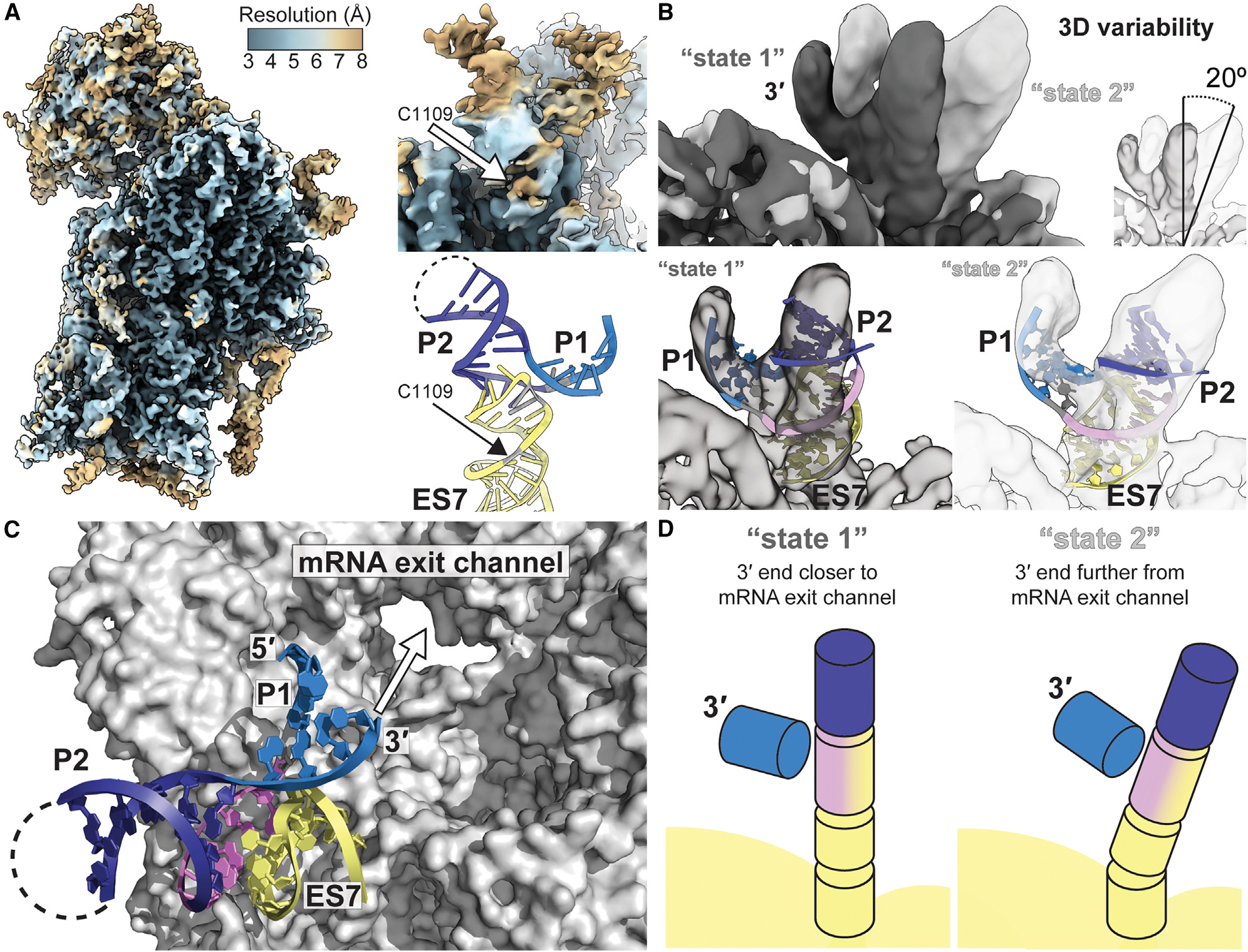
Certain viral RNAs encode proteins downstream of their main open reading frame, expressed through "termination-reinitiation" events. In some cases, structures located upstream of the first stop codon within these viral RNAs bind the ribosome, inhibiting ribosome recycling and inducing reinitiation. We used bioinformatics methods to identify new examples of viral reinitiation-stimulating RNAs and experimentally verified their secondary structure and function. We determined the structure of a representative viral RNA-ribosome complex using cryoelectron microscopy (cryo-EM). 3D classification and variability analyses reveal that the viral RNA structure can sample a range of conformations while remaining tethered to the ribosome, enabling the ribosome to find a reinitiation start site within a limited range of mRNA sequence. Evaluating the conserved features and constraints of this entire RNA class within the context of the cryo-EM reconstruction provides insight into mechanisms enabling reinitiation, a translation regulation strategy employed by many other viral and eukaryotic systems.

Keywords: CP: Molecular biology; RNA structure; cryo-EM; reinitiation; ribosome; viral RNA.


Conflict of interest statement

Declaration of interests The authors declare no competing interests.

Figures

Figure 1.
Figure 1.. Genomic context and phylogenetic distribution of a class of reinitiation-stimulating RNA elements in viruses.
(A) Genomic locations of reinitiation-stimulating elements in viruses in different families. (B) Secondary structure cartoons of the two major types of reinitiation-stimulating elements, located upstream (5′) of the downstream reinitiation start codon. Structures contain either two helical elements (P1 and P2) with the ribosome complementary sequence (RCS) embedded in an asymmetric internal loop (top) or a single helical element (P1) with the RCS located in the apical loop (bottom). (C) Phylogenetic distribution of previously reported and newly identified reinitiation-stimulating RNAs in viruses. (D) Alignment of the sequence, secondary structure (P1–P3), and downstream start codon of all RNAs belonging to the termination upstream ribosome binding site (TURBS) class of reinitiation-stimulating element RNAs. See Table S2.
Figure 2.
Figure 2.. Structure and function of reinitiation-stimulating RNA representatives from diverse viruses.
(A) Design of reinitiation reporter constructs, which encode Rluc in the upstream ORF and Fluc in the downstream ORF. Positive and negative controls and mutant design are described in the main text. The F2A sites (indicated with arrows) cause peptide bond skipping, liberating the Rluc and Fluc proteins from peptides resulting from the inserted viral coding sequences. (B) Translation assays performed in rabbit reticulocyte lysate containing dual luciferase reporters carrying no viral RNA (controls) or putative WT or mutant reinitiation-stimulating sequences from rabbit hemorrhagic disease virus (RHDV), feline calicivirus (FCV), chicken calicivirus (ChCV), turkey calicivirus (TuCV), Newbury agent 1 virus (N1V), St-Valérien calicivirus (SVCV), influenza B virus (IBV), Flanders virus (FLAV), or Wongabel virus (WONV). The ratio of luminescence from the downstream (Fluc) to upstream (Rluc) proteins was normalized to the positive readthrough control. Error bars represent standard deviation. (C) Reactivity to the chemical probing reagent N-methylisatoic anhydride, assayed in vitro by selective 2′ hydroxyl acylation analyzed by primer extension (SHAPE) of putative reinitiation-stimulating RNAs mapped onto the predicted secondary structure model. The full sequence that was probed, including flanking hairpins added to normalize reactivity, can be found in Table S1.
Figure 3.
Figure 3.. Conserved features among all viral TURBS-like RSE RNAs.
(A) Consensus sequence and secondary structure models of all large (left) and small (right) versions of the TURBS class of reinitiation-stimulating elements derived from 43 unique viruses. The sequence and secondary structure of the 18S rRNA from G1104–C1126, a portion of helix 26 also known as ES7, is depicted, with the nucleotides complementary to the RCS within the viral RNA outlined in yellow. See also Table S2. (B) Histogram of the number of base pairs that could form between the viral RNA and rRNA, as determined by the length of consecutive complementary nucleotides, allowing for G•U wobble pairs. (C) Histogram of all possible reinitiation reading frames, comparing the position of the downstream start codon to the upstream stop codon. (D) Histogram of all possible lengths of the linker between the last nucleotide within the P1 stem and the first nucleotide in either the start (black) or stop (gray) codon among all examples of TURBS RNAs.
Figure 4.
Figure 4.. Cryo-EM reconstruction of an RSE-ribosome complex.
(A) Refined map of the full RHDV RNA-80S complex prior to masking. The feature corresponding to RHDV RNA is boxed in red. (B) Refined map of the masked 40S subunit of the RHDV RNA-ribosome complex. The feature corresponding to RHDV RNA is boxed in red. (C and D) Cryo-EM map of the RHDV RNA-ribosome complex, zoomed in on the region boxed in red in (B). An apo-40S structure (C; PDB: 6ZMW) and our model (D; PDB: 9C8K) were each fit into the map, revealing extra density corresponding to the RHDV RNA. (E) A model of the RHDV RNA (top) built into the density of a locally refined map (bottom), including intermolecular base pairs between the RCS of RHDV and the apical loop of ES7. (F) Model of the RHDV RNA with ribosomal ES7. Nucleotides shown in gray (RHDV U6969 and 18S rRNA U1119, A1120, and C1109) could not be unambiguously built into the density. (G) Secondary structure of the RHDV RNA bound to the apical portion of ES7, as determined by the model in (E). Colors match (F).
Figure 5.
Figure 5.. Conformational heterogeneity reveals dynamics related to reinitiation start site selection.
(A) Local resolution of the masked, refined 40S-RHDV RNA map. The position of 18S rRNA C1109 is depicted within the local resolution map and the model with the same view. (B) Maps representing the two extreme states of 3D variability among the particles, which differ in their orientation by 20°. The model of RHDV RNA bound to the apical portion of ES7 rRNA (G1110–C1123) was fit into the two extreme states. (C) Surface view of the 40S with the model of RHDV RNA bound to ES7, with the view looking down into the mRNA exit channel from the decoding groove. (D) Simplified model of the orientation of helices in the two extreme states of 3D variability.
Figure 6.
Figure 6.. Model of the structural bases for reinitiation start site selection within a limited window.
During termination, the stop codon is in the A site. We propose that conformational flexibility and dynamics allow the RHDV RNA to remain bound to ES7 of the 40S while sampling different states. This motion moves the 3′ end of the structure, which could propagate to the downstream mRNA in the decoding groove and allow sampling of nearby RNA, including the nearby reinitiation start codon in the P site. Other factors, including eIF3, could select for certain conformations of RHDV-ES7 and influence start site selection. Met-tRNAi is used in this type of reinitiation, which can be delivered by eIF2, eIF2D, or DENR/MCT1 in vitro, but the mechanisms of delivery during infection have not yet been defined.

Update of

Similar articles

Cited by

References

    1. Aitken CE, and Lorsch JR (2012). A mechanistic overview of translation initiation in eukaryotes. Nat. Struct. Mol. Biol. 19, 568–576. - PubMed
    1. Hinnebusch AG, and Lorsch JR (2012). The mechanism of eukaryotic translation initiation: new insights and challenges. Cold Spring Harb. Perspect. Biol. 4, a011544. - PMC - PubMed
    1. Dever TE, and Green R (2012). The elongation, termination, and recycling phases of translation in eukaryotes. Cold Spring Harb. Perspect. Biol. 4, a013706. 10.1101/cshperspect.a013706. - DOI - PMC - PubMed
    1. Hellen CUT (2018). Translation termination and ribosome recycling in eukaryotes. Cold Spring Harb. Perspect. Biol. 10, a032656. - PMC - PubMed
    1. Young DJ, and Guydosh NR (2022). Rebirth of the translational machinery: The importance of recycling ribosomes. Bioessays 44, 2100269. - PMC - PubMed

Publication types

MeSH terms

Associated data